| Research Article |
Open Access |
|
| Musthaq Ahmed1 and Kaiser Jamil1,2* |
| 1School of Biotechnology, Mahatma Gandhi National Institute for Research and Social Action (MGNIRSA), Street No: 17, Gagan Mahal Road, Hyderabad-500029, Andhra Pradesh, India |
| 2Head of Genetics Department, Bhagwan Mahavir Medical Research Centre, (BMMRC), 10-1-1, Mahavir Marg, Hyderabad-500004, Andhra Pradesh, India |
| *Corresponding author: |
Dr. Kaiser Jamil
Professor and Honorary Director
School of Biotechnology and Bioinformatics
Mahatma Gandhi National Institute for Research and Social Action (MGNIRSA), Gaganmahal Road
Domulguda, Hyderabad, 500029
Andhra Pradesh, India
Tel: +919676872626
Fax: +91-40-66631500
E-mail: kj.bmmrc@gmail.com |
|
| Â |
| Received September 11, 2012; Published October 19, 2012 |
| Â |
| Citation: Ahmed M, Jamil K (2012) BCL-2 as Target for Molecular Docking of Some Neoplastic Drugs. 1:458. doi:10.4172/scientificreports.458 |
| Â |
| Copyright: © 2012 Ahmed M, et al. This is an open-access article distributed under the terms of the Creative Commons Attribution License, which permits unrestricted use, distribution, and reproduction in any medium, provided the original author and source are credited. |
| Â |
| Abstract |
| Â |
| B-Cell Lymphoma (Bcl) is an apoptosis regulator protein which plays an important role in many types of cancers. The Bcl-2 gene has been identified as over expressed in different cancers. In this study we identified the binding affinity of commonly used neoplastic drugs such as Gefitinib, Cisplatin, 5-FU, Gemcitabine and Vinorelbine on Bcl-2 using insilico techniques. Bcl-2 structure (PDB: 1G5M) was used as a target for evaluating the binding efficacy of inhibitors (drugs) using GOLD software. The inhibitor binding positions and affinities were determined using GOLD scoring fitness functions. We identified that amino acid residues ASP10, GLU13, LYS17, GLU42, and SER49 in Bcl-2 were important for inhibitor recognition via hydrogen bonding interactions. These hydrogen bonding interactions play an important role for stability of the target-ligand complex. This technique also determined the comparative efficacy of neoplastic drugs very elegantly using Bcl-2 as its target. The insilico docking techniques can be exploited to build targets and design inhibitors for novel therapeutic agents. Molecular docking helps in understanding the action of neoplastic drugs through affinity binding. |
| Â |
| Keywords |
| Â |
| Cancer; Bcl-2; Gefitinib; Cisplatin; 5-FU; Gemcitabine; Vinorelbine |
| Â |
| Introduction |
| Â |
| Structural studies of Bcl-2 family members have provided many insights into the molecular mechanism of apoptosis and how Bcl-2 family members interact with one another. Bcl-2 and related proteins are key regulators of apoptosis or programmed cell death implicated in human disease including cancer [1]. Bcl-2 was originally identified at the chromosomal breakpoint of t(14;18)-bearing B-cell lymphomas. Although it is not fully understood how Bcl-2 family proteins regulate apoptotic pathways, one possible mechanism is that members of this family engage in various protein-protein interactions to form homoand heterotypic dimers important for their biological functions. Bcl-2 belongs to a growing family of proteins that regulate apoptosis or programmed cell death. The Bcl-2 family includes both death antagonists such as Bcl-2 and Bcl-xL and death agonists such as Bax, Bak, Bid, and Bad. These related proteins share at least one of four homologous regions termed Bcl homology (BH) domains (BH1 to BH4) [2-4]. The antitumor drug Cisplatin with clinical and experimental efficiency is employed as a first-line chemotherapeutic modality in the treatment of epithelial malignancies, including lung, ovarian, testicular, cervix cancer and other cancers [5,6]. Gemcitabine hydrochloride is a deoxycytidine derivative that inhibits DNA elongation through intracellular phosphorylation of ribonucleotide reductase. Studies have proven that Gemcitabine is an anti neoplastic agent that inhibits DNA synthesis, resulting in apoptosis. In addition to its established uses in pancreatic and non-small-cell lung cancer, the drug has been shown in clinical trials to be active against a wide variety of solid tumors [7,8]. Vinorelbine (VNR), a semisynthetic vinca alkaloids derived from vinblastine, is a mitosis-phase specific antineoplastic agent. VNR binds to tubulin, thereby inhibiting tubulin polymerization into microtubules and spindle formation, resulting in apoptosis of susceptible cancer cells. Inhibition of mitotic microtubules correlates with antitumor activity [9]. 5-FU is a cell cycle–specific, S-phase–dependent fluorinated pyrimidine analogue with moderate activity in breast cancer, colon cancer, ovarian cancer, and oral squamous cell carcinoma [10,11]. Gefitinib (Iressa) is a quinazoline derivative that inhibits EGFR tyrosine kinase activity by binding to the adenosine triphosphate pocket within the EGFR catalytic domain [12-14]. In the present study we have attempted to dock the above mentioned neoplastic drugs with Bcl-2 to understand the interactions. |
| Â |
| Methodology |
| Â |
| Obtaining the crystal structure of the target-Bcl-2 |
| Â |
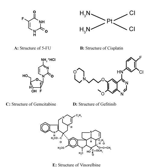
| To prepare the Bcl-2 structure, the crystal structure was taken from the Protein Data Bank (PDB_ID: 1G5M) (Figure 1). Hetero atoms were removed from the binding site and the chain A was selected for docking studies. Hydrogen atoms were added to the enzyme. The molecular docking method was performed using the Gold version 3.0.1 program to study the binding orientation of compounds into the Bcl-2 structure. The docking experiments were performed using the binding site of Bcl-2. |
| Â |
|
|
Figure 1: Sowing the molecular structure of the neoplastic drugs. |
|
| Â |
| Active site identification |
| Â |
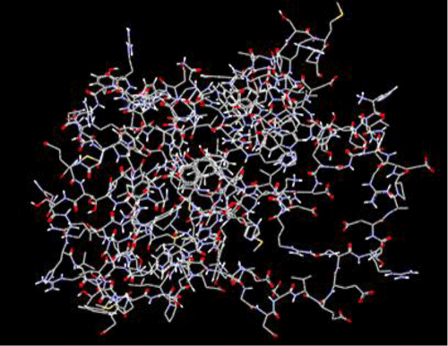
| The binding site identification was carried out using CastP server (Figure 2). The new program CAST, for automatically locating and measuring protein pockets and cavities, is based on precise computational geometry methods, including alpha shape and discrete flow theory. CAST identifies and measures pockets and pocket mouth openings, as well as cavities. The program specifies the atoms lining pockets, pocket openings, and buried cavities; the volume and area of pockets and cavities; and the area and circumference of mouth openings. |
| Â |
|
|
Figure 2: Structure of Bcl-2. |
|
| Â |
| Docking method |
| Â |
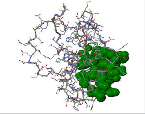
| Docking studies of Gefitinib, Cisplatin, 5-FU, Gemcitabine and Vinorelbine were performed. The structures of these compounds (Figure 3) were constructed and optimized using chemsketch software. Docking was carried out using GOLD (Genetic Optimization of Ligand Docking) software which is based on genetic algorithm (GA). This method allows as partial flexibility of protein and full flexibility of ligand [15]. The compounds are docked to the active site of the Bcl-2. The interaction of these compounds with the active site residues are thoroughly studied using molecular mechanics calculations. The parameters used for GA were population size (100), selection pressure (1.1), number of operations (10,000), number of island (1) and niche size (2). Operator parameters for crossover, mutation and migration were set to 100, 100 and 10 respectively. Default cutoff values of 3.0Ã… (dH-X) for hydrogen bonds and 6.0Ã… for vanderwaals were employed. During docking, the default algorithm speed was selected and the ligand binding site in the alpha glucosidase was defined within a 10Ã… radius with the centroid as CA atom of GLU42. The number of poses for each inhibitor was set 100, and early termination was allowed if the top three bound conformations of a ligand were within 1.5Ã… RMSD. After docking, the individual binding poses of each ligand were observed and their interactions with the protein were studied. The best and most energetically favorable conformation of each ligand was selected. |
| Â |
|
|
Figure 3: Active site of Bcl-2. |
|
| Â |
| Gold score fitness function |
| Â |
| Gold Score performs a force field based scoring function and is made up of four components: |
| Â |
| (i). Protein-ligand hydrogen bond energy (external H-bond); |
| Â |
| (ii). Protein-ligand vanderwaals energy (external vdw); |
| Â |
| (iii). Ligand internal vanderwaals energy (internal vdw); |
| Â |
| (iv). Ligand intramolecular hydrogen bond energy (internal- H- bond). |
| Â |
| The external vdw score is multiplied by a factor of 1.375 when the total fitness score is computed. This is an empirical correction to encourage protein-ligand hydrophobic contact. The fitness function has been optimized for the prediction of ligand binding positions. |
| Â |
| GoldScore = S (hb_ext) + S (vdw_ext) + S (hb_int) + S (vdw_int) |
| Â |
| Where S (hb_ext) is the protein-ligand hydrogen bond score, S (vdw_ext) is the protein-ligand van der Waals score, S (hb_int) is the score from intramolecular hydrogen bond in the ligand and S (vdw_int) is the score from intramolecular strain in the ligand. |
| Â |
| Results and Discussion |
| Â |
| The proteins of the Bcl-2 family are important regulators of apoptosis, or programmed cell death. These proteins regulate this fundamental biological process via the formation of hetero dimmers involving both pro- and anti-apoptotic family members [16]. After collecting the crystal structure, the possible binding sites of Bcl-2 were searched with CASTP server as shown in figure 2. The residues included in active site were TYR 18, TYR 21, LYS 22, GLN 25, ARG 26, ARG 98, GLY 101, ASP 102, PHE 104, SER 105, ARG 106, TYR 108, ARG 109, ASP 111, PHE 112, ALA 113, MET 115, SER 116, GLN 118, LEU 119, ARG 129, THR 132, VAL 133, GLU 136, LEU 137, ARG 146, VAL 148, ALA 149, GLU 152, PHE 153, GLY 155, VAL 156, MET 157, VAL 159, GLU 160. From the binding site analysis of Bcl-2 we identified that, the binding pockets are identical in all chains and the largest binding pocket was taken for further docking studies. The crystal structures of BCL-2 was similar hence we have taken 3G5M (chain A) as representative structure for docking studies. The docking of drugs into the active site of Bcl-2 was performed using the GOLD software and the docking evaluations were made on the basis of GoldScore fitness functions. We preferred Gold fitness score than Chemscore fitness as Gold fitness score is marginally better than Chemscore fitness function. |
| Â |
| Molecular docking study |
| Â |
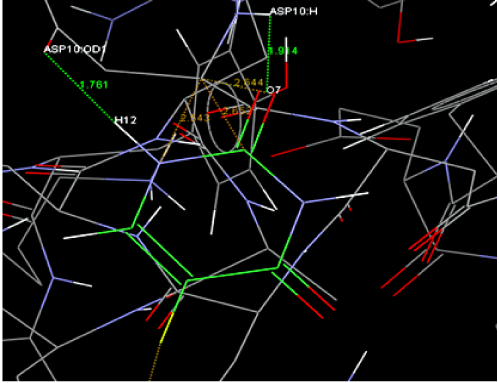
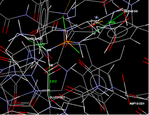
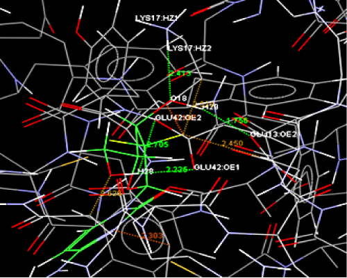
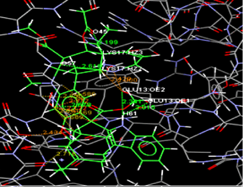
| Structure-based drug design begins with the identification of a molecular target such as a protein such as Bcl-2 in this study. This structure is then used as a blueprint for the drug design of a lead compound. The compounds are modelled for their fit in the active site of the target, considering both steric aspects (i.e., geometric shape) and functional group interactions, such as hydrogen bonding and hydrophobic interactions. The selected docked conformations of Gefitinib, Cisplatin, 5-FU, Gemcitabine and Vinorelbine into the Bcl-2 binding site are shown in figure 4A-4E. The docked conformations revealed that all ligands were located in the hydrophobic binding pocket. In this study, all docked drugs were found to have some interactions between an oxygen atom of the drugs and Bcl-2. Moreover, these docked conformations also formed an H-bonding interaction within the active site (Table 1). In the binding pocket, common H-bonding interactions were formed between all docked drugs and ASP10, GLU13, LYS17, GLU42, and SER49. The specific H-bonding interaction with SER49 was only found in the docked conformation of Cisplatin. In order to explain the binding of these compounds, the H-bonding interactions with the other surrounding residues in the hydrophobic binding pocket were also investigated. In figure 4A, two strong H-bonding interactions between the hydroxyl group (H12) of 5-FU and an oxygen atom of ASP10 and another hydrogen bond between (O7) and Hydroxyl group of ASP10. The docked conformations of other drugs are shown in figure 4B-4E. In the case of docked Cisplatin, three H-bonds with GLU13, LYS17 and SER49 were formed. One Hydrogen bond was observed between hydroxyl group of Bcl-2 (LYS17) and Oxygen (O9) of Gefitinib was observed in figure 4C. A total of four hydrogen bonds were observed in the docking studies of Gemcitabine with Bcl-2 in residues GLU13, LYS17 and GLU42 were involved. From the docking of Vinorelbine into the active site of Bcl-2, we observed total four hydrogen bonds, in which two bonds were formed between the Hydroxyl group (H61) of Vinorelbine and GLU13 of Bcl-2. The other two hydrogen bonds were seen between LYS17 of Bcl-2 and Oxygen atom of Vinorelbine. The atoms involved in bonding with Bcl-2 and their bond lengths along with docking energies were indicated in table 1. The docking results agreed well with the observed in vitro data, which showed that the Bcl-2 inhibitory activity of Gemcitabine (39.01 K.Cal/mol) was higher than those of other drugs. Our investigations shows that 5-FU and Cisplatin has good inhibitory activity on Bcl-2 and this can be helpful for further investigations. The docking results data supports the inhibitory activity of Vinorelbine. |
| |
| Â |
|
|
Figure 4A: Docking studies of 5-FU with Bcl-2. |
|
| Â |
|
|
Figure 4B: Docking of Cisplatin with Bcl-2. |
|
| Â |
|
|
Figure 4C: Docking of Gefitinib with Bcl-2. |
|
| Â |
|
|
Figure 4D: Docking of Gemcitabine with Bcl-2. |
|
| Â |
|
|
Figure 4E: Docking of Vinorelbine with Bcl-2. |
|
| Â |
|
|
Table 1: Showing H-bonding interaction of the neoplastic drugs within the active site; and their bond lengths along with docking energies. |
|
| Â |
| Conclusion |
| Â |
| In this simple and elegant studies we have shown the comparative efficacies of 5 neoplastic drugs which are targetted against Bcl-2 to reduce its expression in vivo and to induce the tumor cell to take the apoptotic pathway – which is inhibited by the over expression of Bcl-2. The docking results agreed well with the observed in vitro data, in which the anti-Bcl activity of the Gemcitabine was higher than other drugs and formed four hydrogen bonds. The docking study revealed the binding orientation of compounds in the Bcl-2 binding pocket surrounding the active site, which resulted in inhibition of enzyme activity. From these results we can conclude that Cisplatin is one of the good inhibitory compounds of Bcl-2. The application of computational sciences to pharmaceutical research is a discipline, which is phenomenal. |
| Â |
| Acknowledgement |
| Â |
| We are grateful to MGNIRSA for the facilities provided. |
| Â |
| |
| References |
| Â |
- Petros AM, Olejniczak ET, Fesik SW (2004) Structural biology of the Bcl-2 family of proteins. Biochim Biophys Acta 1644: 83-94.
- Adams JM, Cory S (1998) The Bcl-2 protein family: arbiters of cell survival. Science 281: 1322-1326.
- Chao D, Korsmeyer SJ (1998) BCL-2 family: regulators of cell death. Annu Rev Immunol16: 395-419.
- Van Delft MF, Huang DC (2006) How the Bcl-2 family of proteins interact to regulate apoptosis. Cell Res16: 203-213.
- Gonzalez VM, Fuertes MA, Alonso C, Perez JM (2001) Is cisplatin-induced cell death always produced by apoptosis? Mol Pharmacol 59: 657-663.
- Jin KL, Park JY, Noh EJ, Hoe KL, Lee JH, et al. (2010) The effect of combined treatment with cisplatin and histone deacetylase inhibitors on HeLa cells.J Gynecol Oncol 21: 262-268.
- Okusaka T, Ishii H, Funakoshi A, Yamao K, Ohkawa S, et al. (2006) Phase II study of single-agent gemcitabine in patients with advanced biliary tract cancer. Cancer Chemother Pharmacol 57: 647-653.
- Okusaka T, Nakachi K, Fukutomi A, Mizuno N, Ohkawa S, et al. (2010) Gemcitabine alone or in combination with cisplatin in patients with biliary tract cancer: a comparative multicentre study in Japan. Br J Cancer 103: 469- 474.
- Hu J, Cheung NK (2009) Methionine depletion with recombinant methioninase: in vitro and in vivo efficacy against neuroblastoma and its synergism with chemotherapeutic drugs. Int J Cancer 124: 1700-1706.
- Yim EK, Lee SB, Lee KH, Kim CJ, Park JS (2006) Analysis of the in vitro synergistic effect of 5-fluorouracil and cisplatin on cervical carcinoma cells. Int J Gynecol Cancer 16: 1321-1329.
- Ch KK, Jamil K, Raju GS (2011) Predicting drug-target interaction in cancers using homology modeled structures of MTHFR gene.Biol Med 3: 70-81.
- Wakeling AE, Guy SP, Woodburn JR, Ashton SE, Curry BJ, et al. (2002) ZD1839 (Iressa): an orally active inhibitor of epidermal growth factor signaling with potential for cancer therapy. Cancer Res 62: 5749-5754.
- Moasser MM, Basso A, Averbuch SD, Rosen N (2001) The tyrosine kinase inhibitor ZD1839 ("Iressa") inhibits HER2-driven signaling and suppresses the growth of HER2-overexpressing tumor cells. Cancer Res 61: 7184-7188.
- Kotra S, Madala KK, Jamil K (2008) Homology models of the mutated EGFR and their response towards quinazoline analogues. J Mol Graph Model 27: 244-254.
- Jamil K, Mustafa SM (2012) Thioredoxin System: A model for determining novel lead molecules for breast cancer chemotherapy. AJMB 4: 3.
- Moroy G, Martin E, Dejaegere A, Stote RH (2009) Molecular basis for Bcl-2 homology 3 domain recognition in the Bcl-2 protein family: identification of conserved hot spot interactions. Journal Biol Chem 284: 17499-17511.
|
| Â |
| Â |