| Research Article |
Open Access |
|
| Murad Jefferson Claudio1*, Ribeiro Jr Ulysses1, Kleber2, Alves Venancio AF2, Pugliese Vincenzo2, Massad E1, Saad William Abraao2, Cecconello I1, Habr-Gama Angelita1 and Gama-Rodrigues Joaquim1 |
| 1Surgery Clinics II, Hospital of Medicine College, University Sao Paulo, SP, Brazil |
| 2Department of Pathology of Medicine College, University São Paulo, SP, Brazil |
| *Corresponding authors: |
Murad Jefferson Claudio
Clinics II - Department of Gastroenterology
University of São Paulo School of Medicine, SP, Brazil
E-mail: muradjc@hotmail.com |
|
| Â |
| Received August 06, 2012; Published October 29, 2012 |
| Â |
| Citation: Murad JC, Ribeiro Jr U, Kleber, Alves VAF, Pugliese V, et al. (2012) Prognostic Predictive Factors in Colorectal Adenocarcinoma with Liver Metastasis. 1:459. doi:10.4172/scientificreports.459 |
| Â |
| Copyright: © 2012 Claudioc MJ, et al. This is an open-access article distributed under the terms of the Creative Commons Attribution License, which permits unrestricted use, distribution, and reproduction in any medium, provided the original author and source are credited. |
| Â |
| Abstract |
| Â |
| Fifty four patients from the Liver Service of the Clinics Hospital of the University of Sao Paulo School Of Medicine, Sao Paulo, Brazil, with hepatic metastases of colorectal adenocarcinoma, without evidence of concomitant metastases in other organs were studied. They were submitted to surgical treatment in the period from 1992 to 2006, and clinical data included: age, sex, size of lesion of colon, stage, histological aspects, molecular markers from lesion of colon and respective liver metastases or larger lesion, compromised regional nodules, margin of free resection of neoplasm and correlation to free interval of disease and prognostic. The significant result was (p<0.05). The intensive desmoplasic reaction around colorectal neoplasia, and high expression of p53 and Ki-67 in colorectal cancer were associated with poor survival. In liver metastasis, the radical surgical resection of hepatic metastasis of colorectal adenocarcinoma with a safety margin higher than 10 mm promotes larger index of survival. Histological factors as the undifferentiated type, lesser peritumoral inflammatory infiltrated, larger desmoplasic reaction seem to be factor of worse prognostic. However the presence of capsule limiting the tumor was associated with worse survival. The high immune expression of p53, Ki-67 and p16 were associated with synchronic lesions and shorter disease-free interval. The lower expression of the TS was associated with better survival in patients who had received postoperative systemic chemotherapy with 5-FU and leucovorin. In conclusion: The significant predictive factors of worse prognostic in patients that have liver metastasis were, in colorectal lesion = desmoplasic reaction, high expression of p53 and Ki-67. In liver metastasis, the predictive factors of better prognosis were = margin of resection higher than 10 mm, well differentiated tumor type, lesser desmoplasic reaction and low expression of p53, Ki-67 and p16. |
| Â |
| Keywords |
| Â |
| Colorectal adenocarcinoma; Liver metastasis; Molecular markers; p53; p16; Thymidylate synthase; Ki-67; Prognosis |
| Â |
| Introduction |
| Â |
| The colorectal cancer is of great importance due to its high incidence in the general population, especially in southeastern and southern Brazil [1]. About two thirds (75%) of the patients with colorectal cancer develop liver metastases, divided into 15% to 25% are synchronous metastases and 40% to 50% are metachronous [2]. Liver resection has resulted in a 5-year survival rate of 15% to 32%, and its results have proved being better than other means of treatment [3]. Recently, advances in molecular biology have brought further information on the biological behavior of different types of cancer, but epigenetic changes that influence the prognosis of patients undergoing liver resection, are still not very well known. |
| Â |
| The p53 (DO-7, DAKO, Carpinteria, USA), is a tumor suppressor gene, which is the most frequently identified genetic alteration in epithelial neoplasms 4. It plays an important role in tumor proliferation and apoptosis [4,5]. The mutant p53 protein promotes the growth of genetically engineered cell populations [6-8]. The Ki-67 (MIB Immunotech, Westbrook, ME, USA) is an antibody which recognizes an antigenic epitopes on a cell nucleus that is exclusively expressed in phases G1 / S / G 2 / M, of the cycle of cell proliferation [9]. The value of Ki-67 as an index of tumor cell proliferation has been greatly studied [9-15]. The p16 is a tumor suppressor gene and promotes hypermethylation [16]. The p16 can be used to increase the sensitivity of detection of small clones of tumor cells in lymph nodes of patients with colorectal cancer. Its detection is related to poorer prognosis and appearance of metastases [17]. |
| Â |
| The thymidylate synthase may be associated with the efficacy of therapeutic response and longer survival of patients with colorectal adenocarcinoma DUKES stage C treated with systemic chemotherapy with 5-fluorouracil and levamisole [18]. In this study, we evaluated the profile of gene expression by immunohistochemistry, in colorectal cancer and its liver metastasis with the aim to study the markers p53, Ki-67, p16 and thymidylate synthase, seeking to establish pattern of tumor behavior and association with prognosis and survival. |
| Â |
| Casuistry |
| Â |
| We studied 54 patients with colorectal adenocarcinoma who developed liver metastasis without concomitant evidence of metastasis in other organs and who underwent surgery from 1992 to 2005. We studied retrospectively the clinical data of these patients to verify age, gender, lesion size in the colon, stage-AstlerColer and Dukes, number of lymph nodes, histological type, inflammatory reaction, desmoplasia, and necrosis. Immunohistochemistry for p53, p16, Ki- 67 and thymidylate synthase was performed in colorectal tumors and its corresponding liver metastasis. We also analyzed the hepatic injury as to its size, number of regional lymph nodes and cancer free margin. The surviving patients were recalled and evaluated clinically and by laboratory and radiological examinations in order to determine the current stage of the disease. We excluded the patients whose liver metastasis could not be histologically confirmed with evidence of neoplasm in other organs besides the intestine and liver at the time of initial surgical treatment and liver metastasis. |
| Â |
| Immunohistochemistry |
| Â |
| The material to be studied was derived from specimens resected by surgery and obtained from paraffin blocks of fixed tissue in patients with metastatic colorectal adenocarcinoma. We studied histological sections of 4 μm thick of tissue fixed in formalin and embedded in paraffin for immune histological analysis. We used the method of Streptavidin-Biotin-Peroxidase, molecular markers for p53, Ki-67, p16 and thymidylate synthase. |
| Â |
| Briefly, detection of the proteins involved the use of histological sections 4 μ thick tissue fixed in formalin and embedded in paraffin. Sections were placed in an oven at 60°C for 24 hours. The slides were washed in xylene and hydrated with alcohol decreasing various shades. The antigen retrieval antibody used varied depending on for the p53, the sections were immersed in sodium citrate (10 mM, pH 6.0) and placed in a microwave oven for 10 minutes, for the Ki-67, p16INK4a and TS (thymidylate synthase), the sections were immersed in sodium citrate (10 nM, pH = 6.0) and put to boil in pressure cooker for four minutes of full pressurization. Endogenous peroxidase activity was removed, using hydrogen peroxide, 4% (p53), and 3% (Ki-67, and p16INK4a TS) in methanol for 30 minutes. They were then washed with Phosphate Buffered Saline (PBS) and incubated with 10% horse serum to block non-specific bindings. After removal of the serum, it was applied to the primary monoclonal antibody (at room temperature for one hour to p53 and 4c. For 16 hours (overnight) to study the Ki-67, TS and p16INK4a). After further washing with PBS, with three changes, the sections were incubated with secondary antibody, LASB + System (Dako Laboratory, K0690, USA) for 30 minutes at 37°C, washed three times and treated with the complex of the streptavidin-biotin peroxidase LSAB + System (Dako Laboratory, K0690, USA). This was followed by another washing with PBS (three changes), then the sections were incubated in diaminobenzidine solution tetrahidrocloride / PBS (Sigma D-5637, USA) 0.05%, 1 ml of DiMethylSulfOxide (DMSO), hydrogen peroxide 6% at 37°C for five minutes., protected from light, and they were washed with distilled water, counter-stained with Harris hematoxylin, dehydrated through alcohol and xylene with subsequent mounting on slides with Entellan (Merck, 107 961, Germany). Histological sections of colorectal adenocarcinoma, previously known to express high levels of p53, Ki-67 were used as positive controls. |
| Â |
| For p16INK4a, we used histological sections of cervix with Cervical Intraepithelial Neoplasm (CIN 3) with known prior immune staining of this protein. To TS (thymidylate synthase) was used as control, cut colon adenocarcinoma previously demonstrated with high immune reactivity. Negative controls corresponded to the histological sections of colorectal adenocarcinoma with the omission of the primary antibody was replaced by PBS. To ensure uniformity of reactions enabling semi-quantitative immune histological analysis, the determination of each antigen was performed in a single reaction. |
| Â |
| Grading of immune histological reactions |
| Â |
| All sections were examined using a pre-defined degree. Specific staining by immunohistochemistry for p53 and Ki-67 was characterized by brown nuclear reactivity. The nuclear reactivity specific for p53, was rated semi-quantitatively on a 0-4 scale for intensity and distribution (intensity: 0=no staining, a stain hardly visible=2= easily visible, however poor, 3=strong, but not as much as the control, staining, 4=as intense as the control; distribution: 0=no staining or less than 5% of the stained sample, 1=positive cells scattered from 5% to 25% of the sample, number=2 areas of positive, corresponding to 26% to 50% of the sample cell 3=positive diffuse interspersed with no staining cells, equivalent to 75% of the cells and 4=almost all. |
| Â |
| Statistical analysis |
| Â |
| Held method of Kaplan-Meyer curves for survival, using log-rank test 28 and χ2 (chi-square) for two values of p, and the Pearson correlation test and Spearman [19]. |
| Â |
| Results |
| Â |
| Clinical characteristics and surgical |
| Â |
| The sample consisted of thirty-two male patients and twenty-two female ones. The mean and standard deviation age was 59.3 + 11.9 years (range 24 years to 77 years). |
| Â |
| What were performed: |
| |
| Â |
| Rectosigmoidectomy in 37 (67%) patients operated on for colorectal tumor. |
| Â |
| Right hepatectomy (14) |
| Â |
| Left hepatectomy (14) |
| Â |
| Segmentectomy right and left (11) |
| Â |
| Nodulectomy (10) |
| Â |
| Biopsy (5) |
| Â |
| Thermal -ablation (1) |
| Â |
| Pathological characteristics |
| Â |
| The dried weight of the liver was equal to 563 g. The number of nodules ranged from one to many (> 10). With a median of 2. The average size of lesions in their major axis was equal to 4.1 cm (range 0.6 to 11 cm). The surgical margin larger than 10 mm was observed in 43 cases. In 11 cases, the margin was less than 1 cm. In 30 cases (55%), liver metastasis was present at the event or colorectal surgery or until six months after such notice, called synchronous metastases. In 24 cases (45%), metachronous metastases appeared until 6 months after colorectal surgery. The value of C.E.A. (Antigen Carcino-embryonic) was registrated prior to surgery of the liver and the last obtained value. The mean preoperative liver surgery was 14 ng / ml (range 0.2 to 400 ng / ml). The mean follow-up was equal to 26.5 months (range 2-127 months) (Figure 1). |
| Â |
|
|
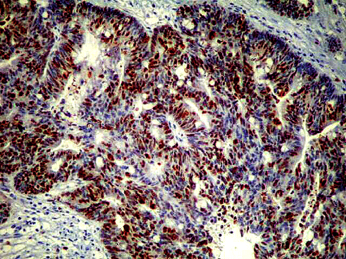
Figure 1: p53 immune expression in liver metastasis. |
|
| Â |
| The synchronism of metastases favored the poorer prognosis in these patients. In the presence of primary tumor desmoplasia intense, high expression of p53 and Ki-67 were associated with poor prognosis. In liver metastasis factors that were associated with poor prognosis were safety margin of less than 10mm. Undifferentiated histological type, the little inflammatory infiltrate, the intense desmoplasia, the presence of peritumoral capsule, the intense expression of p53, p16 and Ki-67 high immune expression are associated with poor prognosis (Figure 2). And the low expression of thymidylate synthase was associated with improved survival in patients receiving 5-FU and leucovorin. |
| Â |
|
|
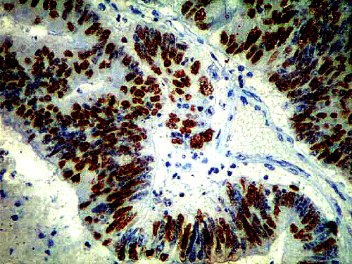
Figure 2: Ki-67 immune expression in liver metastasis. |
|
| Â |
| Discussion |
| Â |
| In this study, there was an association of p53 immune expression with lower disease-free interval (p=0.04) separately in the primary tumor or liver metastasis. The p53 tumor suppressor gene is most commonly identified in epithelial neoplasms [20]. The immune expression of p53 has been accepted as an independent factor for recurrence of colorectal cancer included in the stages of DUKES B and C and also the survival prognosis [21,22]. It was found that p53 has prognostic value in patients with lung adenocarcinoma [20]. It was found that p53 is a prognostic marker for colorectal cancer and its corresponding liver metastasis [17]. |
| Â |
| The lower expression of Ki-67 marker was associated with better overall survival and disease-free interval, the primary tumor and liver metastasis in isolation. This result is in agreement with other authors who studied colorectal cancer and liver metastasis of lung adenocarcinoma [23,15]. The results are in accordance with other authors who studied liver metastasis of colorectal adenocarcinoma [24,25]. The p16INK4a marker in liver metastasis, was associated with survival and disease-free interval (Figure 3). It is the tumor suppressor gene that promotes hypermethylation, acting through the route of microsatellite instability, as an author [26]. To RIBEIRO JR, there was no association with survival and disease-free interval [25]. The thymidilate synthase in the liver metastasis was associated with survival and disease-free interval (Figure 4). Their immune reactivity can identify patients who respond to chemotherapy based on 5-FU, according to other authors [25,27-29]. A higher proportion of increased immune reactivity was found in patients who have undergone pre-operative chemotherapy, before liver resection, which may mean that there is a biochemical resistance. |
| Â |
|
|
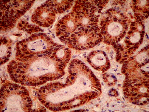
Figure 3: p16ink4a immune expression in liver metastasis. |
|
| Â |
|
|
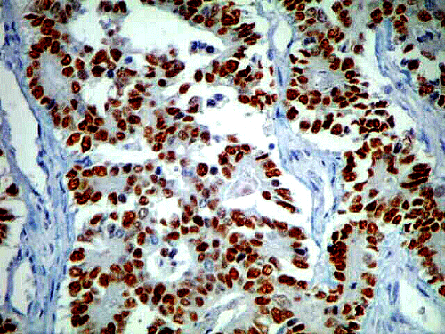
Figure 4: Thymidilate Synthesis immune expression in liver metastasis. |
|
| Â |
| Classically we must comply with the surgical margins of 10mm in radical surgery for resection of liver metastasis, to obtain a higher rate of survival. In some circumstances, where the surgical margin has to be able to slim it will employ a number of molecular markers for genomic analysis of cell constituents and more accurately assess risk and benefit of expanding the margin of resection. Further studies with immunotherapy, anti-CEA antibodies, new with new chemotherapeutic regimens FOLFOX, FOLFIRI, AVASTIN may alter this panel of prognostic factors. |
| Â |
| In the proximal future, new horizons will be achieved early in molecular diagnostics, gene therapy and in the field of immunotherapy for the prevention and treatment of liver metastases and primary tumors of colorectal adenocarcinoma. These developments should benefit decisively treatment of patients with metastatic colorectal adenocarcinoma. |
| Â |
| Conclusion |
| Â |
| Therefore, we have described predictive factors of poor prognosis in patients with colorectal tumors who underwent liver metastases resections. In the primary tumor, the presence of intense desmoplasia, high expression of p53 and Ki-67 were associated with poor prognosis. The safety margin less than 10 mm was associated with poor prognosis, in liver metastasis. The undifferentiated histological type, the little inflammatory infiltrate peritumoral, the intense desmoplasia, the presence of peritumoral capsule and the intense immune expression of p53, p16 and Ki-67 were associated with worse prognosis. The low immune expression of thymidilate synthase was associated with improved survival in patients receiving 5-FU and leucovorin. |
| Â |
| |
| References |
| Â |
- Instituto Nacional do Câncer (2003) Estimativa da incidência e mortalidade por câncer no Brasil.
- Choti MA, Bulkley GB (1999) Management of hepatic metastases. Liver Transpl Surg 5: 65-80.
- Gayowski TJ, Iwatsuki S, Madariaga JR, Selby R, Todo S, et al. (1994) Experience in hepatic resection for metastatic colorectal cancer: analyses of clinical and pathologic risk factors. Surgery 116: 703-710.
- Levine AJ, Momand J, Finlay CA (1991) The p 53 tumor supressor gene. Nature 351: 453-456.
- Allen JI (1995) Molecular biology of colorectal cancer: A clinicianÂ’s view. Perspect Colon Rectal Surg 8: 181-202.
- Clarke AR, Purdie CA, Harrison DJ, Morris RG, Bird CC, et al. (1993) Thymocyte apoptosis induced by p53-dependent and independent pathways. Nature 363: 786-787.
- Oren M (1994) Relationship of p53 to the control of apoptotic cell death. Semin Cancer Biol 5: 221-227.
- Blondal JA, Benchimol S (1994) The role of p53 in tumor progression.Semin Cancer Biol 5: 177-186.
- MURAD JC - Hormônio de crescimento asociado a dieta rica em glutamina na sÃndrome do intestino curto em ratos. São Paulo, 1996. Tese (mestrado) - Universidade Federal de São Paulo - Escola Paulista de Medicina.81p.
- Goldblum JR, Appelman HD (1995) Stromal tumors of the jejunum: A hystologic and immunohistochemical study of the 20 cases. Am J Surg Pathol 19: 71-80.
- Lam KY, Law SY, So MK, Fok M, Ma LT, et al. (1996) Prognostic implication of proliferative markers MIB-1 and PC10 in esophageal squamous cell carcinoma. Cancer 77: 7-13.
- Kawamura T, Goseki N, Koike M, Takizawa T, Endo M (1996) Acceleration of proliferative activity of esophageal squamous cell carcinoma with invasion beyond mucosa: Immunohistochemical analysis of Ki 67 and p53 antigen in relation to histopathologic findings. Cancer 77: 843-849.
- Carrilo R, Candia A, Rodriguez-Peralto JL, Caz V (1997) Prognostic significance of DNA ploidy and proliferative index (MIB-1 index)in gastrointestinal stromal tumors. Hum Pathol 28: 160-165.
- Chang MS, Choe G, Kim WH, Kim YI (1998) Small intestinal stromal tumors: a clinicopathologic study of 31 tumors. Pathology International 48: 341-347.
- Matheus RS (2000) Relevância dos parâmetros morfológicos e biológicos em carcinoma pulmonar dos tipos histológicos de células não pequenas e em suas metástases hematogênicas. . São Paulo, 2000. Tese (mestrado) - Universidade Federal de São Paulo- Escola Paulista de Medicina. 66p.
- Hayashi N, Arakawa H, Nagase H, Yanagisawa A, Kato Y, et al. (1994) Genetic diagnosis identies occult lymph node metastases undetectable by the histopathological method. Cancer Res 15: 3853-3856.
- Sanchez-Cespedes M, Esteller M, Hibi K, Cope FO, Westra WH, et al. (1999) Molecular detection of neoplastic cells in lymph nodes of metastatic colorectal cancer patients predicts recurrence. Clin Cancer Res 5: 2450-2454.
- Bokemeyer C, Hartmann JT, Kanz L (1997) Current aspects of adjuvant and palliative chemotherapy in colorectal carcinoma. Praxis (Bern 1994) 86: 1510-1516.
- Lee ET (2003) Statistical methods for survival data analysis. (3rd Edn.), Wiley Series in Probability and Statistics, California.
- Carvalho PE, Antonãngelo L, Bernardi FD, Leão LE, Rodrigues OR, et al. (2000) Useful panel markers to express the biological status in resected lung adenocarcinomas. Jpn J Clin Oncol 30: 478-486.
- Auvinen A, Isola J, Visakorpi T, Kolvula T, Virtanen S, et al. (1994) Overexpression of p53 and long-term survival in colon carcinoma. Br J Cancer 70: 293-296.
- Diez M, Enriquez JM, Camunas J, Gonzales A, Gutierrez A, et al. (1995) Prediction of recurrence in B-C stages of colorectal cancer by p53 nuclear overexpression in comparison with standard pathological features. Eur J Surg Oncol 21: 635-639.
- Rawet V (1998) Carcinoma colorretal: estadiamento e parâmetros prognósticos. São Paulo, 1998. 194p. Tese de Doutorado - Faculdade de Medicina, Universidade de São Paulo.
- Kyzer S, Gordon PH (1997) Determination of proliferative activity in colorectal carcinoma using monoclonal antibody Ki67. Dis Colon Rectum 40: 322-325.
- RIBEIRO JR U (2002) Contribuição da imunohistoquÃmica na avaliação da resposta e do prognóstico do câncer de reto distal tratado por radioterapia e quimioterapia neoadjuvante. São Paulo, 2002. 117 p. Tese (Livre-Docência) - Faculdade de Medicina da Universidade de São Paulo.
- Myohanen SK, Baylin SB, Herman JG (1998) Hypermethylation can selectively silence individual p16ink4A alleles in neoplasia. Cancer Res 58: 591-593.
- Paradiso A, Simone G, Petroni S, Leone B, Vallejo C, et al. (2000) Thymidilate synthase and p53 primary tumour expression as predictive factors for advanced colorectal cancer patients. Br J Cancer 82: 560-567.
- Corsi DC, Ciaparrone M, Zannoni G, Mancini M, Cassano A, et al. (2002) Predictive value of thymidylate synthase expression in resected metastases of colorectal cancer. Eur J Cancer 38: 527-534.
- Etienne MC, Chazal M, Laurent-Puig P, Magne N, Rosty C, et al. (2002) Prognostic value of tumoralthymidylitesinthase and p53 in metastatic colorectal cancer patients receiving fluorouracil-based chemotherapy phenotype and genotypic analyses. J Clin Oncol 20: 2832-2843.
|
| Â |
| Â |