Make the best use of Scientific Research and information from our 700+ peer reviewed, Open Access Journals that operates with the help of 50,000+ Editorial Board Members and esteemed reviewers and 1000+ Scientific associations in Medical, Clinical, Pharmaceutical, Engineering, Technology and Management Fields.
Meet Inspiring Speakers and Experts at our 3000+ Global Conferenceseries Events with over 600+ Conferences, 1200+ Symposiums and 1200+ Workshops on Medical, Pharma, Engineering, Science, Technology and Business
Case Report Open Access
Failure of Ventricular Capture and Pacemaker Exit Block Secondary to Moderate Hyperkalemia
| Pau Alonso*, Raquel Lopez, Maria Jose Sancho-Tello, Ana Andres, Oscar Cano, Joaquin Osca and Jose Olague | |
| Department of Cardiology, Electrophysiology Section, La Fe University Hospital, Valencia, Spain | |
| Corresponding Author : | Pau Alonso Department of Cardiology Electrophysiology Section, La Fe University Hospital Partida de la Mar nº 15. Almàsser Tel: 0034676037149 E-mail: pau_i_au@hotmail.com |
| Received: October 07, 2015; Accepted: November 18, 2015; Published: November 28, 2015 | |
| Citation: Alonso P, Lopez R, Sancho-Tello MS, Andres A, et al. (2015) Failure of Ventricular Capture and Pacemaker Exit Block Secondary to Moderate Hyperkalemia. Arrhythm Open Access 1:102. doi:10.4172/atoa.1000102 | |
| Copyright: © 2015 Alonso P, et al. This is an open-access article distributed under the terms of the Creative Commons Attribution License, which permits unrestricted use, distribution, and reproduction in any medium, provided the original author and source are credited. | |
| Related article at Pubmed, Scholar Google | |
Visit for more related articles at
Abstract
An 81-year-old woman was admitted to our institution with bradicardia, weakness and presyncope. At admission physical examination revealed a pulse rate of 42 per minute, blood pressure of 95/50 mmHg, and an oxygen saturation of 93% on room air. The interrogation of the device showed a correct ventricular sensing and a threshold for ventricular pacing of 6.5 V at 1 ms pulse width. Analytic test showed a creatinine 3.78 mg/dl, Urea 187 mg/dl, sodium 133 mEq/L, potassium 6.2 mEq/L, and pH 7.37. Our patient showed a moderate acute hyperkalemia, which caused pacemaker exit block and capture failure.
| Keywords |
| Pacemaker failure; Hyperkalemia |
| Case Report |
| Hyperkalemia is a life threatening metabolic condition. When the K level increases, the intraventricular conduction velocity is decreased and the paced QRS complex widens. When the K level reaches 7 mEq/L pacing thresholds could increase, with or without increased latency. Occasionally failure to capture appears at a lower level, especially in the presence of heart disease [1,2]. We report a case where moderate hyperkalemia caused pacemaker exit block and a failure of ventricular pacemaker capture. An 81-year-old woman was admitted to our institution with bradycardia, weakness and presyncope. She had a history of type 2 diabetes mellitus and chronic kidney disease (eGFR of 12 ml/min). Patient underwent bivalvular mechanic mitral- aortic valve replacement 13 years ago. Six months after cardiac surgery the patient underwent a VVI pacemaker implant (Vitatron G20) for permanent atrial fibrillation with slow ventricular response. The patient’s pacemaker was programmed VVI with a lower rate limit 70 beats/min. In the last weeks, congestive heart failure symptoms had appeared, thus loop diuretic dose was increased (Furosemide 100 mg per day). Her medications also included digoxin 0.25 mg per day, warfarin 2 mg per day, and insulin; thus she didn’t take antiarrhythmic drugs at admission physical examination revealed a pulse rate of 42 per minute, blood pressure of 95/50 mmHg, and an oxygen saturation of 93% on room air with a respiratory rate of 24. Signs and symptoms of acute heart failure were present. |
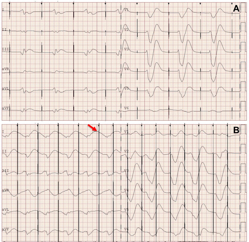
| Transthoracic echocardiography showed a normal left ventricular size, with conserved LVEF and normally functioning prosthetic valves. Left ventricular hypertrophy was detected, with a increased wall thickness (interventricular septum, 14 mm; posterior wall, 13 mm). Left atrium was dilated (30 cm2). The surface Electrocardiogram (ECG) revealed no spontaneous atrial activity and unipolar pacing spikes without capture (Figure 1A), with a ventricular escape rhythm at 40 bpm. The escape rhythm had a marked increase in QRS duration (360 ms) with a slurred morphology. Pacemaker sensing was appropriate, thus the spikes were delivered 850 ms after QRS detection. |
| The reference ECG demonstrated a normal ventricular paced rhythm. The interrogation of the device showed a appropriate ventricular sensing and a capture failure at 2.5 V at 0.4 ms pulse width. The threshold for ventricular pacing was 6.5 V at 1 ms pulse width with a pacing impedance of 400 ohms. Pulse generator output was then programmed at 8 V at 1 ms pulse width (Figure 1B), with a lower rate 70 bpm, which showed a first degree pacemaker exit block (latency after the spike and the QRS).When pacing was performed at shorter cycle length (600 ms) some spikes fall into the refractory period of paced QRS complexes, thus they failed to capture the ventricular myocardium (Figure 1B). Analytic test showed a creatinine 3.78 mg/dl, Urea 187 mg/dl, Glucose 172 mg/dl, sodium 133 mEq/L, potassium 6.2 mEq/L, pH 7.37, and HCO3 24 mEq/L. |
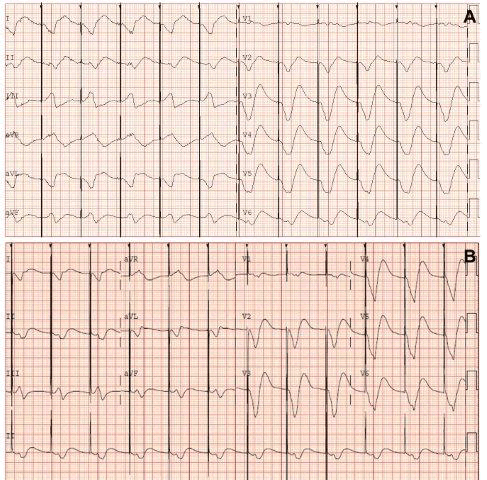
| With these findings the patient was immediately treated with intravenous administration of sodium bicarbonate, calcium gluconate, salbutamol, and glucose and insulin infusions. One hour after the treatment beginning potassium serum level was 5.5 mEq/L. The ECG showed a ventricular paced rhythm with a QRS width of 260 ms, probably due to incomplete cellular ionic homeostasis restoration (Figure 2A). Twenty-four hours after the treatment beginning, potassium serum level was finally normalized (4.3 mEq/L) and the ECG showed a normal ventricular paced rhythm, similar to previous weeks (Fig. 2B). The device was re-interrogated and the threshold for ventricular pacing was 1.25 V at 0.4 ms pulse width. Thus p`ulse generator output was programmed at 2.5 V at 0.4 ms pulse width, with a lower rate 70 bpm. Hyperkalemia is a well-known cause of cardiac toxicity, due to a reduction of the electronegativity of the resting myocardial potential, thus acute hyperkalemia requires urgent intravenous therapy. Hyperkalemia causes a depression in myocardial excitability and in impulse conduction velocity [3,4]. Therefore, pacemaker exit block with increase of latency and loss of myocardial capture can be seen in the context of severe hyperkalemia (serum potassium >7 mEq/L) [5]. It has been described the presence of exit block and loss of capture [6,7] in the context of severe hyperkalemia, but the presence of both due to moderate hyperkalemia has not yet been reported. Our patient showed a moderate acute hyperkalemia (6.2 mEq/L), which caused both, pacemaker exit block and capture failure. One explanation for the greater susceptibility of this patient to hyperkalemia is the presence of severe heart disease with associated diastolic heart failure, mainly due to the rapid onset of ionic imbalance. This case report underlines the importance of prompt medical therapy if the suspicion is high, even in case of moderate hyperkalemia (mostly in patients with underlying heart disease). |
| References |
|
Figures at a glance
 |
 |
| Figure 1 | Figure 2 |
Post your comment
Relevant Topics
Recommended Journals
Article Tools
Article Usage
- Total views: 11495
- [From(publication date):
March-2016 - Sep 22, 2024] - Breakdown by view type
- HTML page views : 10838
- PDF downloads : 657