A Pathological Lesions Study of Bovine Abomasums in Urmia Abattoir
Received: 15-May-2012 / Accepted Date: 18-Jul-2012 / Published Date: 25-Jul-2012 DOI: 10.4172/2161-0681.1000121
Abstract
Abstract
The objective of this study was to evaluate pathologic lesions on abomasums which were collected from 492
cattle in Urmia abattoir, located in North West of Iran, during different seasons from February 2010 to July 2011.
Furthermore, this research was carried out in a 6-month period through 46 times randomly visiting the abattoir.
Additionally, abomasums separated from other parts of the gastrointestinal tract, and macroscopic examination
was started. Subsequently an incision made over greater curvature in order to examining the mucosa; moreover for
microscopic observation some specimens were taken from abomasums and referred to pathological examination. The
results of this study demonstrated that 77 specimens encompassed ulcer and erosions, trichobezoar and indigestible
masses (10.97%), 48 samples had hyperemia (9.7%), 26 samples showed abomasal parasites (5.28%), 14 samples
were with abomasal distention (2.84% ), 5 samples had soil and sand (1.01%) and one developed a tumor (0.2%).
Statistical analysis indicated that there is not significant correlation between onset of ulcers and seasons so that its
occurrence in winter, spring and summer was 16.9%, 13.6% and 17.9% respectively. Although there was a slight
decrease, parasitic infestation increased concurrently with environmental heat which was recorded as 4.3% in winter,
5.02% in spring and 6.8% in summer.
Keywords: Bovine; Pathological Lesions; Urmia abattoir; Ruminants
311814Introduction
Cattle are compound stomach animals belonging to bovidae family. Iran has a large number of livestock which ruminants especially cattle play the most important role in food industry and other associated industries [1]. Ruminants’ digestive tract may be more exposed to digestion activities and close relationship with outer environment such as husbandry management [2,3]. Abomasum is the main stomach of ruminants in lower right quadrant of the abdomen that will be affected primarily or secondarily by infections, parasites and foreign bodies somehow prohibit proper nutrition and prehension of essential substances formetabolism, inducing to anorexia, weakness, emaciation and death [4-6]. The outcomes of these lesions are such as gastric ulcers which occurs at all ages in cattle that occasionally causes acute hemorrhages in this organ that is along with indigestion and melena, and sometimes perforation of abomasum takes place, producing painful acute local peritonitis or acute diffuse peritonitis with sudden death [6,7]. In a study by Jensen et al. [8], it indicated that through necropsy of 1988 cattle 1.6% collapsed due to perforative ulcers and hemorrhages of abomasums. Furthermore, in another study by Aukema and Breukink in 1974 demonstrated that through necropsy of 1200 cattle, 9.1% had abomasal ulcers and also in 141 of them lethal hemorrhagic abomasal ulcers observed though rarely of such lesions can be diagnosed by usual physical examination [9]. For this, they usually can be diagnosed during abattoir inspection. Currently, concurrent with development of intense husbandry methods and achievement of the highest livestock production potency contribute to provide such lesions in the stomach through the stresses, diseases and inappropriate feeding, hence the aim of this study is to evaluate cattle abomasal pathological lesions.
Materials and Methods
Study area
The district Urmia of agro-ecological zone was selected for the present study. Climatically, the study region is subtropical and receives an annual rainfall of about 150-350 mm. The temperature is the highest in June, before the onset of monsoon season. During summer, the daily maximum temperature exceeds 45°C and rarely declines below 22°C. Relative humidity is the lowest during April and May and rises during the monsoon season. One year cycle is divided into four seasons: winter (December-February), spring (March-April), summer (May- September) and autumn (October-November). Summer also includes monsoon season (July-August).
Sample collection and pathological observations
This research has been carried out over 46 days in different months from February 2010 to July 2011, in Urmia abattoir in Iran, which 492 abomasums were randomly examined. Statistical society in this study was Sarabi, Holstein and Azerbaijani slaughtered Cattle, from both male and female sexes that were from 1 month to 6 years of age. For determination the prevalence proportion, according to previous studies, chi-square test and qualitative methods, 492 cattle were allocated to study. Before slaughter the race, age based on teeth and sex of cattle was obtained, and abomasums were appraised for the presence or absence of any pathological lesions. In the presence of any pathological lesions, abomasums were separated from ruminoreticulum and after an incision made on the abomasums by scissors and forceps, the number and type of any pathological lesions were observed. Also then an incision made on abomasum over the longitudinal axis of the greater curvature, and its content evaluated in case of existing foreign body, trichobezoars, soil, sand and indigestible materials. In addition, its mucosal surface gently washed with water in order to diagnose lesions such as hyperaemia, ulcer, inflammation, protrusions and also parasites. After abomasal examining information as date, seasons, location of sampling, quantity, size of each lesion and abnormal abomasal content were recorded. Abomasums mucosal tissue in suffered cattle from the presence of any abnormality was studied as well. In all cases sampling was done from pyloric and ventral ofabomasums to 1.5×1.5 cm dimensions for histopathological studies. Samples were fixed in formalin 10% and sent to the pathology laboratory of Urmia University, veterinary medicine faculty where according to common pathologic cross-sectional method they were dehydrated in a graded alcohol, cleared in xylene and embedded in paraffin at 58-600C, slices with 5-6 μ thickness were provided and then were studied microscopically after hematoxylin and eosin staining. In this study of differentiation between sex diversity from affliction to abomasums lesions and also the effect of age and relationship of the lesions with aging we used qualitative methods and chi-square test. It should be noted that the relationship between presence of lesions and abomasal pathological complications prevalence also by same qualitative methods and chi-square test was analysed. In order to report the frequency we used of frequency table and bar chart.
Statistical Analysis
Consequently, histopathologica results and other data were analysed by qualitative methods.
Results
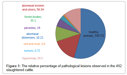
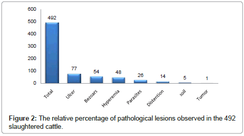
In this research during diverse seasons from February 2010 to July 2011 on 492 slaughtered cattle abomasum in Urmia abattoir, the capital of western Azarbaijan province,were evaluated which 77 samples showed erosions and ulcers (15.65%), 54 samples had foreign bodies, trichobezoars and indigestible masses (10.97%). However, 48 revealed mild to severe hyperemia (9.7%) somewhat the mucosa remained hyperemic even after washing and ulcers detected which were 2 mm to 2 cm that usually were focally in fundus and ventral of greater curvature, furthermore some linear ulcers and sometimes volcanic ulcers observed, made by Theileriosis, with hyperemic and protruded edges. Most of erosions and ulcers in abomasal mucosa, internal layers and muscular layer will not be affected. In 26 samples abomasal parasites detected (5.28%), 14 samples carried abomasal distention (2.84%), and 5 samples contained soil, sand (1.01%) and finally just in one found a tumour mass (0.2%) although in some samples different pathological forms were observed and recorded separately. It would be obvious that there is a close relationship between foreign bodies and ulcers with the proportion of 11, 15 percent respectively (Figure 1, Figure 2). In these samples except some cases, ulcers occurred in the abomasum with foreign bodies and scar tissue. Statistical analysis showed that there was not any significant correlation between ulceration and seasons (p>0.05), somehow in winter 2010 the relative occurrence percentage of abomasal ulcers were 16.9% and 17.0% in summer though in spring a slight reduce took place (13.6%). However, the nutrition conditions and feeding are the most important factor which most of them occur with alteration of nutrition. Parasitic infestation also occurs along with the increase of environmental heat that is 4.3% in winter 5.02% in and 6.8% in summer (Table 1).
| Date | Day | TNOS | AU (%) | C (%) | TNOP (%) | DA (%) | MS (%) | GT (%) | FMHB (%) |
|---|---|---|---|---|---|---|---|---|---|
| February2010 | 4 | 65 | 11(2.23) | 4(0.81) | 2(0.40) | 2(0.40) | _ | _ | 7(1.42) |
| March2010 | 4 | 71 | 12 (2.43) | 6(1.22) | 4(0.81) | 3(0.60) | _ | _ | 4(0.81) |
| April2011 | 3 | 73 | 9 (1.84) | 3(0.60) | 3(0.60) | _ | 1(0.20) | _ | 7(1.42) |
| May2011 | 5 | 90 | 14(2.84) | 11(2.23) | 4(0.81) | 3(0.60 | 2(0.40) | _ | 6(1.22) |
| June2011 | 4 | 76 | 10(2.03) | 8(1.62) | 5(1.02) | 2(0.40 | _ | _ | 12(2.44) |
| July2011 | 6 | 117 | 21(4.28) | 16(3.25) | 8 (1.62) | 4(0.81) | 2(0.40) | 1 (0.20) | 18(3.66) |
| Total | 26 | 492 | 77(15.65) | 48(9.75) | 26(5.28) | 14(2.84) | 5(1.01) | 1(0.20) | 54(10.97) |
TNOS: The number of samples AU: Abomasum ulcer, C: Congestion, TNOP: The number of parasites DA: Distended abomasum, MS: Mud and soil, GT: Gland tumor, FMHB: Foreign material and hair ball
Table1: The results of 492 lesions abomasum.
Discussion
Abomasum is the main stomach of ruminants that may be primarily or secondarily affected especially in cattle at all ages. The results of this research indicated erosion and ulcers (15.65%), foreign body and trichobezoar (10.97%), hyperaemia (9.7%), abomasal parasites (5.28%), abomasal distention (2.84%), soil, sand and mud (1.01%) and tumour (0.2%). In a study in Canada from 46 operated cattle nearly in 88% of cases found ulcer and hemorrhage, 76% trichobezoar and 14.2% soil and mud [10]. Another study reported that from 141 cattle abomasum in 9.1% occurred ulcer and hemorrhage along with hyperemia. Furthermore, in a survey in 1988 from necropsy of 36 cattle revealed that in about 1.6% observed ulcers concurrent with hyperemia in mucosa which such lesions were more in winter than other seasons [11]. However, through an evaluation in Babol abattoir on 400 cattle abomasum was determined that 16.75% had ulcers, so that most of them were in an oval or linear forms in fundus [12]. In a study in Tehran on 499 Buffaloes and cattle abomasums the frequency of scar, erosion and ulcers were recorded as 1%, 3.2% and 0.6% respectively. In another study in Egypt from 1200 cattle nearly in 9.1% were found ulcer and hemorrhage and in 6.3% was observed scar tissue [13]. The comparison of our results with such studies showed that ulcer, erosion and hyperaemia of the abomasum is relatively high in our research, hence the difference of frequencies in such investigation with other researchers may be due to nutrition, stress, parasitic diseases and other predisposing factors. The results demonstrated that the abomasal lesions are usually in the western Azarbaijan province and occur in all seasons due to freestyle or semi-open conditions which in the latter the feeding is done manually. Therefore, the possibility of intaking indigestible materials is fairly high, somehow in 11% were found foreign bodies upon examination. Emerging of abomasal ulcers are common in slaughtered dairy cattle, feedlot cattle and calves that can be diagnosed during inspection. Most of the Urmia dairy cattle were slaughtered after their efficient economic age had been finished. In fact, it would be clear that such sort of lesions were produced by the large amount of grain feeding during the peak of milk production and stress. The cause of ulceration in young calves may be due to weaning and starting to be fed roughage and low quality forage as well. Additionally, the role of stress in inducing ulceration is very important. Another factor is the simultaneous occurrence of foreign bodies and ulcers which in most of cases, similar to other researches, were visible. Most of the ulcers were observed in fundus and the longitudinal axis of greater curvature because the fundus is a region that HCL, ingesta and foreign bodies accumulate in it and move slowly. The parasitic infestation rate alters seasonally with the high reproduction potency of parasites that were 4.3%, 5%, 6.8% in winter, spring and summer respectively. Actually, grazing the pastures usually contain a large number of parasites by such animals produces survival of them in their hosts. Besides, other than factors such as primary diseases, indigestible materials, stress, foreign bodies, and other microbes such as Clostridium perfrigenes type A and some fungal diseases may be involved as well. Furthermore, investigations on bile acids in abomasum have been done recently which showed the reflux of these substances from the intestine can act as a deterrent and damage the mucosa, causes ulcers finally. Regarding differentiate diagnosis, it is important to utilize paraclinical tests in order to confirm the main agent, and exclude hemorrhagic enteritis, omasal impaction, acute intestinal obstruction, TRP (traumatic reticulopretonitis), etc. The human gastric ulcers are similar to that in animals, thus the attitudes concerning prevention and treatment in human can be beneficial for animals though hemorrhagic ulceration in humans are much less than animals [14-17]. Overall, in western Azarbaijan province due to its specific climatic conditions of rearing animals in pastures and stables along with inappropriate nutrition or some deficiencies such as copper in addition to stress, density and other infectious diseases, the abomasal diseases as ulcers and parasites certainly occur [18-20]. However, in most cases the gastrointestinal affections especially its early stages solely could be visible during necropsy, hence such lesions progress and disrupt the production and growth which induces disadvantages for farmers. For this, it would be essential to provide standard and immune conditions for animals’ health and rations in order to protect their gastrointestinal tract.
References
- Kamalzadeh AM, Rajabbagi A, Kiasat A (2008) Livestock Production Systems and Trends in Livestock Industry in Iran. Journal of agriculture and social science 4: 183-188.
- Rossow N, Horvath Z (1985) Internal Medicine of Domestic Animals. (1stedn), VEB Gustav Fischer, Verlag, Jena, 79.
- Perry BD, Randolph TF, Mcdermott JJ, Sones KR, Teornton PK (2002) Investing in Animal Health Research to Alleviate Poverty. ILRI (International Livestock Research Institute), Nairobi, Kenya, pp.148.
- Hailat N, Nouh S, AI-Darraji A, Lafi S, Al-Ani F, Al-Majali A (l996b) Prevalence and pathology of foreign bodies (plastics) in Awassi Sheep in Jordan. Small Ruminant Res 24: 43-48.
- Robbins SL, Cotranit S, Kuma V (1985) Pathologic Basis of Disease. (3rdedn), WS Saimders. Philadelphia.
- Radostitis OM, Blood DC, Gay CC (1994) Veterinary Medicine, A Textbook of the diseases of cattle. sheep, pigs, goats. (8thedn), S. BailliereTindall, pp. 279-284.
- Tanimoto T, Ohtsuki Y, Nomura Y (1994) Rumenoabomasal lesions in steers induced by naturally ingested hair. Vet Pathol 31: 280-282.
- Jensen R, Pierson RE, Braddy PM, Saari DA, Benitez A, et al. (1976) Fatal abomasal ulcers in yearling feedlot cattle. J Am Vet Med Assoc 169: 524-526.
- Aukema JJ, Breukink HJ (1974) Abomasal ulcer in adult cattle with fatal haemorrhage. Cornell Vet 64: 303-317.
- Chicoine AL, Dowling PM, Boison JO (2008) A survey of antimicrobial use during bovine abdominal surgery by western Canadian veterinarians. Can Vet J 49: 1105–1109.
- Roeder BL, Chengappa MM, Nagaraja TG, Avery TB, Kennedy GA (1988) Experimental induction of abdominal tympany, abomasitis, and abomasal ulceration by intraruminal inoculation of Clostridium perfringens type A in neonatal calves. Am J Vet Res 49: 201-207.
- Raoofi A, Mardjanmehr S H, Bokaie S, Hosseinifard SM ( 2001) Prevalence study and pathological examination of abomasal ulcers at Babols abattoir. Journal of the Faculty of Veterinary Medicine, University of Tehran 56: Pe64-Pe68
- Ahmed AF (2001) Pathogenesis and treatment of abomasal ulceration in cattle. Ph.D. dissertation. Department of Veterinary Surgery, Faculty of Veterinary Medicine, Assiut University, Assiut, Egypt, pp.12-54.
- Nickel R (1979) The Viscera of the Domestic Mammals, 2nd revised edition. Verlag.poul. pareyBertlin, Hamburg, pp.101-109.
- Simpson HV (2000) Pathophysiology of abomasal parasitism: is the host or parasite responsible? Vet J 160: 177-191.
- Balic A, Bowles VM, Meeusen EN (2000) The immunobiology of gastrointestinal nematode infections in ruminants. Adv Parasitol 45: 181-241.
- Love SCJ and Hutchinson GW (2003) Pathology and diagnosis of internal parasites in Ruminants. In Gross Pathology of Ruminants, Proceedings 350, Post Graduate Foundation in Veterinary Science, University of Sydney, Sydney; Chapter16, 309-338.
- Soulsby EJL (1982) Textbook of Veterinary Clinical Parasitology. Vol. I. Helminths, Oxford Blackwell Scientific, London,
- Simpson HV (2000) Pathophysiology of abomasal parasitism: is the host or parasite responsible? Vet J 160: 177-191.
- Yacob HT, Basazinew BK, Basu AK (2008) Experimental concurrent infection of Afar breed goats with Oestrus ovis (L1) and Haemonchus contortus (L3): interaction between parasite populations, changes in parasitological and basic haematological parameters. Exp Parasitol 120: 180-184.
Citation: Tehrani A, Javanbakht J, Marjanmehr SH, Hassan MA, Solati A, et al. (2012) A Pathological Lesions Study of Bovine Abomasums in Urmia Abattoir. J Clin Exp Pathol 2:121. DOI: 10.4172/2161-0681.1000121
Copyright: © 2012 Tehrani A, et al. This is an open-access article distributed under the terms of the Creative Commons Attribution License, which permits unrestricted use, distribution, and reproduction in any medium, provided the original author and source are credited.
Select your language of interest to view the total content in your interested language
Share This Article
Recommended Journals
Open Access Journals
Article Tools
Article Usage
- Total views: 16170
- [From(publication date): 9-2012 - Oct 14, 2025]
- Breakdown by view type
- HTML page views: 11255
- PDF downloads: 4915


