Case Report Open Access
Cytological and Histopathology Features of Meibomian Adenocarcinoma in a Dog Terrier Breed
Abbas Tavasoli1, Javad Javanbakht1*, Radmehr Shafiee2, Zahra Kamyabi-moghaddam1 and Mehdi Aghamohammad Hassan31Department of Pathology, Faculty of Veterinary Medicine, Tehran University, Tehran, Iran
2Graduate Faculty of Veterinary Medicine, Tehran University, Tehran, Iran
3Department of Clinical Science, Faculty of Veterinary Medicine, Tehran University, Tehran, Iran
- *Corresponding Author:
- Dr. Javad Javanbakht
Department of Pathology
Faculty of Veterinary Medicine
Tehran University, Tehran, Iran
Tel: +989372512581
E-mail: javadjavanbakht@ut.ac.ir
Received Date: May 14, 2012; Accepted Date: July 15, 2012; Published Date: July 19, 2012
Citation:Tavasoli A, Javanbakht J, Shafiee R, Moghaddam ZK, Hassan MA (2012) Cytological And Histopathology Features of Meibomian Adenocarcinoma in A Dog Terrier Breed. J Clin Exp Pathol 2:120. doi: 10.4172/2161-0681.1000120
Copyright: © 2012 Tavasoli A, et al. This is an open-access article distributed under the terms of the Creative Commons Attribution License, which permits unrestricted use, distribution, and reproduction in any medium, provided the original author and source are credited.
Visit for more related articles at Journal of Clinical & Experimental Pathology
Abstract
Abstract
In March 2012, an 8-year-old neutered terrier which was admitted to the Veterinary Surgery Clinics of Faculty of
Veterinary Medicine, University of Tehran, Iran. The signalment (i.e. description of appearance of the animals such as
sex, breed, age, etc) and anatomical repartition were summarized and focused on pathomorphological explanation.
Macroscopically, the mass was gray-reddish and arising from the right lower eyelid with local spread to the upper
eyelid and covering the entire globe was observed. Following sedation and local anesthesia, the tumor and globe were
expulsed with Cryosurgery. Cytological smears indicated large number of malignant meibomian cells occurring
either individually or in clusters also cytology indicated high cellularity and contained small clusters of polygonal to
elongated tumor cells. Microscopically, the mass was composed of lobules of various sizes separated with a septum of
connective tissue which included with poorly differentiated (grade III), nuclear pleomorphism and polygonal sebaceous
cells, also with acinular and sheet-like architecture, the findings are consistent with minimal mitotic activity, extensive
invasion, low grade and the presence of hyaline cartilage. Finally, histopathological examination revealed an MAC.
Keywords
Adenocarcinoma; Histology; Cytology; Dog; Meibomian
Introduction
Meibomian sebaceous gland adenocarcinoma of the eyelid is a rare fatal tumor. It accounts for less than 1% of all eyelid neoplasms [1]. Glands of the eyelid consist of sebaceous glands such as meibomian (tarsal) and Zeis glands that opens to hair follicles and moll glands that have modified sweat gland characteristics [2-5]. Meibomian glands are found on the tarsal plate and are responsible for the formation of an oily layer over the thin film of tear [6,7]. Moll glands have apocrine secretion and leave their secretions inside the eyelash follicles [5,8]. Meibomian Adenocarcinoma (MAC) is a kind of highly malignant and deadly neoplasm which is originated from eyelid and occurs in it. Meibomian gland/glands adenocarcinoma also occurs in the eyelid of the Zeis glands in some animals. The clinical diagnosis is difficult because it is easy to confuse with chalazion, eyelid conjunctivitis, conjunctivitis and basal cell carcinoma, and it is easy to offend the surrounding tissue via lymphatic and blood metastasis [9]. MAC rarely has typical characteristics of gland, and the surgery and pathological examination material is very significant to no obvious damage expressions of the eyelid goiter. MAC is a rare tumor which is more likely to be invasive and aggressive with local reoccurrence following incomplete excision [10]. Malignant neoplasms such as the adenocarcinomas of the sebaceous glands, basal cell adenocarcinomas, melanomas and mast cell tumors are tumoral masses of lower frequency [8,11]. Meibomian glands are modified sebaceous glands which are located on the inner surface of the eye. The meibomian adenoma, meibomian carcinoma have been reported in human and calf in the world but the MAC is very rare in dog [9,10,12]. This case might be of interest as it is the first case having meibomian gland tumor on the eyelid of a dog; thus we found it appropriate to make the description of these mass.
Case Report
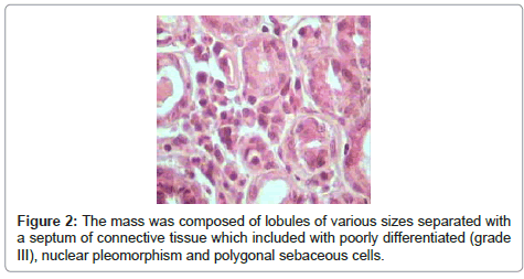
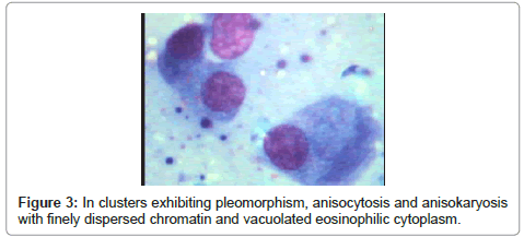
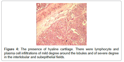
In March 2012, A 8-year-old male dog terrier presented with a manifested painless swelling recurrent right lower eyelid nodule clinically resembling a chalazion to small pets’ hospital of Tehran veterinary college, that in the inner part of its right lower eyelid, an oval or round tumor formation of 1cm x 8mm x 6mm, weighing 8.35 g with red color or gray reddish color and arising from the right lower eyelid with local spread to the upper eyelid and covering the entire globe was observed. Clinical signs were epiphora, photophobia, ocular discharge; conjunctivitis in right eye and after the lesion was completely excised under local anesthesia it submitted for histopathologic examination. The case had a history of meibomian adenocarcinoma. After being fixed with 10% neutral buffered formalin solution, it was embedded in paraffin following the routine processing. 5-micron-thick sections obtained from the paraffin blocks were stained with Hematoxylin- Eosin (HE) technique and then examined under light microscope. Histopathologically, it was seen acinular and sheet patterns, composed of anaplastic cells having hypochromatic nuclei and evidently vacuolated eosinophilic cytoplasm in most of areas (Figure 1, 3 and 4). It was attended that pseudorozette formation which composed of spindle shaped, hyperchromatic nuclei and narrow eosinophilic cytoplasm. A few surviving elements of the acinar elements of the Meibomian glands were observed near the eyelid margin. The mass was composed of lobules of various sizes separated with a connective tissue septum in capillaries which included with poorly differentiated (grade III), nuclear pleomorphism and polygonal sebaceous cells, also with acinular and sheet-like architecture (Figure 4). The nuclei were round or slightly irregular, with small nucleoli. Also H&E stained sections indicated main mass situated in inner right eyelid consisting of lobules of different sizes. The lobules were composed of cells with large hyperchromatic pleomorphic nuclei (Figure 1). The lobules of other cell smears were separated from each other with a septum composed of fibro-vascular connective tissue, and there were several tubular and acinar structures of similar alignment. Acinar and tubular structures were lined with poorly-differentiated cubic to columnar epithelial cells having eosinophilic cytoplasm, with oval or round nuclei. In the center of some of the lobules sebaceous differentiation was observable, and some areas of necrosis and few mitotic figures were seen (Figure 2). Microscopic examination showed that many of the tumor cells were undifferentiated, invasive and mitotic. We noted irregular lobular formations with fibrous septa and extensive pagetoid involvement of the epithelium and the presence of vacuolated cytoplasm suggested sebaceous differentiation (Figure 1). There were lymphocyte and plasma cell infiltrations of mild degree around the lobules and of severe degree in the interlobular and subepithelial fields (Figure 4). The findings are consistent with minimal mitotic activity, extensive invasion, low grade and the presence of hyaline cartilage (Figure 4). Cytological smears revealed large number of malignant meibomian cells occurring either individually or in clusters exhibiting pleomorphism, anisokaryosis and anisocytosis, also cytology indicated high cellularity and contained small clusters of polygonal to elongated tumor cells. The malignant cells possessed round to oval nuclei; a thin nuclear membrane; finely dispersed chromatin; prominent, solitary nucleoli; abundant eosinophilic cytoplasm with rare multinucleated cells and mitotic figures and the background contained a mixed population of inflammatory cells (Figure 3).
Discussion
Neoplastic changes of the sebaceous glands of the eyelid are classified as nodular hyperplasia, epithelioma, adenoma and adenocarcinoma [4]. Malignant meibomian neoplasms may be composed of pavement cells arising from aduct, or may present a basalcell configuration. Although they are modified sebaceous glands of the eyelids, malignant change occurs in them considerably rarely when compared with those in other parts of the body. Adenocarcinoma of the meibomian glands is a neoplasm with atypical glandular features that appear initially as a chalazion formation and then invades the neighbouring tissues. This tumor usually is present in dogs which are old. There is no gender susceptibility, and it can occur in any breed [9,12,13]. Adenocarcinoma of the meibomian glands is the most malignant palpebral tumor. Complete removal surgery may bring a curative effect and histopathology has a key role in the diagnosis of meibomian adenocarcinoma. Histopathology showed that the acini were separated from each other by connective tissue rich in capillaries. The regular arrangement of the cells was lost and there was less orderly cell differentiation. Mitosis was found throughout the neoplastic cells. The less differentiated cells were observed mainly at the periphery and with abundant vacuolated cytoplasmic cells were at the centre of the acini. Most frequently they present an acinar pattern and resemble the normal structure of the gland, but the cells may be arranged in sheets, solid cords or clusters. At least in some part of the tumor large cells with abundant vacuolated cytoplasm were observed, and mitotic features were limited. In poorly differentiated areas these gland tumors imitate a squamous carcinoma and also cytological smears demonstrated large number of malignant meibomian cells, occurring either individually or in clusters exhibiting pleomorphism, anisokaryosis and anisocytosis. Eyelid neoplasms are the most frequently recorded ophthalmic tumors in dogs [9]. Most eyelid neoplasms are benign (73-87%) [6,14] and when malignant, do not typically metastasize and rarely recur [14]. In dogs, 88% of the tumoral masses of the eyelid are benign and 8.2% are reported as being malignant [7,15]. The most commonly observed eyelid tumor of the bovine is squamous cell carcinomas [5,11]. Adenocarcinoma of the Meibomian gland has a tendency for recurrence and metastasis. Metastasis occurs by the lymphatic path and orbit etc. Consequently, in present report, the histopathological examination confirmed the meibomian Adenocarcinoma.
References
- Gokhan N, Sozmen M, Ozba B, Gungor E (2010) Meibomian carcinoma of the eyelid in a Simmental cow. Vet Ophthalmol 13: 336-338.
- Goldschmidt MH, Hendrick MJ (2002) Tumors in theskin and soft tissues. Iowa State Press, Blackwell Publishing Company, Ames, Iowa.
- Dellman HD, Eurell J (1998) Textbook of Veterinary Histology. (5thedn), Lippincott, Williams and Wilkins, Baltimore, Maryland, USA.
- Johnson JS, Lee JA, Cotton DW, Lee WR, Parsons MA (1999) Dimorphic immunohistochemical staining in ocular sebaceous neoplasms: a useful diagnostic aid. Eye (Lond) 13 : 104-108.
- Mc Gavin MD, CarltonWW, Zachary JF (2001) Thomson’s Special Veterinary Pathology. (3rdedn), Mosby Inc., St. Louis, Missouri.
- JunqueiraLC, Carneiro J, Kelly RO (1998) Basic Histology. (8thedn), Beta A.S. Press, Istanbul.
- Mushtak AB, Sinha RD, Prasad R, Prasad J (1990) Comparative histological and histochemical studieson the meibomian glands of goat and sheep. Indian Journal of Animal Science 60: 1085–1087.
- Hirai T, Mubarak M, Kimura T, Ochiai K, Itakura C (1997) Apocrine gland tumor of the eyelid in a dog. Vet Pathol 34: 232-234.
- Bedford PGC (1999) Diseases and surgery of the canine eyelid. (3rdedn), Philadelphia: Lea & Fibige.
- Meuten DJ (2002) Tumors of Domestic Animals. (4thedn), Iowa State Press.
- Moulton JE (1990) Tumors in Domestic Animals. (3rdedn), University of California Press, Los Angeles.
- Gwin RM, Gelatt KN, Williams LW (1982) Ophthalmic neoplasms in the dog. Journal of the American [9]Animal Hospital Association 18: 853-865.
- Morgan RV, Duddy JM, Mcclurg K (1990) Prolapse of the gland of the third eyelid in dogs: A retrospective study of 89 cases. Journal American Animal Hospital Associated 29: 56-62.
- Krehbiel JD, Langham RF (1975) Eyelid neoplasms of dogs. Am J Vet Res 36: 115-119.
- Roberts SM, Severin GA, Lavach JD (1986) Prevalence and treatment of palpebral neoplasms in the dog: 200 cases (1975-1983). J Am Vet Med Assoc 189: 1355-1359.
Relevant Topics
Recommended Journals
Article Tools
Article Usage
- Total views: 19028
- [From(publication date):
September-2012 - Nov 22, 2025] - Breakdown by view type
- HTML page views : 13947
- PDF downloads : 5081


