A Review on Pathogenic Diseases on Corals Associated Risk Factors and Possible Devastations in Future in the Globe
Received: 19-Mar-2019 / Accepted Date: 16-Apr-2019 / Published Date: 22-Apr-2019
Abstract
Coral reefs being highly diverse to marine ecology, yet most of the mystery remain unsolved. Coral reefs are of ecological as well as economic importance. Corals support commercial and subsistence fisheries, tourism, and are of medicinal value. It also creates a barrier against storms, flooding, and erosion by reducing wave action. Further, Corals support the existence of nearly 4,000 species of fish, 800 species of hard corals reef and other oceanic species. They also act as carbon sinkers. Coral polyps take up dissolved CO2 in the water to make their calcareous exoskeleton allowing atmospheric CO2 levels to be reduced. People around the world depend on the coral reefs as it’s used as food, protection, provides employment and attracts tourism. They are attractive sites for recreational activities such as snorkeling and scuba diving. Furthermore, the coral reef ecosystem could represent increasingly significant sources for medical treatments, nutritional supplements, pesticides, cosmetics and other commercial products. Therefore, needs best strategies to improve to protect coral reef.
Keywords: Coral reef; Coral disease; Fishing; Symbiotic zooxanthale; Tourism
Introduction
Corals are major organism in their phylum Cnidarian, also are anthozoans and contains above 6000 known species which includes sea fans, sea pansies and anemones. Stony coral is a largest (Scleractinians) creation of anthozoans, and mainly held responsible for laying the foundations and strengthening of reef structures. The Scleractinians are overseas organisms composed of hundreds to thousands of individuals, called as polyps [1,2]. Corals are having only narrow degree of organ growth as it belongs to Cnidarians. Each consists of three basics tissue layers i.e. which are an exterior, interior and gastro vascular cavity which acts as an internal galaxy for absorption and a film called as mesoglea [1]. In same phylum, all coral polyps are cut two basic structural skins with other membranes. The first is the typical structure of coral, the gastro vascular cavity. It has open only one end which is usually called as mouth where the food is consumed, and certain wastages are expelled. These corals can hold as circle of tentacles and it’s comfortable of body wall which is surround its mouth [1,3].
Although, Coral polyps are having smooth physical body which are unique cellular structure, so-called as Cnidocytes and specific to Cnidarians. Cnidocytes have organelles named as cnidae, it found via tentacles and epidermis those includes nematocysts and its harmful cell. Since, it’s providing mortal toxins; they are essential to capturing target and facilitate coralline agnostic communications [1]. In their gastro dermal cells of coral have symbiotic algae so-called zooxanthellae. The corals allowed staying algae with secure environment and their compounds are required to photosynthesis. It consists of CO2 by product of coral breathing and mineral such as nitrates and phosphates which that metabolic wastages of the coral. In response, algae produce oxygen and help to remove wastes. Furthermost, the byproduct of photosynthesis is used as building blocks for manufactures primary metabolites also synthesis of calcium carbonate. Which are significant to biological data and limestone secreting capacity of coral reef building [1-5].
Often, Zooxanthellae are producing mortal components which are strength of coral reef building. As much as ninety percentage of the organic material are produced by photosynthesis and it’s transferred to the host coral tissue [5]. If algae cells are ineligible by polyps which can undergoes drawn-out physiological stress of colonies, and a host may die shortly. The symbiotic zooxanthellae also consult its shade to the polyp. If the zooxanthellae are displaced, the colony takes on a plain white appearance, which is commonly defined as coral bleaching [2,4] (Figure 1).
While, a stony coral polyp secretes a skeleton of CaCO3 reef structures will formed as large and skinny layer (1-3 mm diameter) but able to grow huge number of colonies. Whereas, all corals are concealing CaCO3, but all are not creators of reef. Especially in fungi species, has own single polyps which can grow as larger than other polyps about 25 cm diameter. Some of coral species cannot produce CaCO3. Some species never depend on the algal metabolites which produced by zooxanthellae and lives in depth of ocean [1,5].
During synthesis of stony coral in lowest polyps, produce a cup is named as calyx and close walls of cup is called as basal plate. Thin, calcareous septa (sclerosepta), is responsible for physical protection and an enlarged surface area of soft tissue of polyps, extend upward from the basal plate and radiate external from its center. Occasionally, a polyp will launch its base and secrete a new floor to its cup, establish a new basal plate above the old one.
This creates a minute chamber in the skeleton. While the colony is alive, the CaCO3 is placed on screen and moves to coral. When polyps are physically stressed, they bond into the calyx so that effectively no part is exposed above the skeletal platform. This protects of the organism from predators and the elements [1,5]. In addition to considerable horizontal component, the polyps of colonial corals are coupled with laterally to their neighbors by a thin parallel sheet of tissue called the coenosarc, which covers the limestone between the calyxes. Together, polyps and coenosarc constitute a thin layer of living tissue over the back of limestone they have secreted. Thus, the living colony lies entirely above the skeleton [4].
Coral Disease
Pathogen on corals
Coral disease is a major provider to declining coral abundance [6-8]. Recently, a research studies showed that an increase a prevalence of diseases such as syndromes in the great barrier reef in wider indopacific region [8,9]. All corals are lives in nearest to symbiotic dino Xagellates which range of endolithic organisms such as Algae, fungi, and bacteria [10,11]. The greatest outstanding rises in predominance is ‘White Syndromes’ (WS), consist of a group of syndromes that are characterized by a distinct line between strong surface of coral tissue and freshly stripped coral skeleton [12]. A rate of wound evolution in range from 1.0 to 20 mm a day in white band disease and from 10 cm2 a day in white pox [13-15].
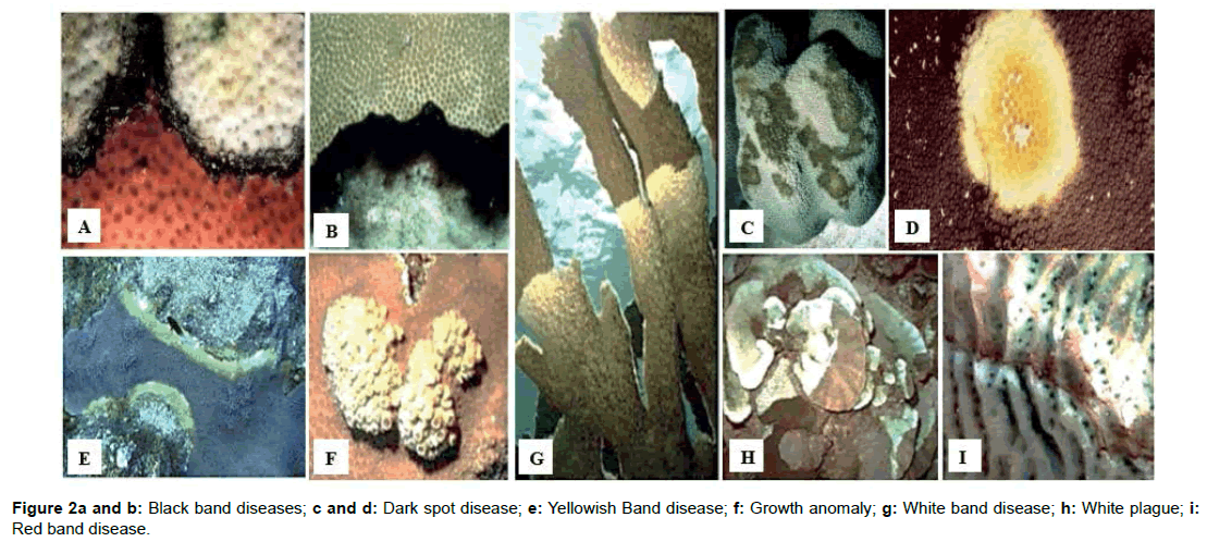
In healthy Caribbean coral, when microbial communities affect the coral were strikingly different from the community on the same species of corals that has infected with black band disease [16]. Molecular studies of Montastraea annularis corals with a white plague with shifts in the microbial community even in healthy tissues [17]. These coralassociated with bacteria, probably help to define a coral’s ecological place on the reef and interruption of this microbial community could make corals susceptible to mortality [18-21]. As the mucolytic activity of pathogens is higher than benign bacteria, they can make the coral sick absorption of almost 109 cells/mL [22,23]. Though the etiological agents are not specific to corals yet are able to pass diseases which is multiple and distinctive to marine organism [24,25] (Figure 2).
Bacterial diseases of corals
According to the Bourne, is being believed as the causative microbial agent of some coral disease widely reported that bacterial populations living in coral mucus [26,27]. White Band Disease (WBD) was first described in the Caribbean in the mid-1970s [28]. It is characterized by a tissue that is peeling off in a consistent band starting at the base and moving up in the branch and with a tissue loss of several millimeters a day, which can eventually kill entire colonies. Acropora palmata populations, along with A. cervicornis populations, have also been affected by White Band Disease (WBD) (28-30]. Further it’s noted that etiology of WBD is not fully understood, yet it assumed that this bacterial infection is of unknown origin [13,28-31].
Also noted that aggregations of gram-negative bacteria have been found in association with Acropora palmata showing signs of WBD Type I [29]. Ritchie and Smith (1998) [32] later proposed Vibrio carchariae as the causal agent of WBD Type II, but it was never confirmed. White Pox Disease (WPD) was first described in 1996 on reefs of Key West, FL and has been observed throughout the Caribbean [33]. When the causal agent Serratia marcescens is the only confirmed agent resulting in WPD lesions in Acropora palmata [15]. Other microbes could also result in similar disease patterns but are yet not described. Its due to an organism in the enteric bacterium (Serratia marcescens) which is commonly found in clinical situations [34,35] and commonly found in water, soil, crops and in wide range of organisms including insects, plants and human feces [34,36,37]. WPD which spread rapidly is notable with white patches of a bare skeleton which can be found on the surfaces or undersides of branches. Sutherland and Ritchie (2004) [8] reported these lesions can grow at an average rate of 2.5 cm2 day- 1 and greatest tissue loss coincides with high temperature. White pox disease is sometimes mistaken for white band disease, bleaching, and predation scars by Coralliophila abbreviata, although its distinct lesion characteristics can be easily discerned by trained eyes. Also, Rogers et al. [38] mentioned it is not clear when WPD first appeared.
An E. cloaca being a family member of Enterobacteriaceae and like most of the other family members is an opportunistic pathogen that causes infections in hospitalized patients [39,40]. Santavy [41] has found, Porites astreoides harbor a bacterial species, possibly Moraxella sp. that forms ovoids within P. astreoides and seems to take part in the normal life cycle of healthy corals. Also, some other studies have proved that certain species of nitrogen-fixing bacteria are associated with corals and/or their skeletons [42,43].
Mucus layer of corals
Mucus is consisting of primary metabolites such carbohydrates, proteins, and lipid which secreted by surface of coral and used defined as Coral Surface Micro-layer (CSM). The Surface Muco-polysaccharides Layer (SML) and Muco-Polysaccharides Layer (MPSL) are secreted by microbial populations which live in surface of corals [44]. Mucus are responsible for significant of coral mortality, mainly in the Caribbean [45]. Mucus layer may both act as a protective physicochemical barrier [8,46,47] and growth media for bacteria which include pathogens [23,48,49]. Particularly, the SML provides hundred times of growth media for bacteria than other layers and here able to act as metabolically in nearby seawater [50].
Phylogenetic Relationships of Coral Pathogens
Molecular and genomic techniques are used to study the depth of coral pathogens. Molecular techniques are used to identify new organisms and new metabolic processes [51]. Unrestricted mete genomic studies provide information on all genomic regions. It enables the functioning of both taxonomic description and potential metabolic of the microorganism within an environment [52]. 16S rDNA have identified that microbial population related with corals are diverse to develop both species-species [53] and generalist associations [52]. The part of the DNA now most commonly used for taxonomic purposes for bacteria is the 16S rRNA gene [54-58]. Identification and classification of the marine higher fungi have followed traditional avenues of the evaluation and significance of morphological characters at the light microscope level. For the Ascomycota, ultra-structural characters in the transmission and scanning electron microscope levels have also been used [59]. In studying phylogenetic relationships of pathogens, the morphology and analyses of DNA sequences are used. Ribosomal DNA has been extensively used for systematic and phylogenetic sides due to exploiting variation in the molecular techniques [60].
Importance of Coral Reef
Variety of fish species and marine life are inhabitants of coral reefs. They decorate the sea floor and create a vast diversity in the ecosystem. Reefs are made up of many groups of coral, which is a tiny living organism that has a larger role in balancing the marine ecosystem. Someone may think that the coral reefs are inanimate, but they are alive and beneficial not only to ocean but for humans live in the land.
Medicine properties
Some compounds found in coral reefs are of medicinal value. They are being used to treat asthma, arthritis, human bacterial infections, Alzheimer’s disease, cardio vascular diseases, Viruses and even type of cancer. It has given a great boost to the medical field [61]. Researchers are continuing to study other medicinal and health benefits coral can provide, including anti-tumor activity [62].
Fishing activity
Coral reefs are habitats to various species of fish and many countries rely on these sites for fishing. According to NOAA (National Oceanic and Atmospheric Administration), fishing activities in coral reefs carries in certain countries which are emerging to produce food for millions of people [63].
Tourism
Coral reef ecosystem is home for variety of marine life such as clown fish, sharks, and sea turtles which attracts tourists all around the world. They are attractive sites for scuba divers or snorkelers as well and thus coral reefs can worth millions of dollars a year, globally in tourism if the reef ’s health is impacted negatively so is the community. Tourism will be disturbed causing a decrease in destinations of income and loss of employment [64].
Coastal protection
Coral reefs act as strong obstacles between the sea and land. Its reefs are so strong that it obstructs the waves during storms hurricanes typhoons and even tsunamis. It saves the property from erosion, flooding and so on of the shore. Coral reef further saves billions of dollars annually by terms of reduced insurances, coastal defense and restoration costs [65].
Conclusion
Coral reefs have highly significant due to their highly applications on this planet such as protecting seashores from hurtful of wave action and tropical storms, be responsible for habitats and shelter for many marine organisms. Coral reefs are one of source of nitrogen and other essential nutrients for ocean food chains and impact of fixation of carbon and nitrogen also assist to nutrient recycling. Therefore, most of marine organism lives in coral reefs.
Coral Reefs are habitats to various species of fish and many countries rely on these sites for fishing. In Australian economy, through fishing and tourism coral reef has make above one half million in annually. The coral reef is cleansing environment and climate over the past million’s years. Lessening biodiversity through the removal of species unescapably this leads to the breakdown in ecosystem health and function.
Enthusiastic environment is needed for natural resources such as nutrition and medications also services are reprocessing and distillation of water and air, the materialization of soil and breaking the pollutants, social cultural and recreational activities and high species diversity. This diversity is providing a larger gene pool and provide good societies as a survival option when climates and environmental conditions are changed. Therefore, death warning for species when there is limit. So, they may play crucial role in an ecosystem, uncertainly removed all organisms are atmosphere effect within community. Currently, real challenges of humankind activities and climate changes are affecting the health and function of species.
Coral reefs are a precious gift that provides humans with many resources in medicine, and fishing. It brings in billions of dollars that benefit the community as well. Furthermore, the coral reef ecosystem could represent increasingly significant sources for medical treatments, nutritional supplements, pesticides, cosmetics and other commercial products. So that needs to develop strategies to protect coral reef. By protecting the ocean, we ensure the health of reefs of thousands of species. A healthy ocean will keep coral beautiful.
Consent for Publication
We certify this manuscript has not been published elsewhere and is not submitted to another Journal.
Competing Interest
The author(s) declare that they have no competing interests.
Acknowledgement
Support given by Senior Lecturer Dr. M.P Kumara, Faculty of Fisheries & Marine Science, Ocean University of Sri Lanka, Tangalle, Sri Lanka is also highly appreciated. The authors wish to thank Mr. D.P.G.S.P Jayasinghe Department of Veterinary Pathobiology, Faculty of Veterinary Medicine and Animal Science is also appreciated.
References
- Barnes RD (1987) Invertebrate Zoology; Fifth Edition. Fort Worth, TX: Harcourt Brace Jovanovich College Publishers. 92-96, 127-134, 149-162.
- Lalli CM, Parsons TR (1995)Biological Oceanography: An Introduction. Oxford, UK: Butterworth-Heinemann Ltd. 220-233.
- Levinton JS (1995) Marine Biology: Function, Biodiversity, Ecology. New York: Oxford University Press. J Mar Biol Assoc UK 306-319.
- Barnes RSK, Hughes RN (1999) An Introduction to Marine Ecology; third edition. Oxford, UK: Blackwell Science Ltd. 117-141.
- Sumich JL (1996) An Introduction to the Biology of Marine Life, sixth edition. Dubuque. Am Biol Teach 255-269.
- Green EP, Bruckner AW (2000) The significance of coral disease epizootiology for coral reef conservation. Biol Conserv 96: 347-361.
- Aronson RB, Precht WF (2001) White-band disease and the changing face of Caribbean coral reefs. Hydrobiologia 460: 24-38
- Sutherland KP, Ritchie KB (2004) White Pox Disease of the Caribbean Elkhorn Coral, Acropora palmata. Coral Health Dis Springer-Verlag, Berlin.
- Willis BL, Page CA, Dinsdale EA (2004) Corals disease on the Great Barrier Reef. Coral Health Dis Springer-Verlag Heildelberg
- Highsmith RC (1981) Lime-boring algae in coral skeletons. J Exp Mar Biol Ecol 55: 267-281.
- Campion-Alsumard T Le, Golubic S, Hutchings P (1995) Microbial endoliths in skeletons of live and dead corals-Porites Lobata (Moorea, French-Polynesia). Mar Ecol Prog Ser 117: 149-157.
- Bythell J, Pantos O, Richardson L (2004) White plague, white band, and other white diseases. Coral Health Dis Springer-Verlag, Berlin, Heidelberg 351-365.
- Antonius A (1981) Coral reef pathology- a review. Agricultural Information Bank for Asia, South-East Asian Regional Center for Graduate Study and Research in Agriculture 2: 3-6.
- Gladfelter WB (1982) White-band disease in Acropora palmate implications for the structure and growth of shallow reefs. Bull Mar Sci 32: 639-643.
- Patterson KL, Porter JW, Ritchie KB, Polson SW, Mueller E, et al. (2002) The etiology of white pox, a lethal disease of the Caribbean elkhorn coral, Acropora palmata.Proceedings of the National Academy of Sciences. 99: 8725-8730.
- Frias-Lopez J, Zerkle AL, Bonheyo GT, Fouke BW (2002) Partitioning of bacterial communities between seawater and healthy, black band diseased, and dead coral surfaces. Appl Environ Microbiol 68: 2214-2228.
- Pantos O, Cooney R, Le Tissier M, Barer M, O’Donnell A, et al. (2003) The bacterial ecology of a plague-like disease affecting the Caribbean coral Montastraea annularis. Environ Microbiol 5: 370-382.
- Ducklow HW, Mitchell R (1979) Bacterial populations and adaptations in the mucus layers on living corals. Limnol Oceanogr 24: 715-725.
- Segel LA, Ducklow HW (1982) A theoretical investigation into the influence of sublethal stresses on coral-bacterial ecosystem dynamics. Bull Mar Sci 32: 919-935.
- Knowlton N, Rohwer F (2003) Multispecies microbial mutualisms on coral reefs: the host as a habitat. Am Nat 162: S51-S62.
- Rohwer F, Kelley S (2004) Culture-independent analyses of coral-associated microbes. Coral Health Dis Heidelberg (Germany): Springer-Verlag 265-275.
- Deplancke B, Vidal O, Ganessunker D, Donovan SM, Mackie RI, et al. (2002) Selective growth of mucolytic bacteria including Clostridium perfringens in a neonatal piglet model of total parenteral nutrition. Am J Clin Nutr 76: 1117-1125.
- Israeli T, Banin E, Rosenberg E. (2001) Growth, differentiation and death of Vibrio shiloi in coral tissue as a function of seawater temperature. Aquat Microb Ecol 24: 1-8.
- Pantos O, Bythell JC (2006) Bacterial community structure associated with white band disease in the elkhorn coral Acropora palmata determined using culture-independent 16S rRNA techniques. Dis Aquatic Organisms 69: 79-88.
- Harvell D, Group CDW (2007) Coral disease, environmental drivers and the balance between coral and microbial associates. Oceanogr 20: 172-195.
- Koren O, Rosenberg E (2006) Bacteria Associated with Mucus and Tissues of the Coral Oculina patagonica in summer and winter. J Appl Environ Microbiol 72: 5254-5259.
- Mitchell R, Chet I (1975) Bacterial attack of corals in polluted sea-water. Microb Ecol 2: 227-233.
- Peters EC (1997) Diseases of coral reef organisms. In Birkeland, C. (ed.), Life and Death of Reefs. New York: Chapman & Hall. 114-139.
- Peters EC, Oprandy JJ, Yevich PP (1983) Possible causal agent of white band disease in Caribbean acroporid corals. J Invertebr Pathol 41: 394-396.
- Bythell JC, Hillis-Starr ZM, Rogers CS (2000) Local variability but landscape stability in coral reef communities following repeated hurricane impacts. Mar Ecol Prog Ser 204: 93-100.
- Aronson RB, MacIntyre IG, Precht WF, Murdoch TJT, Wapnick CM (2002) The expanding scale of species turnover events on coral reefs in Belize. Ecol Monogr 72: 233-249.
- Ritchie KB, Smith GW (1998) Type II white-band disease. Rev Biol Trop 46: 199-203.
- Holden C (1996) Coral disease hot spot in the Florida Keys. Science 274: 2017.
- Grimont PA, Grimont F (1994) Genus VIII Serratia Bizio. Bergey's Manual of Systemic Bacteriology. 477-484.
- Miranda G, Kelly C, Solorzano F, Leanos B, Coria R, et al. (1996) Use of pulse-field gel electrophoresis typing to study an outbreak of infection due to Serratia marcescens in a neonatal intensive care unit. J Clin Microbiol 34: 3138-3141.
- Carbonell GV, Colleta HHMD, Yano T, Darin ALC, Levy CE, et al. (2000) Clinical relevance and virulence factors of pigmented Serratia marcescens. FEMS Immunol Microbiol Mtds 28: 143-149.
- Baya A (1992) Serratia Marcescens: a potential pathogen for fish. J Fish Dis 15: 15-26.
- Rogers CS, Sutherland KP, Porter JW (2005) Has white pox disease been affecting Acropora palmata for over 30 years? Coral Reefs 24: 194.
- Flynn DM, Weinstein RA, Nathan C, Gaston MA, Kabins SA (1987) Patients endogenous flora as a source of “noscomial†Enterobacter in cardiac surgery. J Infect Dis 156: 363-368.
- Gaston MA (1988) Enterobacter: an emerging nosocomial pathogen. J Hosp Infect 11: 197- 208.
- Santavy DL (1995) The Diversity of Microorganisms Associated with Marine Invertebrates and Their Roles in the Maintenance of Ecosystems. 211-229.
- Williams WM, Viner AB, Broughton WJ (1987) Nitrogen fixation (acetylene reduction) associated with the living coral Acropora variabilis. Mar Biol 94: 531-535.
- Shashar N, Cohen Y, Loya Y, Sar N (1994) Nitrogen fixation (acetylene reduction) in stony corals: evidence for coral- bacteria interactions. Mar Ecol Prog Ser 111: 259-64.
- Kellogg C (2004) Tropical Archaea: diversity associated with the surface microlayer of corals. Mar Ecol Prog Ser 273: 81-88.
- Porter JW, Dustan P, Jaap WC, Patterson KL, Kosmynin V, et al. (2001) Patterns of spread of coral disease in the Florida Keys. Hydrobiologia 460: 1-24.
- Santavy D, Peters E (1997) Microbial pests: coral disease in the Western Atlantic. Proc 8th Int Coral Reef Sym 1: 607-612.
- Hayes RL, Goreau NI (1998) The significance of emerging diseases in the tropical reef ecosystem. Rev Biol Trop 5: 173-185.
- Toren A, Landau L, Kushmaro A, Loya Y, Rosenberg E (1998) Effect of temperature on adhesion of Vibrio Strain AK-1 to Oculina patagonica and on coral bleaching. Appl Environ Microbiol 64: 1379-1384.
- Lipp EK, Jarrell JL, Griffin DW, Lukasik J, Jaculiewicz J, et al. (2002) Preliminary evidence for human fecal contamination of corals in the Florida Keys, USA. Mar Pollut Bull 44: 666-670.
- Ritchie KB, Smith GW (2004) Microbial communities of coral surface mucopolysaccharide layers. Coral Health Dis Springer-Verlag. 259-264.
- Beja O, Aravind L, Koonin EV, Suzuki MT, Hadd, (2000) Bacterial rhodopsin: evidence for a new type of phototrophy in the sea. Science 289: 1902-1906.
- Wegley L, Edwards R, Rodriguez BB, Liu H, Rohwer F (2007) Metagenomic analysis of the microbial community associated with the coral Porites astreoides. Environ Microbiol 9: 2707-2719.
- Rohwer F, Seguritan V, Azam F, Knowlton N (2002) Diversity and distribution of coral-associated bacteria. Mar Ecol Prog Ser 243: 1-10
- Bottger EC (1989) Rapid determination of bacterial ribosomal RNA sequences by direct sequencing of enzymatically amplified DNA. FEMS Microbiol Lett 65: 171-176.
- Garrity GM, Holt JG (2001) The road map to the Manual. Bergey’s Manual of Systematic Bacteriology 119-166.
- Kolbert CP, Persing DH (1999) Ribosomal DNA sequencing as a tool for identification of bacterial pathogens. Curr Opin Microbiol 2: 299-305.
- Palys T, Nakamura LK, Cohan FM (1997) Discovery and classification of ecological diversity in the bacterial world: the role of DNA sequence data. Int J Syst Bacteriol 47: 1145-1156.
- Tortoli E (2003) Impact of genotypic studies on mycobacterial taxonomy: the new mycobacteria of the 1990s. Clin Microbiol Rev 16: 319-354.
- Frankl A, Mari M, Reggiori F (2015) Electron microscopy for ultrastructural analysis and protein localization in Saccharomyces cerevisiae. Microb Cell 2: 412-428.
- Latiffah Z, Harikhrisna K, Tan SG, Tan SH, Abdullah F, et al. (2002) Restriction analysis and sequencing of the ITS regions and 5.8S gene of rDNA of Ganoderma isolates from infected oil palm and coconut stumps in Malaysia. Ann Appl Biol 141: 133-142.
- Cooper EL, Hirabayashi K, Strychar KB, Sammarco PW (2014) Corals and Their Potential Applications to Integrative Medicine. Evidence-Based Complementary and Alternative Medicine 2014.
- Galloway SB, Bruckner AW, Woodley CM (2009) Coral Health and Disease in the Pacific: Vision for Action. Coral Disease and Health Constrium 314.
- Teh LSL, Teh LCL, Sumaila UR (2013) A Global Estimate of the Number of Coral Reef Fishers. PLoS One 8: e65397.
- Hilmi N, Safa A, Reynaud S, Allemand D (2012) Coral Reefs and Tourism in Egypt’s Red Sea. Topics in Middle Eastern and African Economies 14.
- Guannel G, Arkema K, Ruggiero P, Verutes G (2016) The Power of Three: Coral Reefs, Seagrasses and Mangroves Protect Coastal Regions and Increase Their Resilience. PLoS One 11: e0158094.
Citation: Premarathnaa AD, Sarvananda L, Jayasooriya AP, Amarakoon S (2019) A Review on Pathogenic Diseases on Corals Associated Risk Factors and Possible Devastations in Future in the Globe. J Marine Sci Res Dev 9: 269.
Copyright: © 2019 Premarathnaa AD, et al. This is an open-access article distributed under the terms of the Creative Commons Attribution License, which permits unrestricted use, distribution, and reproduction in any medium, provided the original author and source are credited.
Select your language of interest to view the total content in your interested language
Share This Article
Recommended Journals
Open Access Journals
Article Usage
- Total views: 3868
- [From(publication date): 0-2019 - Dec 22, 2025]
- Breakdown by view type
- HTML page views: 2972
- PDF downloads: 896


