Axillary Masse, is it an Ectopic Mammary Gland?
Received: 29-Apr-2016 / Accepted Date: 16-May-2016 / Published Date: 30-May-2016 DOI: 10.4172/2572-4118.1000108
Abstract
Ectopic breast tissue or super numerary mammary gland is a congenital condition characterized by the development of mammary tissue along the embryonic milk line. This condition Occurs in 0.4-6% of the general population. The most common site of development of super numerary mammary gland is the axillar region. Axillary ectopic breast tissue may provide a diagnostic challenge, as other benign and malignant lesions occur in this area. We report a case of 18 years-old girl presenting an axillary masse considered firstly as an ectopic mammary gland. Ultrasound aspect and axillary MRI correct the diagnosis and lead to provide specific management of this tumor.
Keywords: Ectopic breast tissue; Nervous tumor; Axillary masse
35810Introduction
The axillar region can be the location of much benign or malignant tumor. The most frequent are lymphadenopathy (lymph node reaction after infectious, inflammatory or malignant diseases). Ectopic breast tissue, the most common breast congenital disease, can be considered as a differential diagnosis of axillary masse [1]. Many other rare tumors must be included. Clinical assessment of axillary region can be unhelpful and ultrasound or MRI imaging can be required to establish the right diagnosis [2].
The Case Report
The reported case is about an 18 years-old girl without any medical history. She presented progressively a right axillary masse. Clinical examination showed an axillary delimited soft masse, movable relative to deep plane. An axillary ectopic breast tissue was suspected (Figures 1 and 2). Breast ultrasound examination and mammogram were normal. Axillary ultrasound showed a homogenous delimited solid tumor measuring 6 cm (Figure 3). This examination wasn't helpful to determine the origin of this tumor. An axillary MRI was performed. It showed a multi lobular encapsulated solid tumor, measuring 6 cm. This tumor was closely near to the humeral head and to nervous structure (Figures 4 and 5). We decided to proceed to a surgical biopsy through an axillary incision. The histological examination showed an axillary shwannoma, a peripheral benign nervous tumor (Figures 6 and 7). The patient underwent a tumoral excision in the department of plastic and hand surgery. The after-surgery follow-up was without complications.
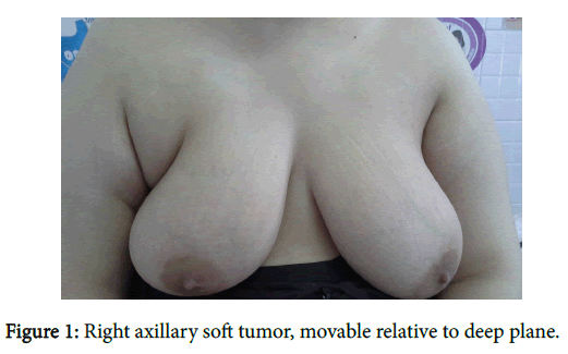
Figure 1: Right axillary soft tumor, movable relative to deep plane.
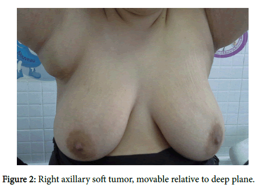
Figure 2: Right axillary soft tumor, movable relative to deep plane.
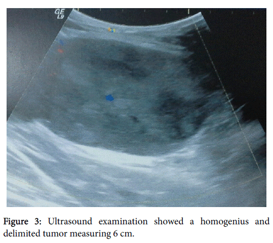
Figure 3: Ultrasound examination showed a homogenius and delimited tumor measuring 6 cm.
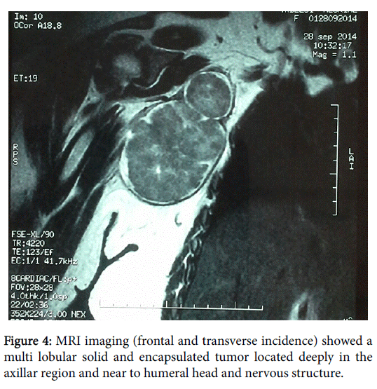
Figure 4: MRI imaging (frontal and transverse incidence) showed a multi lobular solid and encapsulated tumor located deeply in the axillar region and near to humeral head and nervous structure.
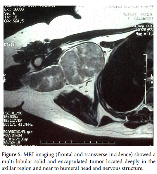
Figure 5: MRI imaging (frontal and transverse incidence) showed a multi lobular solid and encapsulated tumor located deeply in the axillar region and near to humeral head and nervous structure.
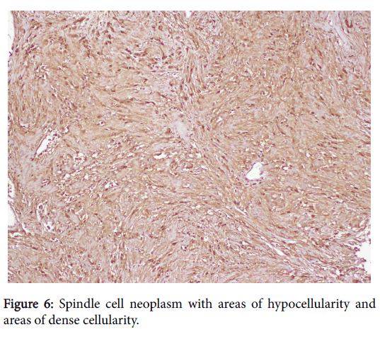
Figure 6: Spindle cell neoplasm with areas of hypocellularity and areas of dense cellularity.
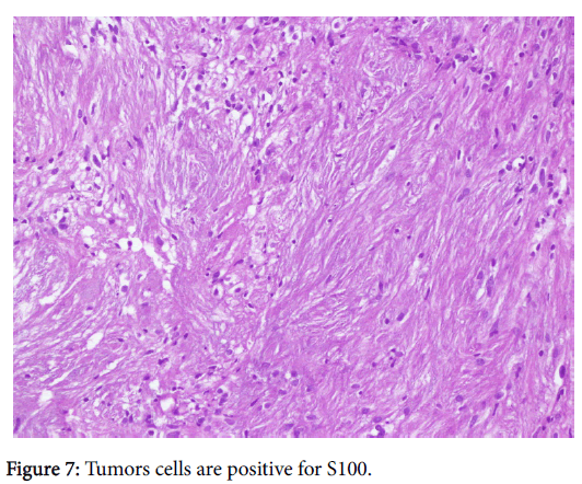
Figure 7: Tumors cells are positive for S100.
Discussions
Ectopic breast tissue is a rare congenital condition occurring in 0.4 to 6% of general population. It is the most common breast congenital abnormality. This entity is divided to two histologic subgroups: supernumerary breast and aberrant breast tissue.
The difference between those subtypes is that the first one consists on an organized ductal system communicating with its overlying skin, whereas the latter is characterized by an unorganized secretory system which has no communication to the overlying skin [3,4]. Ectopic breast tissue is more frequently localized in the axilla [5] but other localizations have been reported in parasternal, subclavicular, submammary, vulvar and anal regions [4-6].
The ectopic breast tissue can be complicated as a normally lactated breast tissue especially by benign and malignant tumors such as carcinoma, intraductal papilloma, fibroadenoma and fibrocystic disease [7,8].
In our case, we had a diagnostic challenge related to the unusual clinical presentation of the described tumor (consistency, clinical characteristic), a ultrasound examination and MRI were needed showing an atypical and unusual aspect of a multi lobular encapsulated solid tumor, measuring 6 cm. histological examination was the only way to establish the diagnosis, it was an axillary shwannoma.
This is a benign tumor of schwann cells, produces a wellcircumscribed, slowly growing lesion. It is the most common benign peripheral nerve lesion. Nerve tumors comprise less than 5% of all upper extremity tumors. Schwannoma usually occurs in the fore arm and hand, but cervical and axillary tumors have been reported [9,10].
Concerning axillary masses, the most common diagnosis is palpable lymph node (secondary to infectious, inflammatory or malignant malformation, lipoma, cysts, hidradentitis suppurativa and ectopic breast tissue. Schwanomma should be included in the differential diagnosis of axillary masses because it has also been reported in the axilla. In addition, schwanoma often is confused with other soft tissue masses, and inadvertent nerve biopsy may lead to permanent disability. Seventeen percent of peripheral nerve trauma is due to iatrogenic injury [11]. Injury to the spinal accessory nerve has been documented after [12] nerve injury after axillary lymph node biopsy has not been reported previously. In our case surgical biopsy was performed and fortunately no complications or nerve injury were seen. Preoperative magnetic resonance imaging may help delineate peripheral nerve sheath tumors in the axilla if the diagnosis is suspected. In addition, clear visualization of the mass is necessary before excision to prevent significant nerve injury.
This case is important to study because of difficulties and challenge to diagnose axillary masse. Many types of tumor occur in this region. In a Department of gynecologic and breast surgery, the mostly frequent axillary masse is related to breast disease such lymph node involvement in case of breast cancer or ectopic mammary gland. MRI is a good tool to have an idea about the origin and the management of axillary masses.
When shwannoma is suspected, a specialist intervention, investigation and management are required. Biopsy is not allowed because of the risk of iatrogenic nerve injuries. Nervous tumor especially shwannoma are to keep in mind when we are confronted to deal with axillary tumor.
Conflict of Interest Statement
We declare that we have no conflict of interest.
References
- Famà F, Gioffrè Florio MA, Villari SA, Caruso R, Barresi V, et al. (2007) Breast abnormalities: a retrospective study of 208 patients. ChirItal 59: 499-506.
- Velanovich V (1995) Ectopic breast tissue, supernumerary breasts, and supernumerary nipples. South Med J 88: 903-906.
- Georgiade NG, Georgiade GS, Riefkohl R, McCarty KS Jr, Carpenter SA (1990). The breast: embryology, anatomy and physiology. In: Georgiade NG, Georgiade GS, Riefkohl R, eds. Aesthetic Surgery of the Breast 1st ed. Philadelphia: WB Saunders3-17.
- Marshall MB, Moynihan JJ, Frost A, Evans SR (1994) Ectopic breast cancer: case report and literature review. SurgOncol 3: 295-304.
- Roodra AK, Hansen JP, Rider JA, Huang S, Rider DL (2002) Ectopic breast cancer: special treatment consideration in postmenopausal patients. Breast J 8: 286-289.
- Chan NG, Penswick JL, Labelle E, Driman DK (2007) Ectopic breast tissue presenting as an anal polyp. Can J Surg 50: E23-24.
- Coras B, Landthaler M, Hofstaedter F, Meisel C, Hohenleutner U (2005) Fibroadenoma of the axilla. Dermatol Surg 31: 1152-1154.
- Amsler E, Sigal-Zafrani B, Marinho E, Aractingi S (2002) Ectopic breast cancer of the axilla. Ann Dermatol Venereol 129: 1389-1391.
- Maiuri F, Donzelli R, Benvenuti D, Sardo L, Cirillo S (2001) Schwannomas of the brachial plexus--diagnostic and surgical problems. Zentralbl Neurochir 62: 93-97.
- Boutsen Y, DeCoene B, Hanson P, Deltombe T, Gilliard C, et al. (1999) Axillary schwannoma masquerading as cervical radiculopathy. Clin Rheumatol 18: 174-176.
- Kretschmer T, Antoniadis G, Braun V, Rath SA, Richter HP (2001) Evaluation of iatrogenic lesions in 722 surgically treated cases of peripheral nerve trauma. J Neurosurg 94: 905-912.
- de Souza FM, Hudson AR (1989) Surgical exploration of enlarged lymph nodes at the root of the neck. J Otolaryngol 18: 112-115.
Citation: Aymen FA, Atef Y, Majed G, Nadia B, Fethi BA, et al. (2016) Axillary Masse, is it an Ectopic Mammary Gland?. Breast Can Curr Res 1: 108. DOI: 10.4172/2572-4118.1000108
Copyright: ©2016 Aymen FM et al. This is an open-access article distributed under the terms of the Creative Commons Attribution License, which permits unrestricted use, distribution, and reproduction in any medium, provided the original author and source are credited
Select your language of interest to view the total content in your interested language
Share This Article
Recommended Journals
Open Access Journals
Article Tools
Article Usage
- Total views: 16392
- [From(publication date): 6-2016 - Jul 06, 2025]
- Breakdown by view type
- HTML page views: 15420
- PDF downloads: 972