Capsule Based Gastric Rhythm Analyzer: A Tool for Diagnosing Gastric Diseases
Received: 13-Dec-2017 / Accepted Date: 21-Dec-2017 / Published Date: 27-Dec-2017 DOI: 10.4172/2161-069X.1000544
Abstract
The proposal of this revolutionary project is a mission which has never been thought by any human kind and which is now on the verge of production by us. Electrogastrography and capsule endoscopy are contributing in the diagnosis of Gastric diseases. On their individual functioning, as a diagnostic tool, they are not capable of producing appropriate results such as the position of tumor in the GI tract. The recording of electrical rhythmic activity can mimic the disability of gastric cells which can’t be diagnosed by endoscopic procedures. Integrating the gastric rhythmic information and capsule endoscopy can lead to an ultimate diagnosis of gastric diseases. The structural information which is captured by a capsule consisting of a camera, LEDs and transmitter provides a visual diagnosis of the gastric disorders/infection. The functional information is obtained by recording the electrical rhythmic activity of the gastric cell via surface electrodes. By mapping the electrical rhythmic signal with the image acquired from the capsule provides the accurate localization of the abnormality.
Keywords: Positioning of the tumor; Gastric rhythms; Capsule endoscopy; Surface electrodes; Electro gastrogram
Introduction
Gastropathy is a general term used for gastric diseases. Many gastric diseases are associated with infection. Historically, it was widely believed that the highly acidic environment of the stomach would keep the stomach immune from infection. The gastric diseases are the third fatal diseases in the world; those diseases are needed to be diagnosed properly for sustainment. The common gastric diseases are ulcers, gastritis, cancer, dyspepsia, nausea, tachygastria and bradygastria. Possible diagnostic procedures include endoscopy, capsules, ultrasound, and barium X-ray scan etc. The normal endoscopic procedure is a completely invasive method, which will lead the patient to take more medications and cause discomfort. To overcome these difficulties and hospitalization, the capsule endoscopy is introduced. The capsule endoscopy is a way to capture images of the digestive tract to diagnose whether it is pathologically stable. The imaging by capsule is only confined to the path it travels and the camera facing, through which it can neglect the information that is available aside which will lead to erroneous diagnosis.
A gastric slow wave potential is a rhythmic electrophysiological event in the gastrointestinal tract. The normal conduction of slow waves is one of the key regulators of gastrointestinal motility [1]. Slow waves are generated and propagated by a class of pacemaker cells called the interstitial cells of Cajal (ICC), which also act as an intermediate between nerves and smooth muscle cells [2]. These slow waves then propagated to the surrounding cells and cause peristalsis which helps in the movement of bolus and breaks it down into small particles. These slow waves or gastric rhythms are very sensitive and can easily be affected by the movement of limbs or body, respiration or by vocalization. These artifacts can be minimized by signal conditioning unit. Thus, the integration of information acquired from capsule and the functional information from the electrical activity of gastric cells can provide a best way to overcome the cons of former diagnostic procedures.
Integration
The integration has the theme of merging two interdisciplinary diagnostic information such as gastric rhythmic information and structural information by aphelion software.
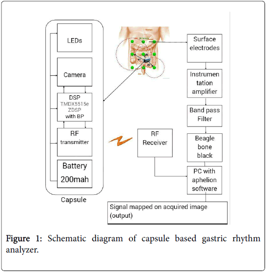
The unique objective is to provide a quality diagnosis which can be made through this integration. The role of the aphelion is to acquire image from capsule and processing it to map with the functional information obtained from the Beaglebone black processor. The schematic diagram (Figure 1) describes the integration which has two main sub systems i.e. capsule unit and gastric rhythm analyzer unit.
Capsule unit
It captures the images of the digestive tract for diagnosis in medicine.
• Capsule: The capsule is the size and shape of a pill which contains a miniaturized camera, optical dome, lens holder, lens, LEDs, Transmitter, Battery and a processing unit [3]. After a patient swallows the capsule, the process begins.
• Camera (image sensor): It captures the image of the inner regions of GI tract and it is highly sensitive and produces high quality images also detects object as small as 0.1 mm.
• LED: It illuminates to provide a clear picture and supports the camera and it is placed around the lens and camera. There are 4 LED’s are arranged in donut shape.
• RF Transmitter: It transmits the image which is obtained from the camera through radio waves to the RF receiver which is placed on the abdomen.
• Battery: It is button shaped and in 2 numbers. Silver oxide as primary battery, such battery has even discharge voltage, disposable and does not cause harm to body. This battery life is eight hours.
• Optical dome: This shape results in easy orientation along central axis of small intestine and also propels the capsule forward easily.
• Lens holder: It is a part of it which accommodates the lens to fix tightly so that it does not get dislocated at any time.
• Lens: These are arranged behind the light receiving window.
Rhythm analyzer unit
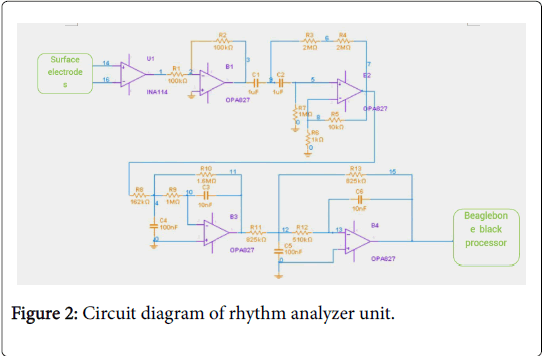
It is an integral unit of electrodes, amplifier, filters and a processor to record the electrical activity of the gastric cells (Figure 2).
• Surface electrodes: These are cutaneous electrodes which taps the electrical impulses produced by the gastric cells.
• Instrumentation amplifier (INA114): The normal amplitude range for a gastric signal is 100-500 micro volts. In order to process by further circuitries, it should be amplified with the gain of 10000dB to provide excellent accuracy in the diagnosis of the gastric diseases.
• Band pass filter (OPA827): The desired frequency range of normal gastric signal is 0.05-0.11 Hz, in order to eliminate the contamination of the signals fourth order band pass filter with appropriate cut-off frequencies is used.
• Beaglebone black processor: It is a microprocessor which receives the signal from the band pass filter through ADC (ADS1499-4PAG) and processes it to provide the diagnosis about the gastric diseases. Python programming language is used to code the processor for arrhythmia detection by considering the frequency and amplitude information of the gastric signals.
Functional Information
Gastric rhythm activity
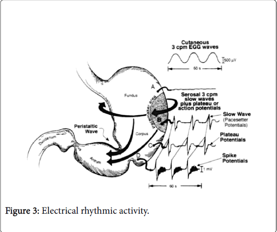
Human gastric slow wave or pacesetter potential activity generated by the ICCs occurs at a rate of 3 cycles per minute (cpm). Pacesetter potential activity is illustrated in Figure 3. Electrodes sewed onto the serous of the stomach records the depolarization and repolarization waves of the pacesetter potentials. The electrical wave front travels around the circumference of the stomach at a high rate of speed and migrates slowly towards the antrum at an increasing velocity. The plateau and spike potential are formed when pacesetter potentials migrated.
As a slow wave disappears in the distal antrum, another slow wave originates in the pacemaker area and begins to migrate toward the antrum approximately every 20 seconds. When there is little smooth muscle contractility (phase I or phase II of inter digestive state, described later), these electrical events reflect depolarization and repolarization of the ICCs and some small degree of contractility of the circular muscle cells. From an in vivo electrical viewpoint, the fasting pacesetter potential activity is relatively weak compared with the gastric myoelectrical activity during the postprandial period, when luminal contents and other stimuli augment gastric neuromuscular activity [4-6].
Electrode placement
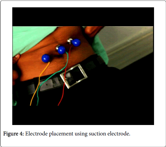
Electrodes are placed on the skin surface of the epigastria over the general area of the corpus and antrum of the stomach. Gastric rhythms are obtained with the electrodes arranged for bipolar recordings.
A reference electrode is approximately positioned on the right side of the patient's abdomen which is relative to the umbilicus and ribs. One active electrode should be placed approximately 10 cm cephalad from the umbilicus and 6 cm to the patient's left. It is important to place these electrodes below, and not on the lowest rib to avoid respiratory signals.
The second active electrode should be placed approximately 4 cm above the umbilicus (mid-way between the umbilicus and Xiphoid) on the midline of the abdomen. The reference electrode is placed 10 to 15 cm to the right of the midline electrode, usually along the midclavicular line and 2 to 3 below the lowest anterior rib on the right side [7,8].
Procedure and its pre-requirements
For clinical studies, patients should fast after midnight and then ingest a 200 Kcal breakfast of two pieces of toast and 4 oz. of apple juice 2 hours before the test. By controlling the pretest meal, more standardized baseline gastric rhythms are obtained. The best electrical recordings are obtained if the patient reclines about 30 to 45 degrees in a comfortable chair [6,9].
Signal acquisition
The electrodes placed on the abdomen captures the electrical signal and provide the functional information of the GI tract. The signal acquired will be produced by the rhythmic activity of the gastric cells (slow waves) is shown in the Figure 4 [10].
Structural Information
Capsule imaging
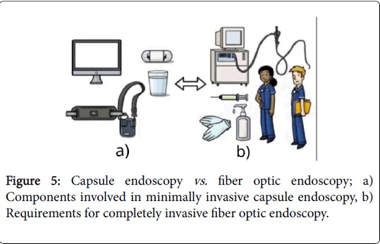
The structural information in the form of images can be captured by viewing it internally. This adopts the endoscopy. Though the fiber-optic endoscopy is invasive, it is widely used by neglecting its disadvantages such as medications and the frustrating procedure.
Capsule endoscopy evolves the procedure ultimately by eliminating hospitalization and being more comfortable [11,12]. The evolvement of endoscopy from wired to wireless enables the patient to carry their normal day-by-day activities are depicted via Figure 5.
Image acquisition
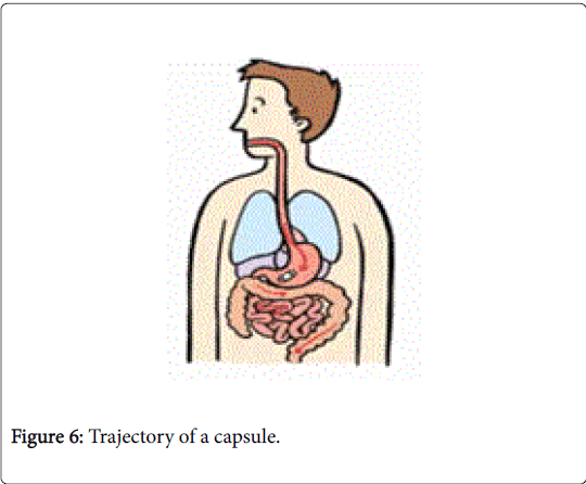
Capsule is swallowed by the patient like pill only after at least 3 hours of fasting and positioned with right lateral. During procedure patient will sip 15 ml of water for every 30 sec. then it takes images as it is propelled (Figure 6) forward by peristalsis [13,14].
Capsule contains light to illuminate digestive system, a camera to take images and an antenna that sends those images to recorder which worn on patient waist as a belt. A computer processes data and produces a continuous images and given to aphelion software.
Observation
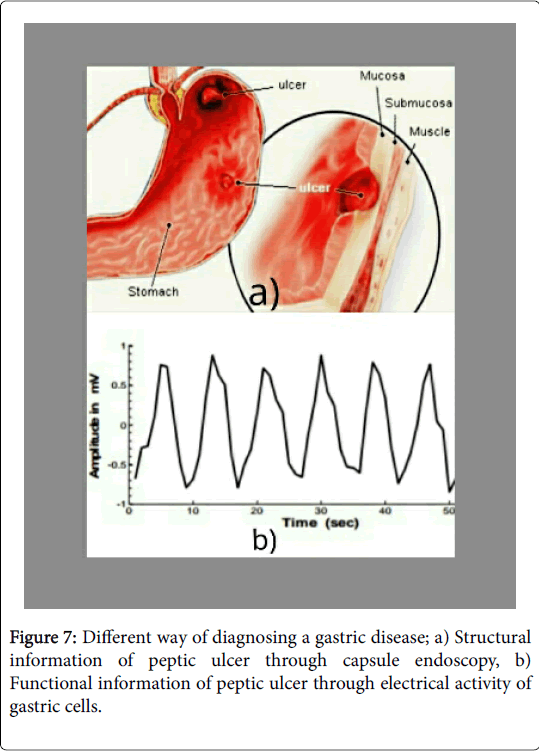
The integration of these two procedures can provide an ultimate diagnosis by using the individual information from both subsystems by using software (Figure 7) [15-17].
Conclusion
The gastrointestinal disease causes substantial mortality and morbidity. This paper provides the effective diagnostic information to the personnel through which the treatment standard has been improved. The revolutionary product serves as a prognostic tool which can predict the epidemiology of the whole GI tract without including any tedious processes. This eliminates the usage of different scanning procedures for different kinds of pathological or abnormal physiological conditions and also prevents prolonged hospitalization through ineffective diagnosis. The product may provide advanced and accurate diagnosis in case of earlier detection and localization of the tumors.
References
- Huizinga JD, Lammers WJ (2008) Gut peristalsis is governed by a multitude of cooperating mechanisms. Am J Physiol Gastrointest Liver Physiol 296: G1-8.
- Hanani M, Farrugia G, Komuro T (2004) Intercellular coupling of interstitial cells of cajal in the digestive tract. Int Rev Cytol 242: 249-282.
- Yuce MR, Dissanayake T (2012) Easy-to-swallow wireless telemetry. IEEE Microwave Magazine 13: 90-101.
- Thuneberg L, Peters S (2001) Toward a concept of stretch-coupling in smooth muscle: Anatomy of intestinal segmentation and sleeve contractions. The Anat Rec 262: 110-124.
- O'Grady G, Du P, Cheng LK, Egbuji JU, Lammers WJEP, et al. (2010) Origin and propagation of human gastric slow-wave activity defined by high-resolution mapping. Am J Physiol Gastrointest Liver Physiol 299: G585-592.
- Vianna EPM, Weinstock J, Elliot D, Summers R, Tranel D, et al. (2006) Increased feelings with increased body signals. Scan 1: 37-48.
- Ercolani M, Baldaro B, Trombini G, Trombini, G (1989) Effects of two tasks and two levels of difficulty on surface electrogastrograms. Percept Mot Skills 69: 99-110.
- Vianna EPM, Tranel D (2006) Gastric myoelectrical activity as an index of emotional arousal. Int J Psychophysiol 61: 70-76.
- Koch KL, Stern RM (2003) Handbook of Electrogastrography. (1st edn) Oxford university press, USA.
- Smout AJPM, Jebbink HJA, Samsom M (1994) Acquisition and analysis of electrogastrographic data: The Dutch experience. In: Chen JZ, McCallum RW Electrogastrography, New York: Raven Press pp: 3-30.
- Tokmakçi M (2007) Analysis of the electrogastrogram using discrete wavelet transform and statistical methods to detect gastric dysrhythmia. J Med Syst 31: 295-302.
- Bhattarai M, Bansal P, Khan Y (2013) Longest duration of retention of video capsule: A case report and literature review. World J Gastrointest Endosc 5: 352-355.
- Jung ES (2006) Design and implementation of the telemetry capsule for measuring of electrogastrography. Innsbruck, Austria, pp: 209-213.
- Colli A, Gana J, Turner D, Yap J, Adams-Webber T, et al. (2014) Capsule endoscopy for the diagnosis of oesophageal varices in people with chronic liver disease or portal vein thrombosis.
- Yuce MR, Alici G, Than TD (2014) Wireless endoscopy. Wiley encyclopedia of electrical and electronics engineering.
- Dickens EJ, Hirst GD, Tomita T (1999) Identification of rhythmically active cells in guinea-pig stomach. J Physiol (Lond) 514: 515-531.
Citation: Gokul M, Durgadevi N, Kumar CR (2017) Capsule Based Gastric Rhythm Analyzer: A Tool for Diagnosing Gastric Diseases. J Gastrointest Dig Syst 7: 544. DOI: 10.4172/2161-069X.1000544
Copyright: © 2017 Gokul M, et al. This is an open-access article distributed under the terms of the Creative Commons Attribution License, which permits unrestricted use, distribution, and reproduction in any medium, provided the original author and source are credited.
Select your language of interest to view the total content in your interested language
Share This Article
Recommended Journals
Open Access Journals
Article Tools
Article Usage
- Total views: 5902
- [From(publication date): 0-2017 - Nov 24, 2025]
- Breakdown by view type
- HTML page views: 4935
- PDF downloads: 967

