Common Benign Bone Tumor in Uncommon Sites: A Case Series
Received: 25-May-2025 / Manuscript No. JOO-25-168005 / Editor assigned: 28-May-2025 / PreQC No. JOO-25-168005 (PQ) / Reviewed: 12-Jun-2025 / QC No. JOO-25-168005 / Revised: 01-Jul-2025 / Manuscript No. JOO-25-168005 (R) / Published Date: 28-Jul-2025 QI No. / JOO-25-168005
Abstract
Introduction: Osteochondromas are common benign bone tumors typically found around the growing ends of long bones, such as the lower end of the femur and the upper end of the tibia. However, their occurrence in smaller bones is rare. Uncommon presentations can also be seen in flat bones, the pelvic body, scapula, skull and the smaller bones of the hands and feet. The clinical presentation can vary depending on the location of the tumor. In our study, we encountered 7 cases of osteochondromas located in unusual sites, specifically in the metatarsals, talus and proximal tibia and fibula.
Conclusion: Osteochondromas can occur at atypical locations. Therefore, it is crucial to thoroughly evaluate all patients who present with swelling and pain in bony regions to ensure accurate diagnosis and appropriate management of osteochondromas.
Keywords
Metatarsal; Talus; Fibula; Tibia; Benign tumor; Bony swelling; Unusual sites
Introduction
In this study, we observed 7 cases of osteochondromas at different sites in our institute. Among these cases, 2 patients had osteochondromas in the talus, 3 in the metatarsals and 2 in the proximal tibia; all patients were symptomatic and treated with surgical excision.
Osteochondromas, also referred to as osteocartilaginous exostoses, represent the majority of primary benign bone tumors. Generally asymptomatic, they typically present as a painless, slow growing mass on the affected bone. The World Health Organization (WHO) defines this lesion as a cartilage capped bony projection arising on the external surface of bone, containing a marrow cavity that is continuous with that of the underlying bone. While many osteochondromas are asymptomatic, symptoms may arise due to the compression of adjacent neurovascular structures, fractures, osseous deformities, bursa formation or malignancy. Most osteochondromas are incidentally diagnosed, with malignant transformation being the most severe complication. However, deformities and interference with the function of major joints are the most common complaints, especially in patients with hereditary multiple osteochondromas.
Case Presentation
Case 1
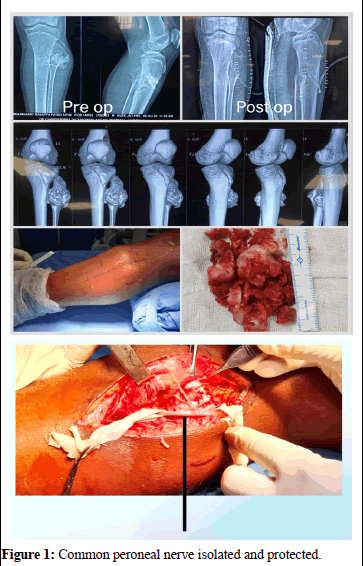
A 14-year-old boy presented with swelling on the anterolateral aspect of proximal part of left leg that had been progressively developing over the past 6 months. There was no history of trauma, fever or surgical interventions. The swelling was non-fluctuant, immobile, non-translucent and non-tender. Sensation and distal pulses were intact and no neurological deficits were observed. The examination revealed no scars, sinuses or distended veins. X-ray imaging of the leg demonstrated an exostosis arising from the posterolateral aspect of the proximal tibia (Figure 1). Under tourniquet control, a posterolateral approach was used for surgical dissection, during which the common peroneal nerve was carefully retracted. The exostosis was removed using a bone chisel and sent for histopathological examination, which confirmed the diagnosis of osteochondroma. Surgical excision was performed to prevent further progression and potential neurovascular compromise. The patient was immobilized with an above-knee slab for 3 weeks before being allowed to bear weight. The parents were informed about the potential risks of recurrence and malignant transformation in the future [1-3].
Case 2
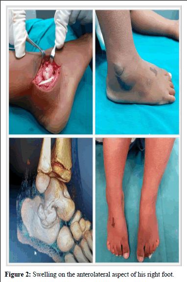
An 8-year-old boy presented with swelling on the anterolateral aspect of his right foot that had developed insidiously over the past 9 months. There was no history of trauma, fever or previous treatments. The examination revealed a hard, non-mobile swelling and no neurological deficits were evident. After a comprehensive workup including laboratory and radiological investigations, the patient underwent surgery. A lateral approach was utilized to excise the tumor arising from the body of the talus, which was then sent for biopsy. The patient was immobilized with a below knee slab for 4 weeks before being allowed to bear weight. The attendees were informed about the potential for recurrence. Follow-up at 3 months, 6 months and 1 year showed that the patient was comfortable, with no signs of recurrence noted during the follow-up visits (Figure 2) [4].
Case 3
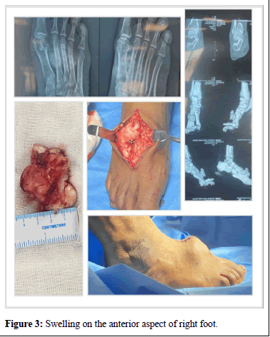
A 46-year-old female housewife presented with swelling on the anterior aspect of her right foot that had been progressively worsening over the past 2 years. Patient was having difficulty in wearing foot wear because of the swelling. There was no history of trauma or other swellings. The swelling was non-fluctuant, immobile and non-tender, with intact sensation and distal pulses. No neurological deficits were found and there were no visible scars, sinuses or distended veins. Xray of the foot (AP oblique view) revealed an exostosis arising from the anterior aspect of the second metatarsal (Figure 3). Under tourniquet control, the surgical team performed dissection through an anterior approach, where the exostosis was found and excised [5,6].
Results and Discussion
Schmale, et al. reported that the occurrence rates of Hereditary Multiple Exostoses (HME) in the bone are 70% in the distal femur, 70% in the proximal tibia, 50% in the proximal humerus and 40% in ribs [7]. The reported incidence of malignant degeneration of the exostoses varies greatly, ranging from 3% to 25% [8]. Osteochondromas are the most often asymptomatic, but complications can arise, in particular, if the tumor is voluminous or if it is located in an at-risk anatomic site. Three types of complications occur:
• Extrinsic, secondary to compression or irritation of an anatomical structure neighboring the exostosis
• Intrinsic, related to a fracture of the base of the pedicle or a malignant transformation
• Mixed, related to bone deformations and interference with joint clearance, most often encountered in multiple exostoses disease [9].
In our study, osteochondroma arising from the talus is excised as it was causing pes valgus deformity and to avoid neurovascular jeopardy and tarsal tunnel syndrome if left untreated [10]. Osteochondroma arising from the trochlea was excised to regain the functional range of movement of the elbow [11]. Asymptomatic 11th rib osteochondroma was left undisturbed as it was not leading to any potential deformity or functional impairment [12].
Conclusion
A suspicion of an osteochondroma at unusual locations is mandatory in narrowing down any differential diagnoses. Depending upon the site, neurovascular involvement, functional impairment status, intra-articular extension, visceral involvement and deformity causing potential of benign osteochondroma, it may need surgical excision or can be treated conservatively if asymptomatic and if no morbidity is anticipated.
References
- Calafiore G1, Calafiore G, Bertone C (2021) Osteochondroma. Report of a case with atypical localization and symptomatology. Acta Biomed 72: 91–96.
- Chrisman OD, Goldesberg RR (1968) Untreated solitary osteochondroma: Report of two cases. JBJS 50: 508-512.
[Crossref] [Google Scholar] [PubMed]
- Kitsoulis P, Galani V, Stefanaki K (2008) Osteochondromas: A review of the clinical, radiological and pathological features. In vivo 22: 633–646.
- Tepelenis K, Papathanakos G, Kitsouli A, Troupis T, Barbouti A, et al. (2021) Osteochondromas: An updated review of epidemiology, pathogenesis, clinical presentation, radiological features and treatment options. In vivo 35: 681-691.
- Venuta F, Rendina EA (2008) Combined pulmonary artery and bronchial sleeve resection. Oper Techn Thoracic Cardiovas Surg 13: 260-273.
- Saglik Y, Altay M, Unal VS, Basarir K (2006) Manifestations and management of osteochondromas: A retrospective analysis of 382 patients. Acta Orthop Belg 72: 748.
[Google Scholar] [PubMed]
- Schmale GA, Conrad 3rd EU, Raskind WH (1994) The natural history of hereditary multiple exostoses. JBJS 76: 986-992.
[Crossref] [Google Scholar] [PubMed]
- Gordon SL, Buchanan JR, Ladda RL (1981) Hereditary multiple exostoses: Report of a kindred. J Med Gen 18: 428-430.
- Lee KC, Davies AM, Cassar-Pullicino VN (2002) Imaging the complications of osteochondromas. Clin Radiol 57: 18-28.
[Crossref] [Google Scholar] [PubMed]
- Suranigi S, Rengasamy K, Najimudeen S, Gnanadoss J (2016) Extensive osteochondroma of talus presenting as tarsal tunnel syndrome: Report of a case and literature review. Arch Bone Joint Surg 4: 269.
[Crossref] [Google Scholar] [PubMed]
- Shariatzadeh H, Jafari D, Taheri H, Jamshidi K, Pahlevansabagh A (2010) Intra-articular osteochondroma of the elbow: A case report. J Shoulder Elbow Surg 19: 1-4.
[Crossref] [Google Scholar] [PubMed]
- Maeda K, Watanabe T, Sato K, Takezoe T (2017) Two cases of asymptomatic rib exostosis treated by prophylactic surgical excision. J Pedia Surg Case Rep 20: 24-28.
Citation: Sachin HG, Kumar KY, Murthy KD, Karthik D, Ramesh LJ (2025) Common Benign Bone Tumor in Uncommon Sites: A Case Series. J Orthop Oncol 11: 315.
Copyright: © 2025 Sachin HG, et al. This is an open-access article distributed under the terms of the Creative Commons Attribution License, which permits unrestricted use, distribution, and reproduction in any medium, provided the original author and source are credited.
Select your language of interest to view the total content in your interested language
Share This Article
Recommended Journals
Open Access Journals
Article Usage
- Total views: 466
- [From(publication date): 0-0 - Dec 07, 2025]
- Breakdown by view type
- HTML page views: 384
- PDF downloads: 82


