Complete Small Bowel with a Dysplastic Foci in Mature Cystic Teratoma of the Ovary: Case Report
Received: 05-Oct-2020 / Accepted Date: 19-Oct-2020 / Published Date: 26-Oct-2020
Abstract
Introduction: Mature cystic teratomas otherwise called dermoid cysts, are the most common ovarian tumors and account for a vast majority of germ cell tumours with highest incidence in women of reproductive age group. They contain elements originating from all three embryonic germ cell layers.
Case report: A 14 years old female presented with complaints of abdominal pain and vomiting since 1 week. On further investigations, USG showed features suggestive of dermoid cyst-right ovary. Right sided oophorectomy was performed and the specimen was sent for histopathological examination. Grossly, the specimen showed multiloculated cyst filled with pultaceous material and tuft of hair. Microscopy showed fibro-collagenous cyst wall lined by stratified squamous epithelium containing adnexal structures and complete small intestine composed of all four layers of mucosa, submucosa, muscularis propria and serosa. Foci of well-formed mucosa showing mild dysplastic changes was also noted.
Conclusion: Mature cystic teratoma of the ovary frequently contains intestinal-type tissues, but these rarely organise into complete intestinal wall with all layers. We hereby report a case of mature cystic teratoma with a rare presentation of complete small intestinal structure showing mild dysplastic changes.
Keywords: Mature cystic teratoma; Dermoid cyst; Ovary neoplasm; Dysplasia; Small intestine
Introduction
Mature Cystic Teratomas (MCT) otherwise called dermoid cysts, are the most common ovarian tumors. They account for vast majority of germ cell tumors and occur with highest incidence in women of reproductive age group [1,2]. MCT contain elements originating from all three embryonic germ cell layers, accounting for 20% of all ovarian tumors and 95% of all germ cell tumors [3]. Though 7% to 13% of MCT present with intestinal epithelium [4.5], MCT with complete intestinal structure is an extresme rare presentation. We hereby report a case of MCT containing all segments of well-formed small intestine with a dysplastic focus.
Case Report
A 14 years old female presented to our hospital with complaints of abdominal pain and vomiting for the past 1 week. Her menstrual history was insignificant with regular cycles (3-5/30 days) and age at menarche being 12 years. On examination, her general conditions were fair. Per abdominal examination revealed soft abdomen with tenderness in the right iliac fossa. For further evaluation, USG abdomen was done which read as follow-large, well-defined, cystic lesion measuring approximately 15.5 × 8.5 × 5 cm noted in the right side of pelvis with internal septations, echogenic component and calcific foci-likely dermoid cyst of the ovary. Left sided ovary, bilateral fallopian tubes and uterus had normal observations.
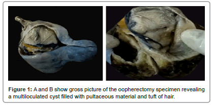
Right sided oophorectomy was performed and we received a specimen of right ovary grossly measuring 8 × 7 × 5 cms. External surface appeared globular, congested and smooth. Cut surface revealed a multiloculated cyst filled with pultaceous material and tuft of hair surrounded by a capsule of varying thickness. The cyst wall showed a solid nodule arising from it called the Rokitansky protuberance which contained cartilaginous bone and tooth measuring 4 × 3 cms. A tubular structure was also seen attached to the cyst wall and measured 2.5 cms shown in Figure 1.
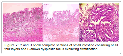
Light microscopy examination with H & E stain showed fibro collagenous tissue which was lined by stratified squamous epithelium and contained adnexal structures, pilosebaceous unit, many thickwalled blood vessels, adipocytes, nerve bundles, scattered melanin pigments, melanophages and complete small intestinal structure. The small intestine was composed of well-formed mucosa, submucosa, muscularis propria and serosa. Adjacent areas also showed gastric mucosa, respiratory epithelium, mature cartilage and salivary gland tissue. Focal areas within the intestinal mucosa exhibited features of mild dysplasia. With the above findings, mature cystic teratoma containing complete small intestinal structure showing mild dysplastic changes was made. The patient is doing good on follow-up without recurrence until date shown in Figure 2.
Results and Discussion
MCT has a wider range of age distribution and can be seen in any individual between infancy and postmenopausal age group. These arise from the totipotent cells of the ovary and have the capacity to differentiate into fully differentiated ectodermal, mesodermal and endodermal tissue. 6 Tissues of ectodermal origin are present in 99%- 100% of the cases with tissues of mesodermal and endodermal origin constituting 73%-93% and 32%-72% of the tissues respectively [4-8]. They usually are slow growing tumors with an average growth rate of 1.8 cm per year [9]. Though intestinal type tissue is frequent constituents of MCT, the organizing into complete intestinal wall with all layers is a rare finding [3]. Only five cases have been reported in the literature so far.
Two cases by Fujiwara, one by Takao, one by Ki EY and one by Tang, Fujiwara reported two cases of MCT with complete segments of intestinal wall showing neoplastic transformation [10]. One case showed benign mucinous cystadenoma of appendiceal type and the other contained intestinal-type adenocarcinoma infiltrating into the neoplastic bowel wall, both of which were confirmed by histopathological examination and immunohistochemical analysis. Takao reported a case of MCT with complete intestinal structure harboring intestinal-type adenocarcinoma. Ki EY reported a case of MCT containing complete colonic wall without evidence of dysplasia or malignant transformation [11]. Tang reported a case of mucinous cystadenoma in MCT associated with complete colonic structure.
Two percent of the MCT undergo malignant transformation. Although tumor markers, presence of solid component under radiological examination and old age may help predict malignant transformation of MCT it’s the histopathological examination that confirms the diagnosis [12]. Literature studies also show that 80% of the malignant transformation occurred in women of reproductive age group [13]. Taking these facts into account, it is important that we have a high index of suspicion with regard to malignant transformation of MCT, irrespective of the age group of the patient, especially when a solid focus in present. In our case as well a focus of dysplastic change was identified.
Conclusion
Though the chances of malignant transformation in MCT remain relatively low, its occurrence cannot be completely ignored because incomplete removal of such tumors may warrant radiotherapy and regular follow-up is required for early pick up of recurrence and better patient outcome. It is therefore important to subject the teratomas, especially those occurring at a younger age to extensive histopathological examination, to rule out any immature component or malignant transformation.
References
- Alotaibi MO, Navarro OM (2010) Imaging of ovarian teratomas in children: A 9-year review. Can Assoc Radiol J 61: 23-28.
- Templeman CL, Fallat ME, Lam AM, Perlman SE, Hertweck SP (2000) Managing mature cystic teratomas of the ovary. Obstet Gynecol Surv 55: 738- 745.
- Fujiwara K, Ginzan S, Silverberg SG (1995) Mature cystic teratomas of the ovary with intestinal wall structures harboring intestinal-type epithelial neoplasms. Gynecol Oncol 56: 97-101.
- Marcial-rojas RA, Medina R (1958) Cystic teratomas of the ovary; a clinical and pathological analysis of two hundred sixty-eight tumors. AMA Arch Pathol 66: 577-589.
- Blackwell WJ, Dockerty MB, Masson JC, Mussey RD (1946) Dermoid cysts of the ovary: Their clinical and pathologic significance. Am J Obstet Gynecol 51: 151-172.
- Wan KM, Foroughi F, Bansal R, Oehler MK (2019) Intestinal adenocarcinoma arising from a mature cystic teratoma. Am J Obstet Gynecol 57: 162-165.
- Caruso PA, Marsh MR, Minkowitz S, Karten G (1971) An intense clinicopathologic study of 305 teratomas of the ovary. Cancer 27: 343-348.
- Takao M, Yoshino Y, Taguchi A, Uno M, Okada S (2018) A case of mature cystic teratoma with intestinal structures harboring intestinal type low grade mucinous neoplasm. Int Cancer Conf J 7: 59-64.
- Caspi B, Appelman Z, Rabinerson D, Zalel Y, Tulandi T (1997) The growth pattern of ovarian dermoid cysts: A prospective study in premenopausal and postmenopausal women. Fertil Steri 68: 501-505.
- Ki EY, Jang DG, Jeong DJ, Kim CJ, Lee SJ (2016) Rare case of complete colon structure in a mature cystic teratoma of the ovary in menopausal woman: A case report. BMC Womens Health 16: 70.
- Tang P, Soukkary S, Kahn E (2003) Mature cystic teratoma of the ovary associated with complete colonic wall and mucinous cystadenoma. Ann Clin Lab Sci 33: 465-470.
- Belaid I, Khechine W, Abdelkader AB, Bedioui A, Ezzairi F (2016) Adenocarcinoma of intestinal type arising in mature cystic teratoma of ovary: A diagnostic dilemma. Int J Path35: 352-356.
- Park CH, Jung MH, Ji YI (2015) Risk factors for malignant transformation of mature cystic teratoma. Obstet Gynecol Sci 58: 475-480.
Citation: Vinola F (2020) Complete Small Bowel with a Dysplastic Foci in Mature Cystic Teratoma of the Ovary: Case Report. J Clin Exp Pathol 10: 384.
Copyright: © 2020 Vinola F. This is an open-access article distributed under the terms of the Creative Commons Attribution License, which permits unrestricted use, distribution, and reproduction in any medium, provided the original author and source are credited.
Select your language of interest to view the total content in your interested language
Share This Article
Recommended Journals
Open Access Journals
Article Usage
- Total views: 3033
- [From(publication date): 0-2020 - Dec 22, 2025]
- Breakdown by view type
- HTML page views: 2222
- PDF downloads: 811


