Make the best use of Scientific Research and information from our 700+ peer reviewed, Open Access Journals that operates with the help of 50,000+ Editorial Board Members and esteemed reviewers and 1000+ Scientific associations in Medical, Clinical, Pharmaceutical, Engineering, Technology and Management Fields.
Meet Inspiring Speakers and Experts at our 3000+ Global Conferenceseries Events with over 600+ Conferences, 1200+ Symposiums and 1200+ Workshops on Medical, Pharma, Engineering, Science, Technology and Business
Short Communication Open Access
Dosimetry Verification of Treatment Plans in Dynamic Techniques in Radiotherapy
| Welnogorska K1, Chmiel A2, Wasilewska-Radwanska M1* and Dankowska A2 | |
| 1Faculty of Physics and Applied Computer Science, AGH University of Science and Technology, 19 Reymonta Street, 30-059 Kraków, Poland | |
| 2Department of Children’s and Adults Radiotherapy, University Children’s Hospital of Cracow, 265 Wielicka Street, 30-663 Kraków, Poland | |
| Corresponding Author : | Marta Wasilewska-Radwanska, PhD, DSc Prof. AGH (Retired), Faculty of Physics and Applied Computer Science AGH University of Science and Technology Al. Mickiewicza 30-059 Krakow, Poland Tel: +48 12 6173002 Fax: +48 12 6340010 E-mail: radwansk@newton.fis.agh.edu.pl |
| Received October 01, 2015; Accepted October 16, 2015; Published October 21, 2015 | |
| Citation: Welnogorska K, Chmiel A, Wasilewska-Radwanska M, Dankowska A (2015) Dosimetry Verification of Treatment Plans in Dynamic Techniques in Radiotherapy. J Clin Oncol & Pract 1:103. doi:10.4172/jcop.1000103 | |
| Copyright: © 2015 Welnogorska K, et al. This is an open-access article distributed under the terms of the Creative Commons Attribution License, which permits unrestricted use, distribution, and reproduction in any medium, provided the original author and source are credited. | |
| Related article at Pubmed, Scholar Google | |
Visit for more related articles at
Abstract
The goal of this work was verification of treatment dose distribution in the VMAT method assuming gamma index minimization and dependence between the points with γ>1 and the position of the gantry’s arm.
| Keywords |
| Dynamic radiotherapy; Treatment plans; Dosimetry verification; Gamma index |
| Introduction |
| The development of dynamic irradiation techniques (Intensity Modulated RadioTherapy [IMRT] and Volumetric Modulated Arc Therapy [VMAT]) is a reason of changes in dosimetric verification methods of treatment plans. These methods are characterized by rapid changes in dose distribution across the irradiated organs. That is why we cannot verify the dose delivered to the patient during treatment using in-vivo methods, which is used to control 3D-CRT plans [1]. |
| Experimental verification of the planned dose distribution in dynamic techniques is carried out by determining the gamma index [2]. This factor is defined as a difference between calculated and measured dose in a phantom. It connects a percentage acceptance criterion between dose values for measured and received from the treatment plan data (Diff %) and distance criterion (mm). The plan verification passes if the gamma index <1 for at least 95% of compared points. |
| Methodology Measurements |
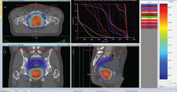
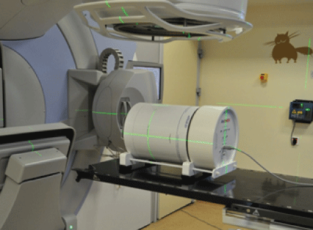
| There were prepared 90 treatment plans (Figure 1) using Monaco® 5.0 treatment planning system. This system to optimize a treatment plan using the Monte Carlo method. For each case there were calculated verification plan in QA SPL Monaco module. Cylindrical water-equivalent phantom ArcCHECK (Figure 2) [3] was used to verify measurements. The phantom was placed on the therapeutic table in accordance with lasers and light field of the accelerator. The dose measurements were performed applying a 6 MV (MeV) photon beam from Elekta Synergy linac accelerator with multileaf collimator (MLC) Agility (160 leafs) [4]. |
| Results and Discussion |
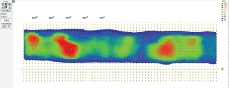
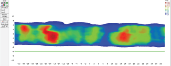
| In SNC PatientTM compares measured (Figure 3) and calculated (Figure 4), the dose distribution. Tested four criteria: 3% 3 mm 3% of 2 mm, 2% 3 mm and 2% 2 mm (threshold 10%). Obtained map gamma (Figure 5). |
| Statistical analysis was performed. The gamma factor was determined respectively: 98.53 ± 0.14% for the criterion of 3% 3 mm, 97.29 ± 0.23% for 3% 2 mm, 95.19 ± 0.37% for 2% 2 mm and 91.64 ± 0.56% for 2% 2 mm. |
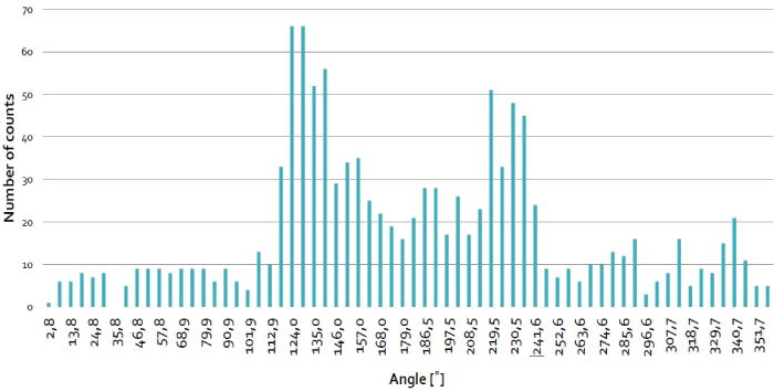
| 1169 points with γ>1 for the acceptance criterion 3% 3 mm, were analyzed. Points were counted for each position of the gantry’s arm and shown in Figure 6. |
| There was also found that most of points with γ>1 were located in places where the beam passes through the sides of the therapeutic table. |
| Conclusions |
| The results of statistical analysis indicate we can use lower acceptance criteria. The use of lower criteria enables more precise implementation of the treatment plan during therapy sessions. |
| Most of erroneous points were located in places where the beam passes through the sides of the therapeutic table. The planning system did not take into account accurate electron density used to irradiate phantom through the therapeutic table. These results show how it is important to set accurate electron density of the table. |
| References |
|
Figures at a glance
 |
 |
 |
| Figure 1 | Figure 2 | Figure 3 |
 |
 |
 |
| Figure 4 | Figure 5 | Figure 6 |
Post your comment
Relevant Topics
Recommended Journals
Article Tools
Article Usage
- Total views: 12298
- [From(publication date):
November-2015 - Aug 17, 2025] - Breakdown by view type
- HTML page views : 7896
- PDF downloads : 4402