Epithelioid Hemangioendothelioma of the Rib: A Rare Tumour with a Confusing Localization
Received: 20-Oct-2013 / Accepted Date: 07-Feb-2014 / Published Date: 11-Feb-2014 DOI: 10.4172/2161-0681.1000159
Abstract
Background: Epithelioid hemangioendothelioma is a rare tumour whose prognosis ranges from benign form similar to hemangioma to aggressive tumors like angiosarcoma. Its localization in the rib is very rare and less than 10 cases of costal localized forms have been reported in the literature. The authors describe another case of costal epithelioid hemangioendothelioma illustrated by microscopic findings.
Case presentation: We present the case of a 80-year-old man who reported a pain in the shoulder. Radiologic findings showed a hypervascularized tumour and microscopic findings diagnosed an epithelioid hemangioendothelioma. The treatment was the en-bloc resection of second to fourth posterior left ribs and the wedge resection of the left lung nodule and the patient didn’t present complications after a 6-year- follow-up period.
Conclusion: This case emphasizes on the challenging diagnosis of epithelioid hemangioendothelioma and the importance of surgical resection in localized forms.
Keywords: Epithelioid hemangioendothelioma; Surgery; Pathology; Rib; Elderly
313226Background
Epithelioid hemangioendothelioma is a rare tumour accounting for less than 1% of bone tumors [1]. The term ‘‘Epithelioid Hemangioendothelioma (EH)’’was first applied by Weiss and Enzinger to a soft tissue vascular tumor of borderline malignancy [2].
Its terminology results from its behavior considered between hemangioma and angiosarcoma. These tumors are challenging concerning their diagnosis and their management. The authors present another case of epithelioid hemangioendothelioma localized to the rib. To the best of our knowledge, there are less than 10 cases of local epithelioid hemangioendothelioma of the rib reported in the English literature.
Observation
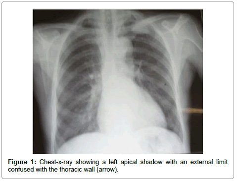
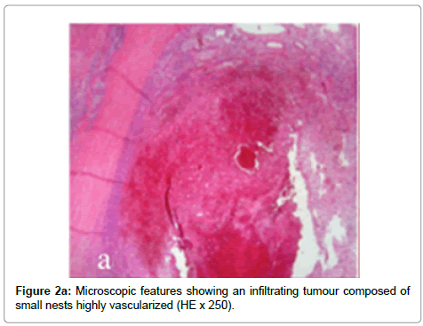
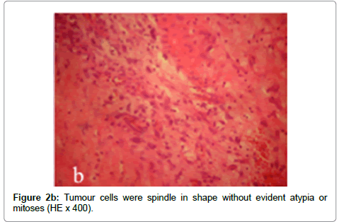
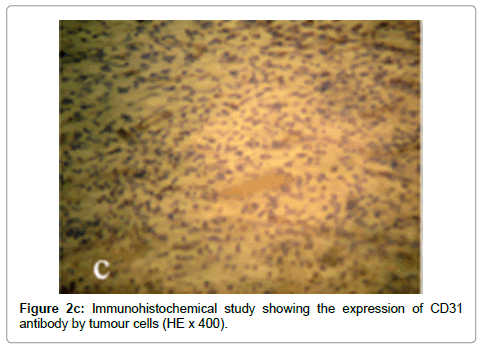
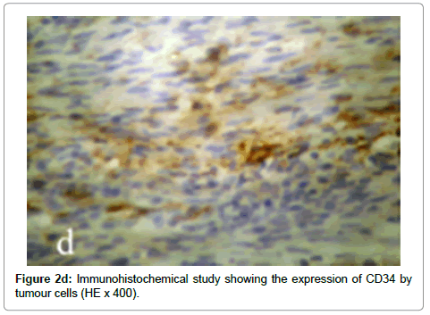
We present the case of an 80-year-old man with an antecedent of hypertension, who presented a 2-year lasting pain of the shoulder associated to a recent weight loss. Physical examination was normal. Chest-X-ray showed a left apical shadow with an external limit confused with the thoracic wall (Figure 1). Thoracic CT-scan showed a left posterior parietal mass developed upon the posterior arch of the third rib. This tumour was hypervascularized mimicking a hemangioma. A 1-centemeter mass of the inferior left lobe was also observed. An abdominal and cerebral CT-scan was also performed and showed no abnormalities elsewhere. An en-bloc resection of the posterior arch of the second, third and fourth ribs was performed in association to a wedge resection of the left lung nodule. Microscopic examination of the costal tumour showed an infiltrating tumour composed of small nests with immature intracytoplasmic lumina. Tumour cells were spindle in shape without evident atypical or mitoses (Figure 2a). Immunohistochemical study using CD31 and CD34 antibodies showed the positivity of tumour cells and highlighted their vascular origin (Figure 2b). The small nodule consisted in a benign tumor diagnosed as a hamarto chondroma. The patient presented no complications after a 6-month follow-up period (Figure 2c, 2d).
Discussion
This case deals with an epithelioid hemangioendothelioma particular by its costal localization in an elderly patient. These tumors are rarely reported in the rib with less than 10 cases reported in the literature. Besides, they’ve been rarely reported in elderly patients [3]. These tumors are more frequently observed in women with a mean age between the fourth and fifth decade [4]. The radiologic features were suggestive of a hemangioma due to the high vasculature of the tumour. In other cases reported in the literature, the radiologic findings were suggestive of malignant process, infectious disease, fibrous dysplasia or fibrous histiocytic lesions [5,6]. Positive diagnosis was based on microscopic findings, which consisted in single files, cords and small nests, typically lacking well-formed vascular channels, with only immature, intra-cytoplasmic lumina being observed. Other distinctive features include an infiltrative growth pattern and the presence of a matrix varying from a light blue (myxoid, myxochondroid) to a deep pink (hyaline) extra-cellular stroma. The cells have an eosinophilic cytoplasm containing vacuoles, which deform the cytoplasm (blister cells). Some vacuoles contain fragmented erythrocytes. In most cases the cells show a low nuclear grade. These tumors cause a real pitfall with angiosarcoma. Distinctive features consist in the less number of mitoses and atypical cells in epithelioid hemangioendothelioma. Hemangioendothelioma of the soft and bone tissue have been recently divided into 2 groups, which are low grade and high grade according to their size and mitotic rate [7,8]. Patients with tumors larger than 3 cm in diameter and having more than 3 mitoses/50 HPFs have a 5-year disease-specific survival of 59% in contrast to 100% survival in patients whose tumors lacked these features [9]. The tumour reported in this case is classified into the low-grade group. Some authors reported a translocation t [1,3] as a diagnostic marker of these tumors but recent studies showed the existence of other translocations which don’t seem to be therapeutic targets [9-11]. Surgical resection plays a key role in the management of these tumors especially in localized forms. Shinamura et al. [12] reported an epithelioid hemangioendothelioma of the chest wall with metastases to the rib, which was treated only by surgical resection and the patient presented no complication. This case made us wonder about the inefficacy of chemotherapy in multiple forms but further studies are necessary to assess this finding because 20-30% of tumors metastasize, and about 15% of patients die of their disease [13,14]. The Table 1 illustrates the cases of epithelioid hemangioendothelioma reported in the literature.
| Author | Year | Presentation | Treatment | Follow-up |
|---|---|---|---|---|
| Shimamura J, et al. [12] | 2014 | Tumour of the chest wall with metastases to the rib | Surgical resection | - |
| Sirikulchayanonta V, et al. [5] | 2010 | Tumour of 3 contiguous bones | Surgical resection | Stable after 3 years |
| Chirieac LR, et al. [15] | 2006 | Tumour of the rib and intercostal lymph nodes | Surgical resection | - |
| Mohan H, et al. [6] | 2001 | Tumour of the rib | Surgical resection | - |
| Our case, et al. | 2014 | Tumour of the rib | Surgical resection | Stable after 6 months |
Table 1: The cases of costal epithelioid hemangioendothelioma published in the English literature.
Conclusion
This case of rib limited EH emphasizes the challenging diagnosis of these tumors which is based on microscopic examination. Surgeons and clinicians should be aware of those lesions with an unusual radiological and clinical behavior. Besides, it highlights the key-role of surgical resection in localized forms.
References
- Koch M, Nielsen GP, Yoon SS (2008) Malignant tumors of blood vessels: angiosarcomas, hemangioendotheliomas, and hemangioperictyomas. J Surg Oncol 97: 321-329.
- Weiss SW, Enzinger FM (1982) Epithelioid hemangioendothelioma: a vascular tumor often mistaken for a carcinoma. Cancer 50: 970-981.
- Marquez-Medina D, Samam-PrezVargas JC, Tuset-DerAbrain N, Montero-Fernandez A, Taberner-Bonastre T, et al. (2011) Pleural epithelioid hemangioendothelioma in an elderly patient. A case report and review of the literature. Lung Cancer 73: 1169.
- Miettinen M (2010) Modern Soft Tissue Pathology: Tumors and Non-Neoplastic Conditions. (1stedn), Cambridge University Press, Cambridge, UK.
- Sirikulchayanonta V, Jinawath A, Jaovisidha S (2010) Epithelioid hemangioma involving three contiguous bones: a case report with a review of the literature. Korean J Radiol 11: 692-696.
- Mohan H, Palta A, Nada R, Punia RS, Jogai S, et al. (2001) Epithelioid haemangioendothelioma of the rib--a case report. Indian J Pathol Microbiol 44: 375-377.
- Deyrup AT, Tighiouart M, Montag AG, Weiss SW (2008) Epithelioid hemangioendothelioma of soft tissue: a proposal for risk stratification based on 49 cases. Am J Surg Pathol 32: 924-927.
- Fletcher CD, Unni K, Mertens F (2002) WHO.Pathology & Genetics.Tumours of Soft Tissue and Bone. . Lyon: IARC Press.
- Errani C, Zhang L, Sung YS, Hajdu M, Singer S, et al. (2011) A novel WWTR1- CAMTA1 gene fusion is a consistent abnormality in epithelioid hemangioendothelioma of different anatomic sites. Genes Chromosomes Cancer 50: 644-653.
- Tanas MR, Sboner A, Oliveira AM, Erickson-Johnson MR, Hespelt J, et al. (2011) Identification of a disease-defining gene fusion in epithelioid hemangioendothelioma. Sci Transl Med 3: 98ra82.
- Errani C, Sung YS, Zhang L, Healey JH, Antonescu CR (2012) Monoclonality of multifocal epithelioid hemangioendothelioma of the liver by analysis of WWTR1-CAMTA1 breakpoints. Cancer Genet 205: 12-17.
- Shimamura J, Kiyoshima M, Suzuki H, Kitahara M, Asato Y, et al. (2014) Epithelioid hemangioendothelioma originating from the chest wall with rib metastases. Gen Thorac Cardiovasc Surg.
- Weiss SW, Enzinger FM (1982) Epithelioid hemangioendothelioma: a vascular tumor often mistaken for a carcinoma. Cancer 50: 970-981.
- Mentzel T, Beham A, Calonje E, Katenkamp D, Fletcher CD (1997) Epithelioid hemangioendothelioma of skin and soft tissues: clinicopathologic and immunohistochemical study of 30 cases. Am J Surg Pathol 21: 363-374.
Citation: Mlika M, Ismail O, Marghli A, Boudaya S, Kilani T, et al. (2014) Epithelioid Hemangioendothelioma of the Rib: A Rare Tumour with a Confusing Localization. J Clin Exp Pathol 4:159. DOI: 10.4172/2161-0681.1000159
Copyright: © 2014 Mlika M, et al. This is an open-access article distributed under the terms of the Creative Commons Attribution License, which permits unrestricted use, distribution, and reproduction in any medium, provided the original author and source are credited.
Select your language of interest to view the total content in your interested language
Share This Article
Recommended Journals
Open Access Journals
Article Tools
Article Usage
- Total views: 15161
- [From(publication date): 3-2014 - Aug 17, 2025]
- Breakdown by view type
- HTML page views: 10527
- PDF downloads: 4634

