Gastroprotective Effect of Sargassum wightii Fractions on Indomethacin Induced Gastric Ulcer in Wistar Albino Rats
Received: 05-Dec-2023 / Manuscript No. JGDS-23-122152 / Editor assigned: 08-Dec-2023 / PreQC No. JGDS-23-122152 (PQ) / Reviewed: 26-Dec-2023 / QC No. JGDS-23-122152 / Revised: 27-Jan-2025 / Manuscript No. JGDS-23-122152 (R) / Published Date: 03-Feb-2025
Abstract
Seaweeds or marine macroalgae have been reported to have various biological activities including antioxidant, antiviral, antibacterial, antiprotozoal, antifungal, anti-inflammatory, antitumor, antidiabetic, antineoplastic, anti cholinesterase, anti-tyrosinase anticoagulant, hypolipidemia and hypercholesterolemia. Sargassum wightii is brown seaweed which is used for our research, the fractions were isolated, showed a good anti-ulcer activity in wistar rat model system. The results showed that 250 mg/kg of the seaweed extract a good anti-ulcer activity as compared with the standard drug (20 mg/kg Omeprazole). From this study Sargassum wightii can be used for the treatment of Gastric ulcer.
Keywords: Sargassum wightii, Fractions, Wistar rat model system, Anti-ulcer
Introduction
Inflammation which is the host defence mechanism that protects the body by causing the affected area to become red, hot, swollen, painful, and lose its ability to function. This natural process aids in the removal of microbes and assists in re-establishing tissue homeostasis. However, gastrointestinal ulcers are brought on by inflammation in the gastrointestinal tract's mucosa. Millions of individuals worldwide suffer with gastrointestinal ulcers which has become a common severe illness of the digestive system. The common approaches for its therapy are known to be suppression of inflammatory cell activity or inhibition of inflammatory mediator production [1].
In order to treat these disorders, Steroidal Anti-Inflammatory Drugs (SAID) and Non-Steroidal Anti-Inflammatory Medications (NSAID) are frequently employed. The most typical side effects of aspirin or indomethacin include nausea, vomiting, and stomach pain. Additionally, anti-inflammatory and antiulcerogenic medications are primarily utilised to manage disease symptoms rather than to treat or stop the inflammatory and ulcerogenic processes. Nevertheless, a lot of natural compounds have analgesic and anti-inflammatory benefits, and their side-effect rates are often low. Numerous research on marine species show that a variety of marine life-produced chemicals have beneficial pharmacological properties. Both biological and chemical variety is abundant in the marine environment. The seaweeds, or macroalgae, are thought to be a plentiful supply of bioactive substances with potential for medicinal and medical uses. The growing trend of new macroalgal compounds encourages marine science toward the prospective field of medication discovery research. Seaweeds are the popular name for marine macroalgae that are commercially available. Due of their widespread abundance, algae might be a source of novel medicinal chemicals [2].
Red algae has been reported to contain bioactive compounds that can reduce inflammation of the gastrointestinal tract, prevent and treat gastric ulcers and cancers brought on by oxidative stress, and reduce inflammatory activities by inhibiting the production of inflammatory mediators and inducing cancer cell apoptosis in the stomach and colon.
Since edible algae have been consumed as food and utilised in traditional medicines for ages, natural chemicals generated from them would be less dangerous to use as anti-inflammatory and gastrointestinal anti ulcerogenic treatments.
Marine algae are known to possess several activities such as antimicrobial, antiviral, antimalarial, fungicidal, bactericidal, bacteriostatic, antimycobacterial. These are a rich source of bioactive compounds such asterpenoids, amino acids, acrylic acid, phenolic compounds, steroids, phlorotannins, halogenated compounds, alkanes, cyclic polysulphides, fatty acids, polyunsaturated fatty acids, polyphenols, peptides, carotenoids and sulfated polysaccharides. The compounds such as acrylic acid, terpenoids, phlorotannins and steroids are the major ones responsible for antimicrobial activity of marine algae. These also show anti-tumor activity mainly due to the presence of fucose-containing sulfated polysaccharides [3].
Seaweeds or marine macroalgae have been reported to have various biological activities including antioxidant, antiviral, antibacterial, antiprotozoal, antifungal, anti-inflammatory, antitumor, antidiabetic, antineoplastic, anti-cholinesterase, anti-tyrosinase anticoagulant, hypolipidemia, hypercholestrolemia. Seaweeds areutilised as fertiliser, food ingredient, source of sodium alginate, and for animal feed.
Nowadays, seaweeds being low-calorie foods are used in the preparation of soups and salads in the Asian continent. Seaweeds are found in oceans all over the world and classified into three types on the basis of their colours as red seaweed (Rhodophyta), brown seaweed (Phaeophyta) and green seaweed (Chlorophyta). Fucoxanthin, a dominant xanthophyll pigment that hides the existence of other pigments and gives the Phaeophyta their brown colour, whereas phycoerythrin, a pigment that reflects red light and absorbs blue light, gives the Rhodophyta their red colour and the presence of chlorophyll a and b are responsible for the green colour of the Chlorophyta.
Brown seaweed has anti-proliferative properties which is due to the presence of abundant sulfated polysaccharides and fucoidans. These also has antioxidant property. Sargassum (Sargassaceae) belonging to the brown seaweed has free radical scavenging, anticancer and antiviral, antioxidant and anticholinesterase effects. Among them, Sargassum wightii which is a dark-brown macroalgae is reported to have antioxidant and anticholinesterase activity. The presence of alginic acid in S. wightii is reported to have anti-inflammatory effects. In many parts of Asia, Sargassum wightii is found in abundance in the shorelines while in India, it occurs mainly in the southern coasts of Tamilnadu. In the present study, the antiulcer potential of the two fractions obtained from Sargassum wightii has been evaluated using wistar rats [4].
Objective: The goal of the current study is to evaluate the anti ulcer property of fractions from Sargassum wightii.
Materials and Methods
Seaweed collection
Sargassum wightii, a brown alga, was found in the subtidal area of the Mandapam coast (Lat. 09°17' N; Long. 79°07' E, Gulf of Mannar, India). It weighed around 1 kg. The brown seaweeds were cleaned with distilled water to get rid of the salt, sand, and epiphytes and then dried in the shade for five days in a room with air conditioning. A mixer grinder was used to chop and powder the shade-dried specimen [5].
Seaweed extracts
The powdered seaweed samples were soaked in water for 48 hours at room temperature before being filtered through Whatman filter paper under vacuum. The filtrate was then concentrated at 50℃ using a rotary vacuum evaporator made by Superfit in India. Until analysis, the ethanolic extract fractions were kept at -20°C.
Acute toxicity studies (LD50)
The following assumption was made while applying the LD50 estimation method, which makes use of 13 wistar rats. The determination of the toxic range is the initial step. The rat was divided into three groups (n=3) and given doses of 10 mg/kg, 100 mg/kg, and 1000 mg/kg solubilized in a 3%, v/v Tween 85 solution. For 24 hours, the number of deaths in the treated rat was monitored. The second phase's dose is based on the previous phase's death pattern. A new batch of four rats will receive 100 mg/kg, 250 mg/kg, or 500 mg/kg of the extract if there were no fatalities in the initial phase. For 24 hours, the treated animals were monitored for lethality or symptoms of acute intoxication. The geometric mean of the greatest non-lethal dose and the least toxic dose is known as the LD50. Using the below formula, the LD50 was determined [6].
LD50=√(Do × D100)
Do=Maximum dose that resulted in zero mortality
D100=Lowest dose that resulted in mortality
Animal husbandry
The animal house, Department of biochemistry, Karpagam academy of higher education, Coimbatore, provided 35 wistar rats (weighing between 120 and 150 g). Before the trial began, they were kept for two weeks and fed grower mash (vital feed, grand cereal), allowing them to acclimate to the animal house settings (a cross-ventilated room with a temperature between 25°C and 28°C, and a 12 h light/12 h dark cycle). The study was carried out in accordance with the ethical committee's guidelines and ethical approval (KAHE/IAEC/ 2021/11-09/007), the ARRIVE recommendations for reporting in vivo experiments, and the CPCSEA, Government of India, guidelines for the care and use of laboratory animals.
Evaluation of ulcer activity using wistar rats
Experimental design: Seven groups of five rats each were formed from the thirty-five (35) wistar rats as shown in Table 1, who were fasted for 18 hours before to extract delivery. The rats in groups 3 and 4 were treated with petroleum ether fraction of seaweed at doses of 100 mg/kg and 250 mg/kg respectively for 21 days. The rats in groups 5 and 6 were treated with chloroform fraction of seaweed at doses of 100 mg/kg and 250 mg/kg respectively for 21 days. Group 1 was regarded as the control group and treated with 1 ml/kg water. Group 2 was the standard group that received Omeprazole for 21 days and Indomethacin as inducer. Group 7 was the negative control that received only Indomethacin as inducer. All the groups except group 1 were treated with indomethacin as inducer. After eight hours, chloroform was used to sacrifice all the rats. Therefore, the ulcer index will be larger the more severe the drop, as it will take longer for a stock to recover and reach its previous record. It was observed and scored how many and how severe the lesions were. For the purpose of assessing the antiulcerogenic action, the levels of Lipid Peroxidation (LPO), Catalase (CAT), Superoxide Dismutase (SOD) and glycoproteins (total hexoses, hexosamine, fucose, and sialic acid) were determined using the appropriate techniques [7].
Test tubes were filled with the gastrointestinal contents, which were then centrifuged for 10 minutes at 3000 rpm. We used a digital pH meter to determine the pH of the supernatant, ml/100 g body weight was used to calculate the volume of the supernatant [8].
| Group 1 | Control group healthy wistar albino rats receive vehicle alone |
| Group 2 | Standard drug group–omeprazole for 21 days+indomethacin as inducer |
| Group 3 | Treatment group–animal would receive a single dose of extract (fraction 1(petroleum ether) 100 mg/kg) for 21 days by gavage+indomethacin as inducer |
| Group 4 | Treatment group–animal would receive a single dose of extract (fraction 1(petroleum ether) 250 mg/kg) for 21 days by gavage+indomethacin as inducer |
| Group 5 | Treatment group–animal would receive a single dose of extract (fraction 2(chloroform) 100 mg/kg) for 21 days by gavage+indomethacin as inducer |
| Group 6 | Treatment group–animal would receive a single dose of extract (fraction 2(chloroform) 250 mg/kg) for 21 days by gavage+indomethacin as inducer |
| Group 7 | An ulcerated control group(negative control)–animals would receive only Indomethacin as inducer |
Table 1: Experimental design indicating seven group of Wistar rat.
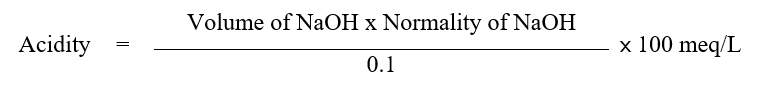
Determination of free and total acidity in gastric juice
Volumetric analysis was used to calculate the amount of free and total acid in the gastric juice taken from rats with ligated pyloruses.
One millilitre of gastric juice was pipetted into a 100 millilitre conical flask along with two to three drops of Topfer's reagent (HiMedia Pvt., Ltd., Mumbai), which was then titrated with 0.01 N NaOH (previously standardised with 0.01 N oxalic acid) until all traces of the red colour vanished and changed to a yellowish orange colour. It was noticed that the alkali volume corresponds to the free acidity. Following the addition of 2 to 3 drops of phenolphthalein solution, the titration procedure was repeated until a distinct red tinge returned. It was once more noted that the total amount of added alkali corresponds to the total acidity. using the following formula, the free and total acidity of gastric juice was determined;
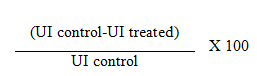
Determination of Ulcer Index (UI) and percentage inhibition
In order to conduct preliminary research on the effectiveness of an optimised chemical in an animal model, ulcer index and % inhibition were computed in ethanolic extract fraction of-induced rats. Lesions were typically circular, but they might also be linear. The scoring system was used to calculate the ulcer index.
Score 0: Represents no ulcer.
Score 1: For dilated vessels and pointed ulcers.
Score 2.5: Small ulcers: 4 mm or lesser in length.
Score 5: Big ulcers: 5 mm or greater in length.
The mean ulcer score was used to compute the UI for each animal. The following formula was used to compute the percentage of inhibition:
Histopathology procedure
The various stomach components underwent cleaning, fixing, and dehydration procedures before being cleared in xylene, cleared in neutral buffered formalin (10%), and embedded in paraffin wax. Hematoxylin and Eosin (H and E) and cresol violet stain were used to stain the tissues after sectioning them at a thickness of 5 m with a rotary microtome [9].
Estimation of glycoproteins
Using a modified version of approach, the glycoproteins detected in the stomach juice were extracted. 5 mL of 95 percent ethanol and 0.1 mL of gastric juice were combined, and they were centrifuged at 3000 rpm for 30 minutes. In order to obtain a glycoprotein precipitate, the supernatant was removed. This precipitate was used to calculate mucopolysaccharides like sialic acid, hexose, fucose, and hexosamine.
Assay of sialic acid
The gastric juice's glycoprotein precipitate was hydrolyzed using 0.1 N H2SO4. The hydrolysate was then treated with thiobarbituric acid to liberate a colour complex, which was subsequently extracted with butanol and detected at 550 nm after being reduced with periodic acid.
Assay of total hexose
The orcinol-H2SO4 reagent was supplied to the gastric juice glycoprotein precipitate and heated at 80°C for 15 minutes in order to obtain a colour that could be seen at 540 nm [10].
Assay of fucose
Before using cystein hydrochloride, the gastric juice glycoprotein precipitate was first treated with H2SO4. The content of fucose in the sample was determined using the difference between the absorbance measurements made at 393 nm and 440 nm.
Assay of hexosamine
The precipitation of glycoprotein was dissolved with HCl. After being treated with acetylacetone, this hydrolysate was coloured with Erlich reagent and ethanol and measured at 530 nm.
Assay of total non-amino polysaccharide
First, ethanol was used to precipitate 0.1 mL of stomach liquid. The precipitate was then treated with tryptophan and bromosulfuric acid reagent before being placed in a water bath to create colour, which was measured at 520 nm.
Estimation of lipid peroxidation
Lipid Peroxidation (LPO) was measured using the earlier reported method. MDA at concentrations ranging from 3 to 12 nM was added to 100 mL of gastric juice, along with 0.2 mL of sodium dodecyl sulphate, 1.5 mL of acetic acid, and 1.5 mL of TBA. Up to 4 mL of this mixture was made with distilled water and kept at 90°C for an hour in a boiling water bath. The pink hue produced was measured at 532 nm against a reagent blank after 1 mL of distilled water was introduced and allowed to cool to room temperature [11].
Estimation of protein
Rats with pylorus ligations were measured for the amount of protein level in their stomach juice.
Estimation of superoxide dismutase activity
The activity of Superoxide Dismutase (SOD) (EC 1.15.1.1) in gastric was assessed using the technique suggested. Diethylenetriamine Pentaacetic Acid (DTPA), Ethylenediamine Tetraacetic Acid (EDTA), and Tris-HCl buffer were combined in a volume of 0.1 mL, 0.5 mL, and 2.5 mL, respectively. 0.5 mL of pyrogallol was added to the mixture to measure the rate of auto oxidation, and the increase in absorbance was observed at 420 nm for 3 minutes. To 0.1 mL of gastric juice collected in a different tube, 2.5 mL of Tris-HCl buffer, 0.1 mL of EDTA, and 0.5 mL of DTPA were added. This combination received 0.5 mL of pyrogallol, and the increase in absorbance was monitored for three minutes at 420 nm. This measurement was used to calculate the rate at which gastric juice inhibited the pyrogallol auto-oxidation.
Estimation of catalase activity
Catalase (CAT) (EC 1.11.1.6) activity in gastric juice was calculated as described in the method. The ideal pH for catalase appears to be pH 7.5. 1 mL of phosphate buffer was added to 0.1 mL of gastric juice. 0.5 mL of H2O2 was then added to start the reaction. By adding 2 mL of dichromate-acetic acid reagent, the reaction was stopped at 0, 30, and 60 s intervals. In separate tubes, 1.6 mL of buffer and 2 mL of dichromate-acetic acid reagent were combined to create the reagent blank. For 10 minutes, the test tubes and blanks were cooked in a boiling water bath to create purple hue. When the tubes reached room temperature, they were tested for colour intensity at 570 nm in comparison to the blank.
Statistical analysis
In order to analyse the data of the present study, the Statistical Package for Social Sciences was used (SPSS-21). The average and Standard Error of the Mean (SEM) for the sample groups were used to represent the results. One-way Analyses Of Variance (ANOVA) were performed on the raw data before the post hoc Turkey's test.
Results
Acute toxicity studies (LD50)
The acute toxicity test carried out showed that the aqueous extract of Sargassum wightii was highly safe having a high safety margin when compared to the standard range when given orally and this can be seen in the Tables 2 and 3. No deaths were recorded after 24 hours of administration of the various doses (150, 250 and 500 mg/ kg body weight) of the aqueous extract of Sargassum wightii. The LD50 was determined as 500 mg/kg [12].
Estimation of catalase activity
Catalase (CAT) (EC 1.11.1.6) activity in gastric juice was calculated as described in the method. The ideal pH for catalase
| S. no | Dose (mg/kg) | Mortality |
| 1 | 150 mg/kg | 0/3 |
| 2 | 250 mg/kg | 0/3 |
| 3 | 500 mg/kg | 0/3 |
Table 2: Acute toxicity study of the aqueous crude extract of Sargassum wightii.
| Group | Ulcer index | Total acidity (m Eq/L) |
| Control | 18.2 ± 0.3 | 4 ± 1.22 |
| Standard | 42.8 ± 3.5 | 6 ± 0.41 |
| Fraction 1 (100 mg/kg) | 40 ± 3.2 | 4.5 ± 0.26 |
| Fraction 1 (250 mg/kg) | 16.6 ± 0.5 | 3.5 ± 0.32 |
| Fraction 2 (100 mg/kg) | 16.6 ± 0.5 | 15 ± 0.21 |
| Fraction 2 (250 mg/kg) | 40 ± 3.2 | 6 ± 0.23 |
| Inducer | 11.6 ± 0.5 | 13 ± 1.07 |
Table 3: Ulcer studies on S. wightii fractions.
According to the results of sialic acid given in the Table 4, in the groups treated with omeprazole, there was an increase in sialic acid content 52 µg/mL than the control 47 µg/mL. Animals treated with indomethacin only showed 42 µg/mL while those treated with the petroleum ether fraction 1 (100 mg/kg and 250 mg/kg) showed 53 µg/mL and 67 µg/mL respectively and those undergone treatment with chloroform fraction 2 (100 mg/kg and 250 mg/kg) showed 62 µg/mL and 57 µg/mL respectively. From the data represented in the Table 4, in the groups treated with omeprazole, there was an increase in fucose content 53 µg/mL than the control 50 µg/mL. Animals treated with indomethacin only showed 46 µg/mL while those treated with the petroleum ether fraction 1 (100 mg/kg and 250 mg/kg) showed 58 µg/mL and 73 µg/mL respectively and those undergone treatments with chloroform fraction 2 (100 mg/kg and 250 mg/kg) showed 68 µg/mL and 63 µg/mL respectively (Figure 1) [13].
| Determinant (µg/mL) | Control | Standard | Petroleum ether fraction 1 (100 mg/kg) | Petroleum ether fraction 1 (250 mg/kg) | Chloroform fraction 2 (100 mg/kg) | Chloroform fraction 2 (250 mg/kg) | Inducer |
| Sialic acid | 47 ± 0.003 | 52 ± 0.005 | 53 ± 0.004 | 67 ± 0.004 | 62 ± 0.002 | 57 ± 0.003 | 42 ± 0.004 |
| Hexosamine | 47 ± 0.003 | 51 ± 0.005 | 55 ± 0.005 | 71 ± 0.006 | 66 ± 0.005 | 61 ± 0.0045 | 41 ± 0.004 |
| Total hexose | 46 ± 0.004 | 50 ± 0.004 | 54 ± 0.005 | 71 ± 0.0045 | 64 ± 0.005 | 59 ± 0.0047 | 41 ± 0.005 |
| TNAP | 48 ± 0.004 | 52 ± 0.003 | 60 ± 0.005 | 70 ± 0.007 | 65 ± 0.0035 | 61 ± 0.005 | 43 ± 0.0075 |
| Fucose | 50 ± 0.0061 | 53 ± 0.003 | 58 ± 0.006 | 73 ± 0.0075 | 68 ± 0.004 | 63 ± 0.0055 | 46 ± 0.0075 |
Table 4: Levels of glycoproteins in gastric juice.

Figure 1: Effect of lipid peroxidation on fractions of S. wightii.
The lipid peroxidation level in untreated rat was 0.154 nmoles/mg of protein. The chloroform fraction of Sargassum wightii showed 0.112 and 0.124 nmoles/mg of protein at 100 and 250 mg/kg respectively. The petroleum ether fraction S. wightii showed comparatively similar to the standard drug, Omeprazole (conc). The induced drug, Indomethacin (conc) showed a higher lipid peroxidation effect 0.164 nmoles/mg of protein than the standard 0.145 nmoles/mg of protein. The control rat showed 0.154 nmoles/mg of protein which was comparatively similar to the standard.
Effect of Sargassum wightii on antioxidant status in gastric mucosa
The levels of activities of enzymatic antioxidants in gastric mucosa of experimental animals are represented in Figures 2 and 3. The activity level of enzymatic antioxidants, SOD and CAT was significantly (p<0.001) increased in indomethacin induced wistar rat compared to control rats. A significant (p<0.001) increase in the levels of SOD and CAT were observed in induced treated with petroleum ether fraction and chloroform fraction compared with induced. No significant changes were registered in standard only administered with omeprazole treated group [14].

Figure 2: Effect of superoxide dismutase on fractions of S. wightii.

Figure 3: Effect of catalase on fractions of S. wightii.
Effect of protein on seaweed treatment
The levels of protein in gastric mucosa of experimental animals are represented in Figure 4. The level of protein was significantly (p<0.001) increased in indomethacin induced wistar rat compared to control rats. A significant (p<0.001) increase in the levels of protein were observed in induced treated with petroleum ether fraction and chloroform fraction compared with induced. No significant changes were registered in standard only administered with omeprazole treated group [15].

Figure 4: Effect of protein on fractions of S. wightii.
Histological analysis
In Figure 5, A) Shows the histological examination of stomach of control and experimental group of rats. The gastric mucosa of control rats showed glandular architecture andlamina propria showing mild lymphocytic infiltration. Submucosa and muscularis propria appears unremarkable. No evidence of ulceration or neutrophilic activity noted. B) The gastric mucosa for standard rats showed gastric mucosa with maintained glandular architecture andlamina propria showing mild lymphocytic infiltration. Submucosa and muscularis propria appears unremarkable. No evidence of ulceration or neutrophilic activitynoted. C1) The gastric mucosa for treated rats (Indomethacin induced petroleum ether fraction 1 (100 mg/kg)) showed glandular architecture andlamina propria showing mild mixed inflammatory cell infiltration. Submucosa and muscularis propria appears unremarkable. No evidence of ulceration orneutrophilic activity noted. C2) The gastric mucosa for treated rats (Indomethacin induced petroleum ether fraction 1 (250 mg/kg)) showed glandular architecture andlamina propria showing mild mixed inflammatory cell infiltration. Submucosa and muscularis propria appears unremarkable. No evidence of ulceration orneutrophilic activity noted. D1) The gastric mucosa for treated rats (Indomethacin induced chloroform fraction 2 (100 mg/kg)) showed glandular architecture andlamina propria showing mild mixed inflammatory cell infiltration. No evidence of ulceration orneutrophilic activity noted. Submucosa and Muscularis propria appears unremarkable. D2) The gastric mucosa for treated rats (Indomethacin induced chloroform fraction 2 (250 mg/kg)) showed glandular architecture andlamina propria showing mild mixed inflammatory cell infiltration. No evidence of ulceration orneutrophilic activity noted. Submucosa and muscularis propria appears unremarkable. E) The gastric mucosa for induced (Indomethacin) rats showed gastric mucosa with maintained glandular architecture andlamina propria showing mixed inflammatory cell infiltration. Submucosa and muscularis propria appearsunremarkable [16].

Figure 5: Magnification X 100, hematoxylin and eosin staining. A) It shows gastric mucosa with maintained glandular architecture andlamina propria showing mild lymphocytic infiltration. No evidence of ulceration or neutrophilic activity noted. Submucosa and muscularis propria appears unremarkable; B) It shows gastric mucosa with maintained glandular architecture andlamina propria showing mild lymphocytic infiltration. No evidence of ulceration or neutrophilic activitynoted. Submucosa and muscularis propria appears unremarkable; C1) It shows gastric mucosa with maintained glandular architecture andlamina propria showing mild mixed inflammatory cell infiltration. No evidence of ulceration orneutrophilic activity noted. Submucosa and muscularis propria appears unremarkable; C2) It shows gastric mucosa with maintained glandular architecture andlamina propria showing mild lymphocytic infiltration. No evidence of ulceration or neutrophilic activitynoted. Submucosa and muscularis propria appears unremarkable; D1) Section shows gastric mucosa with maintained glandular architecture andlamina propria showing mild mixed inflammatory cell infiltration. No evidence of ulceration orneutrophilic activity noted. Submucosa and muscularis propria appears unremarkable; D2) Section shows gastric mucosa with maintained glandular architecture andlamina propria showing mild mixed inflammatory cell infiltration. No evidence of ulceration orneutrophilic activity noted. Submucosa and muscularis propria appears unremarkable; E) It shows gastric mucosa with maintained glandular architecture andlamina propria showing mixed inflammatory cell infiltration. Submucosa and muscularis propria appearsunremarkable.
Discussion
The acute toxicity test carried out showed that the aqueous extract of Sargassum wightii, no deaths were recorded after 24 hours of administration of the various doses (150, 250 and 500 mg/kg body weight) of the aqueous extract of Sargassum wightii. The LD50 was determined as 500 mg/kg [17].
Our study has shown that omeprazole, petroleum ether fraction and chloroform fraction have a positive effect on the content of glycoprotein fractions in gastric mucosa. In indomethacin treated in wistar albino rats showed decrease in fucose, sialic acid, hexosamine, total hexose, TNAP were observed compared with untreated group. Almost similar results were observed in group treated with petroleum ether fraction and chloroform fraction of Sargassum wightii and omeprazole.
Stress induced ulceration involves damage by ROS apart from acid and pepsin related factors. During stress, LPO and enzyme activities like SOD, CAT were significantly increased. The increase in SOD and CAT were due to increase in ROS generation during mucosal damage. This evidently caused increased lipid peroxidation and mucosal damage as seen from increase in ulcer sours in comparison to the control group shown by Miller TA in 1987. The proton pump inhibitors suppress the acid aggression and also performs other unknown mechanisms because of which these are known to have cytoprotective effect [18].
We found a positive effect of omeprazole on the synthesis of insoluble glycoprotein and on the number of functioning mucus reducing cells. The use of indomethacin causes reduction in the number of functioning mucus-producing cells and also significantly suppresses the insoluble glycoprotein synthesis of the mucosal barrier. The gastroprotective effect of aqueous extract of Sargassum wightii was evident from significant reduction in the lesion index in S. wightii treated rats as against ulcerated rats. The preliminary phytochemical analysis revealed the presence of various components such as steroids, phenols, tannins, flavonoids, S. wightii aqueous extract (data not shown) confirming the reports of previous studies. Flavonoids are reported to possess the properties of preventing the formation of lesions by various necrotic agents. In the present study petroleum ether fraction showed the presence of flavonoids. Hence the antiulcer activity of petroleum ether fraction may be due to the presence of flavonoid.
Gastric acid has been shown to play an important permissible role in NSAID associated mucosal injury. Increase in mucosal resistance and decrease in aggressive factors mainly acid and pepsin are associated with gastric protection offered by prostaglandins. Prostaglandins E2 and I2 are the predominant prostaglandins synthesized by gastric mucosa and are known to inhibit the secretion of gastric acid and stimulate the secretion of mucus, bicarbonate and hydrophobic surfactant like phospholipids secretion in gastric epithelial cells. The possible involvement of S. wightii on enhancing mucosal resistance could have offered gastroprotection.
Yukie Niwa in 1989 reported the lipid peroxidation levels in the serum were significantly increased at the site of skin lesions and thereafter released into the serum and provided to the liver where glutathione peroxidase are induced to be increased in order to metabolise or degrade lipid peroxides. Similar results were also observed in our research where the peroxide value was found to be increased in the wistar rat induced with Indomethacin. In standard, petroleum ether fraction and chloroform fraction, lipid peroxidation decreases and also there is a decrease in lesion formation [19].
SOD scavenges the super oxide radical O2−, one of the Reactive Oxygen Species (ROS) responsible for lipid peroxidation. This reaction leads to an increase in the generation of peroxyl radical H2O2−, which is also capable of producing more oxidative damage. CAT and other peroxidases further reduce H2O2−. In the present study, the antioxidant activity of S. wightii in gastric mucosal homogenates observed from decrease in LPO may be due to increase in SOD and CAT activities. It is well known that antioxidant activity is commonly related with gastroprotective and cytoprotective activities. Literature reports that flavonoids can be related to the antiulcer activity, playing a major role in some mechanisms of action. Not only flavonoids, but also other phenolic substances are known for their antioxidant activities, which can be related to some antiulcer mechanisms. The potent antioxidant property of S. wightii could have resulted in its ulcer healing activity [20].
Conclusion
The present study explores several parameters that claim the significant gastro-protective activity of petroleum ether and chloroform fraction for Sargassum wightii. This may help to develop economical phytochemical alternatives for the inervations of chemical-induced gastric ulcers. This was found to be the first report on the antiulcer activity of Sargassum wightii. Since Sargassum wightii is a functional food, the authors claim that Sargassum wightii can be used as a nutraceuticals.
Acknowledgement
The authors are thankful for the financial support granted by KAHE under Seed Money Project No. (KAHE/R-Acad/A1/SeedMoney/036). The second author is grateful to the authorities of Karpagam Academy of Higher Education for the financial assistance as University Research Assistant for Herbal project. All the authors are grateful to the authorities of Karpagam Academy for providing the necessary facilities to carry out this work.
Conflicts of Interest
The authors declare no conflict of interest.
References
- Aly A (1987) Prostaglandins in clinical treatment of gastroduodenal mucosal lesions: a review. Scand J Gastroenterol Suppl 22: 43-49.
[Crossref] [Google Scholar] [PubMed]
- Jiménez-Escrig A, Sánchez-Muniz FJ (2000) Dietary fibre from edible seaweeds: Chemical structure, physicochemical properties and effects on cholesterol metabolism. Nutr Res 20: 585-598.
- Kahraman A, Erkasap N, Köken T, Serteser M, Aktepe F, et al. (2003) The antioxidative and antihistaminic properties of quercetin in ethanol-induced gastric lesions. Toxicology 183: 133-142.
[Crossref] [Google Scholar] [PubMed]
- Aminoff D (1961) Methods for the quantitative estimation of N-acetylneuraminic acid and their application to hydrolysates of sialomucoids. Biochem J 81: 384.
[Crossref] [Google Scholar] [PubMed]
- Syad AN, Shunmugiah KP, Kasi PD (2013) Antioxidant and anti-cholinesterase activity of Sargassum wightii. Pharm Biol 51: 1401-1410.
[Crossref] [Google Scholar] [PubMed]
- Audibert L, Fauchon M, Blanc N, Hauchard D, Ar Gall E (2010) Phenolic compounds in the brown seaweed Ascophyllum nodosum: distribution and radical‐scavenging activities. Phytochem Anal 21: 399-405.
[Crossref] [Google Scholar] [PubMed]
- Havsteen BH (2002) The biochemistry and medical significance of the flavonoids. Pharmacol Ther 96: 67-202.
[Crossref] [Google Scholar] [PubMed]
- Boligon AA, de Freitas RB, de Brum TF, Waczuk EP, Klimaczewski CV, et al. (2014) Antiulcerogenic activity of Scutia buxifolia on gastric ulcers induced by ethanol in rats. Acta Pharm Sin B 4: 358-367.
[Crossref] [Google Scholar] [PubMed]
- Bruce SO, Okoye CL, Orji CE, Ezeonyi EI, Ezewudo EM (2022) Pharmacognostic, Phytochemical and Antiulcer Properties of Ethanol Crude Extract and Fractions of the Leaves of Picralima nitida Durand and Hook (Apocynaceae). World J Pharm Res 11: 20-40.
- Sarithakumari CH, Renju GL, Kurup GM (2013) Anti-inflammatory and antioxidant potential of alginic acid isolated from the marine algae, Sargassum wightii on adjuvant-induced arthritic rats. Inflammopharmacology 21: 261-268.
[Crossref] [Google Scholar] [PubMed]
- Chandranath SI, Bastaki SM, Singh J (2002) A comparative study on the activity of lansoprazole, omeprazole and PD‐136450 on acidified ethanol‐and indomethacin‐induced gastric lesions in the rat. Clin Exp Pharmacol Physiol 29:173-180.
[Crossref] [Google Scholar] [PubMed]
- Deepika C (2018) FTIR, SEM, EDS and GCMS metabolite profiling of macroalgae–Sargassum wightii. Int Re J Eng Technol 6: 6791-6797.
- Chen J, Wang YC, Yu QR, Bi YH, He PM, et al. (2016) Molecular phylogenetic analysis of floating Sargassum horneri associated with green tides in coastal area of Qingdao. J Biol 33: 39-42.
- Das D, Bandyopadhyay D, Bhattacharjee M, Banerjee RK (1997) Hydroxyl radical is the major causative factor in stress-induced gastric ulceration. Free Radic Biol Med 23: 8-18.
[Crossref] [Google Scholar] [PubMed]
- Dashputre NL, Naikwade NS (2011) Evaluation of anti-ulcer activity of methanolic extract of Abutilon indicum Linn leaves in experimental rats. Int J Pharm Sci. Drug Res 1: 97-100.
- Deepika RC (2017) Fucose-containing sulfated polysaccharides from Sargassum wightii: extraction technology and anticancer activity assessment. IJPCBS 7.
- Dhargalkar VK, Pereira N (2005) Seaweed: promising plant of the millennium.
- Dias PF, Siqueira JM, Vendruscolo LF, Neiva TD, Gagliardi AR, et al. (2005) Antiangiogenic and antitumoral properties of a polysaccharide isolated from the seaweed Sargassum stenophyllum. Cancer Chemother Pharmacol 56: 436-446.
[Crossref] [Google Scholar] [PubMed]
- Farzaei MH, Khazaei M, Abbasabadei Z, Feyzmahdavi M, Mohseni GR (2013) Protective effect of Tragopogon graminifolius DC against ethanol induced gastric ulcer. Iran Red Crescent Med J 15: 813.
[Crossref] [Google Scholar] [PubMed]
- Gallé JB, Attioua B, Kaiser M, Rusig AM, Lobstein A, et al. (2013) Eleganolone, a diterpene from the French marine alga Bifurcaria bifurcata inhibits growth of the human pathogens Trypanosoma brucei and Plasmodium falciparum. Mar Drugs 11: 599-610.
[Crossref] [Google Scholar] [PubMed]
Citation: Suganyadevi P, Manokiruthika V, Varshini R, Hariprasath L, Muthusami S (2025) Gastroprotective Effect of Sargassum wightii Fractions on Indomethacin Induced Gastric Ulcer in Wistar Albino Rats. J Gastrointest Dig Syst 15: 846.
Copyright: © 2025 Suganyadevi P, et al. This is an open-access article distributed under the terms of the Creative Commons Attribution License, which permits unrestricted use, distribution and reproduction in any medium, provided the original author and source are credited.
Select your language of interest to view the total content in your interested language
Share This Article
Recommended Journals
Open Access Journals
Article Usage
- Total views: 317
- [From(publication date): 0-0 - Dec 02, 2025]
- Breakdown by view type
- HTML page views: 221
- PDF downloads: 96