Prosthetic Rehabilitation in a Partially Edentulous Patient with Magnetic Dentures
Received: 04-Dec-2017 / Accepted Date: 19-Dec-2017 / Published Date: 26-Dec-2017 DOI: 10.4172/2376-032X.1000221
Abstract
Since their introduction in the field of dentistry, magnets have been gaining popularity. In the field of prosthodontics, they can be used to enhance the retention of the denture in a completely edentulous patient as well as in partially edentulous patient in form of magnetically retained overdenture. This article demonstates the rehabilitation of a partially edentulous patient with a magnetic assembly mandibular overdenture and conventional maxillary complete denture.
Keywords: Partial edentulism; Overdenture; Magnets; Rehabilitation; Trauma
Introduction
An overdenture is defined as any removable dental prosthesis that covers and rests on one or more remaining natural teeth, the roots of natural teeth and/or dental implants [1]. This concept is in compliance with the Devan’s dictum of preserving the remaining dental structures. These overdentures as stated by the definition can be either tooth retained or implant retained. While the latter is a surgical procedure which cannot be performed in every case, the former treatment modality is comparatively an easy, economical and less time consuming process. Also, it helps in transferring the occlusal forces to the alveolar bone through the periodontal ligament of the retained tooth roots thus, preventing bone resorption [2].
To furthur enhance the retention, various attachment systems are available which can be attached to the remaining teeth. These attachments also help in distribution of masticatory forces thus, minimizing trauma to abutments and soft tissues and attenuating ridge resorption [3].
There are various attachment systems available in the market among which dental magnetic attachment systems have gained popularity these days particularly since the development of hard magnetic substances such as samarium-cobalt [4-6] and iron-neodynium-boron magnets [7-9]. The present case report presents rehabilitation of a partially edentulous patient with mandibular overdenture utilizing magnetic attachments and conventional maxillary complete denture.
Case Presentation
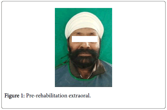
A 49 year old male patient reported to our Department with the chief complaint of missing teeth since past 4 months and inability to chew food (Figure 1).
His intraoral examination showed completely edentulous maxillary arch and multiple missing teeth in the mandibular arch with only two teeth remaining (34 and 44). The radiographic examination of the remaining teeth showed no bone loss. Based on the clinical examination, the different treatment options possible for the patient were:
• Extraction of the remaining teeth in the mandibular arch followed by conventional complete denture.
• Extraction of the remaining teeth followed by implant supported prosthesis.
• Tooth retained overdenture with or without attachments.
As the remaining two teeth were in sound condition and at favourable location and based on the patient’s desire to preserve the remaining teeth for as long as possible, it was decided to retain the teeth and fabricate an attachment retained overdenture for the mandibular arch and conventional complete denture for the maxillary arch.
Procedure
Intentional root canal treatment was performed on 33 and 43 followed by decoronation slightly above the gingival margin with chamfer finish line. Thereafter, two third of the root canal filling material was removed using the Dyna spiral drill. The seat was then prepared using the Dyna seat drill.
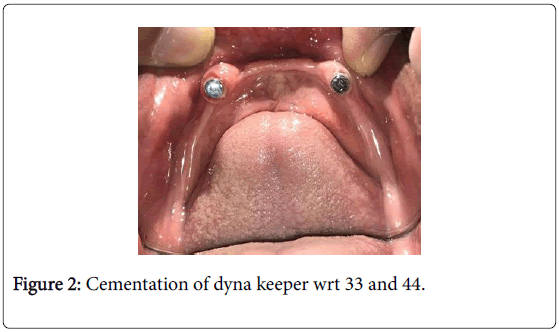
Once the space was created for the prefabricated Dyna direct keeper (4.8 mm), they were cemented using Resin bonded cement (U200, 3M ESPE) and finished to prevent any rough surface or edges (Figure 2).
Primary impression of both the maxillary and mandibular arch were taken followed by fabrication of special tray. Border moulding was then done in the conventional manner using low fusing impression compound and secondary impression with Zinc oxide eugenol impression material. The procedures from the jaw relation till the processing of the dentures were done in the conventional manner.
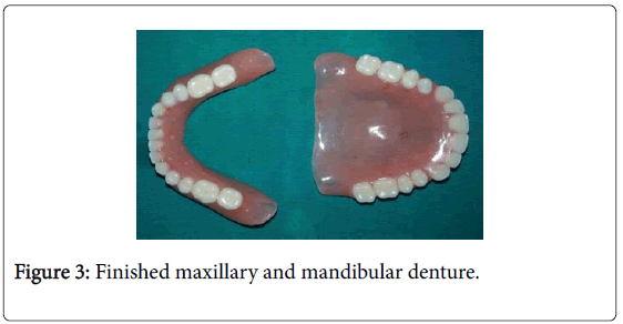
After finishing and polishing (Figure 3), the patient was recalled for the denture insertion appointment and the magnet was incorporated in the mandibular denture using chairside method.
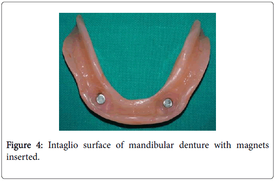
For this, first the keeper was marked with the indelible pencil and this marking signifying the keeper position was transferred on the denture. Then, a space was created in the intaglio surface of the mandibular denture in the area of the marking.
This area was filled with autopolymerizing resin and Dyna magnets were properly positioned after which the denture was inserted in the patient’s mouth. The denture was removed from the patient’s mouth before complete polymerization of the resin.
It was rechecked for proper positioning by repeated insertion and removal of the denture. After complete polymerization, any sharp edge around the magnet around the magnet was rounded off (Figure 4).
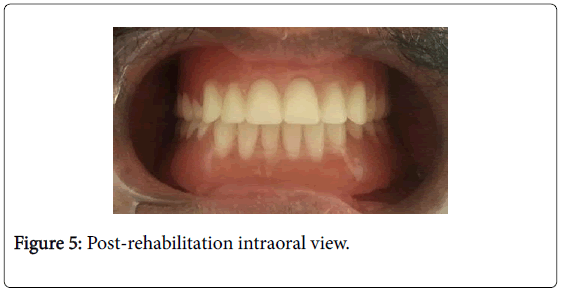
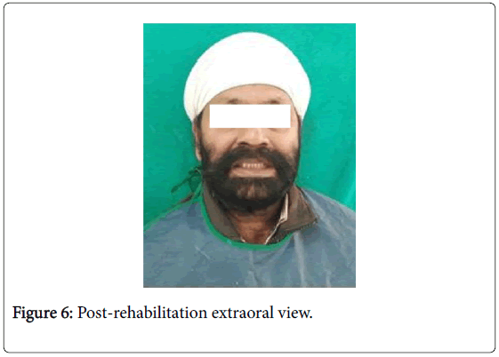
The prosthesis was then delivered to the patient and the instructions were given (Figures 5 and 6).
Discussion
In the present case, magnetically retained mandibular overdenture was used as magnets transfer no detrimental lateral forces to the supporting tooth by dissipating lateral functional forces [10] thus, helping in maintaining a favourable clinical situation. They are easy to incorporate in the denture and provide automatic reseating and constant retention with number of cycles [11]. The magnetic assembly chosen for the present case is a new generation attachment system i.e., Dyna (Direct) System which can be applied directly by the dentist, is biocompatible [12], corrosion free and very easy to use. Also, it has the advantage that it may be used on extremely compromised elements.
The magnetic systems may be an open-field or a closed-field systems [13]. Dyna magnetic system is based on open field magnetic system. Thus, the magnetic flux field radiate into the surroundings and in comparison to closed field magnets, they do not require close contact of the magnet with the object to attract it.
An overdenture during functional movements has a certain degree of mobility which implies that the magnets are not always in contact with the keeper or abutments. It is by using open field magnets that the magnetic attraction may be retained even in such situations and patients themselves experience open-field magnets as more “comfortable”. This system is simple to use and reliable which offers both the dentist and the patient very firm and unique denture retention especially in compromised situations [14,15].
Conclusion
Magnetic assembly used in this case is a new-generation magnetic attachment system which provides predictable retention and offers long-term durability. By preserving natural abutment teeth, these dentures offer has better proprioception and also provide psychologically satisfaction as the patient had not undergone extraction.
References
- Zheng R (2005) The glossary of prosthodontic terms. J Prosthet Dent 94: 10-92.
- Anupam P, Anandakrishna GN, Vibha S, Suma J, Shally K (2014) Mandibular overdenture retained by magnetic assembly: A clinical tip. J Indian Prosthodont Soc 14: 328-33.
- Arora SJ, Arora A, Sangwan R, Jain P (2017) Attachment retained overdenture. Int J Recent Scientific Res 8: 16187-16189.
- Strnat KJ (1972) The hard magnetic properties of rare earth-transition metal alloys. IEEE Trans Magn 8: 511-516.
- Tawara Y, Strnat KJ (1976) Rare earth-cobalt permanent magnets near the 2-17 composition. IEEE Trans Magn 12: 954-958.
- Sagawa M, Fujimura S, Yamamoto H, Matsuura Y, Hiraga K (1984) Permanent magnet materials based on the rare earth-iron-boron tetragonal compounds. IEEE Trans Magn 20: 1584-1589.
- Sagawa M, Fujimura S, Togawa N, Yamamoto H, Matsuura Y (1984) New material for permanent magnets on a base of Nd and Fe. J Appl Phys 55: 2083-2087.
- Croat JJ, Herbst JF, Lee RW, Pinkerton FE (1984) Pr-Fe and Nd-Fe-based materials: A new class of high-performance permanent magnets. J Appl Phys 55: 2078-2082.
- Smith GA, Laird WR, Grant AA (1983) Magnetic retention units for overdenture. J Oral Rehab 10: 481-488.
- Riley MA, Walmsley AD, Harris IR (2001) Magnets in prosthetic dentistry. J prosthet Dent 86: 137-142.
- Gonella AM, Amedeo MR, Cannas M, Pezzoli M (1989) Magnetic materials used in dentistry. Preliminary data on in vitro biocompatibility. Minerva Stomatologica 38: 1053-1058.
- Janya S, Gubrellay P, Purwur A, Khanna S (2013) Magnet retained mandibular overdenture: A multidisciplinary approach. J Interdisciplinary Dent 3: 43-46.
- Gendusa NJ (1988) Magnetically overlay dentures. Quintessence Int 19: 265-271.
- Coca I, Wisser W (2002) A clinical follow up study of magnetically retained overdentures. Eur J Prosthet Rest Dent 10: 73-78.
Citation: Upadhayaya V, Yadav A, Jain P, Kushaldeep K (2017) Prosthetic Rehabilitation in a Partially Edentulous Patient with Magnetic Dentures. J Interdiscipl Med Dent Sci 5: 221. DOI: 10.4172/2376-032X.1000221
Copyright: © 2017 Upadhayaya V, et al. This is an open-access article distributed under the terms of the Creative Commons Attribution License, which permits unrestricted use, distribution, and reproduction in any medium, provided the original author and source are credited.
Select your language of interest to view the total content in your interested language
Share This Article
Recommended Journals
Open Access Journals
Article Tools
Article Usage
- Total views: 6300
- [From(publication date): 0-2017 - Dec 21, 2025]
- Breakdown by view type
- HTML page views: 5338
- PDF downloads: 962

