Xylene/Chloroform Free Microwave Tissue Processing and Staining a Non-hazardous and Time Effective Alternative
Received: 06-Mar-2018 / Accepted Date: 14-May-2018 / Published Date: 21-May-2018 DOI: 10.4172/2161-0681.1000344
Abstract
Introduction: Tissue processing using xylene/chloroform has been employed in histopathology reporting for the past 100 years. Microwave technique has not only reduced the processing time from 1 day to one hour but also doesn’t use xylene/chloroform and has been found to be on par with conventional processing. Xylene is expensive and detrimental to human health. The present study replaces chloroform with isopropanolol in tissue processing and xylene with liquid dish wash solution (LDW) in staining which is not only cheap but also non bio hazardous.
Aim: To assess the efficacy of xylene free processing (microwave) and staining versus conventional tissueprocessing and hematoxylin and eosin are staining.
Materials and methods: Two Tissue bits from 15 consecutively submitted samples at RL Jalappa Hospital after 1st Jan 2015 each of breast, cervix, lymph node, fat, thyroid, skin, alimentary tract, muscle, salivary gland, liver and kidney were taken and one was processed and stained using conventional method, other using xylene/chloroform free processing and conventional staining and other with both xylene/chloroform free processing and staining. The 495 sections were evaluated and scored by two pathologists independently for nuclear staining, cytoplasmic staining, uniformity, clarity and crispness.
Results: In the samples evaluated xylene free processing and staining seems to be on par with conventional xylene processed and stained sections terms of nuclear and cytoplasmic detail, clarity and crispness. However Xylene cleared sections showed slightly better uniform staining (p value->0.05)
Conclusion: Xylene/chloroform free processing and staining is not only a rapid but also safe alternative to conventional processing and staining using xylene. However more extensive studies need to be done on other types of tissues for validation
Keywords: Xylene; Alternative; Microwave processing; Liquid dish wash; Xylene free staining
Introduction
Tissue processing using chemicals like xylene/chloroform has been employed in histopathology reporting for the past 100 years. It not only takes almost a day but uses hazardous chemicals like xylene/ chloroform. These hazardous chemicals are used in clearing in tissue processing and deparrafinisation before staining. Use of these chemicals is not only toxic and hazardous but is also expensive. Microwave tissue processing technique was first used by Boon and Kok from Netherlands [1]. Microwave ovens generate heat from within by rotation and collision of charged molecules. Microwave processing not only reduced the processing time from 10-12 h to one hour but also doesn’t use chemicals such as Xylene/Chloroform which are hazardous to our health [2]. Studies have shown that microwave processing is on par with conventional processing [3-7]. Xylene used as a deparaffinising agent is an excellent clearing agent but has detrimental effect on human health and known to cause hepatitis, pneumonia, depression etc [8]. Though xylene has been known to be toxic and regulatory bodies have made it compulsory to monitor exposure levels to these toxic chemicals and to follow strict guidelines for their disposal, in developing countries most laboratory follow neither of the above measures to ensure safety. The Occupational Safety and Health Administration(OSHA) in United States has laid down strict guidelines restricting maximum exposure of 100 ppm for xylene, no such regulatory body monitors the same in our country. Many studies have been done in the past and shown that liquid dish wash solution is an excellent alternative to xylene in staining and is not only cost and time effective but also eco-friendly achieving satisfactory sections for diagnosis in more than 95% of cases in using xylene free technique [9-14]. Few other studies have been done using oils such as groundnut oil/coconut oil and others using kerosene mixed with xylene to reduce the toxic effects of xylene [15,16]. However no method has been standardized so that it can be adopted in laboratories and most studies have been on small sample size. Many studies have been done using microwave processing to speed up the tissue processing and few studies on using alternatives for xylene in staining. However the present study aims at achieving both and eliminating xylene/ chloroform from both tissue processing and staining thus not only reducing the health hazards in laboratory technicians but also reducing overall turnaround time as it uses microwave processing. In the present study matched specimens will be used to test the method against various tissues so that the standardized procedure can be adopted in laboratories for routine processing. The present study was done using microwave processing using domestic microwave which can be a time effective, cheap, safe and fast alternative for conventional processing. Along with omission of these chemicals from tissue processing the present study aimed to omit them from staining too. Hence in the present study staining of microwave processed sections was done using xylene free hematoxylin and eosin (H&E) staining using liquid dish wash solution.
Objectives
To assess the quality of staining in Xylene/chloroform free microwave processed sections stained by conventional and xylene free H&E staining.
To compare the quality of staining of Xylene/chloroform free microwave processed conventional H&E stained sections versus the conventional tissue processed and H&E stained sections using xylene/ chloroform versus Xylene/chloroform free microwave processed with xylene free H&E stained sections.
Sample size
Sample size was estimated based on the values obtained from the study by Kango et al., diagnosis of lesion in conventional processing was 94% and in microwave processing 100% [5]. Using this values sample size was estimated by using the formula of difference in proportions.
Sample size of 158+158=316 was obtained at α=0.05, β at 0.20 (Power at 80)
Sample
Tissue bits from 15 consecutively submitted resection specimens at RL Jalappa Hospital after 1st Jan 2015 each of breast, cervix, lymph node, fat, thyroid, skin, alimentary tract, muscle, salivary gland, liver and kidney will be taken.
Methods
The present study was a laboratory exploratory study in which two tissue bits from 15 consecutively submitted samples at Department of Pathology, RL Jalappa Hospital and research centre attached to Sri Devaraj Urs Medical College between January to June 2015 each of breast, cervix, lymph node, fat, thyroid, skin, alimentary tract, muscle, salivary gland, liver and kidney was taken and one was processed by conventional tissue processing and other by xylene free microwave tissue processing. Both will then embedded in paraffin wax and sections of 4 micron thickness will be cut and stained using routine hematoxylin and eosin staining. One set of sections from microwave processed blocks were stained using xylene free staining using liquid dish wash solution as clearing agent. The 495 sections of the 330 blocks will be evaluated and scored by two pathologists independently for nuclear staining, cytoplasmic staining, uniformity, clarity and crispness. Scoring system scored the parameters as 0 and 1 with scores between 3-5 considered as satisfactory. Scoring system used was the one used by Ankle et al. [11] Procedure for Microwave processing was from a study by Nangia et al. [17] Procedure for staining using Liquid dish wash solution was from a study by Falkeholm et al. [10]. Ethical Clearance was obtained from Institutional Ethics Committee of Sri Devaraj Urs Medical College for conducting and publishing the study.
Scoring system
Nuclear Staining: Adequate-Score 1 Inadequate-Score 0
Cytoplasmic Staining: Adequate-Score 1 Inadequate-Score 0
Clarity: Present-Score 1, Absent-Score 0
Uniformity of Staining: Present-Score 1, Absent-Score 0
Crispness: Present-Score 1, Absent-Score 0
Statistical analysis
Percentage for individual component scoring and sensitivity and specificity of Xylene free staing was calculated. P value was calculated. Agreement between scoring by two pathologists was assessed by Cohens Kappa was calculated.
Results
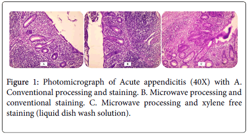
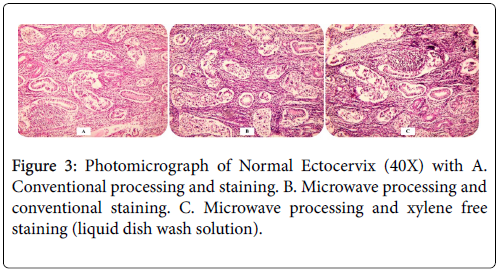
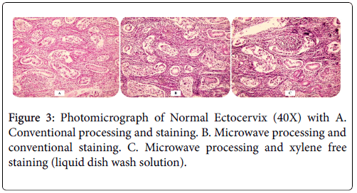
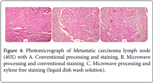
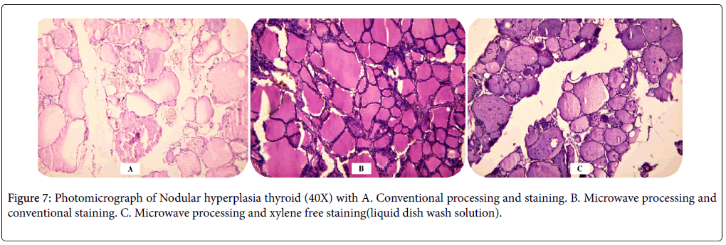
Significant reduction in tissue processing and staining time was noted between conventional and Xylene free processing and staining. In the present study, conventional staining using chloroform took around 10 h for processing and 55-60 min for staining whereas xylene free microwave processing took only 60 min and only 25-30 minutes for xylene free staining with liquid dish wash solution. Majority of the cases were adequate for diagnosis with their staining characteristics depicted in Figures 1-7 where A represented tissues processed and stained using conventional methods. B represented tissues processed using microwave processing and stained using conventional staining whereas C represented tissues processed using microwave processing and stained using xylene free staining using liquid dish wash solution.
Total score 3-5 and 3.1% was inadequate for diagnosis in xylene/ chloroform free processing and staining versus 97.5% cases adequate for diagnosis and 2.5% inadequate for diagnosis in conventional processing and staining. The cases that were inadequate for diagnosis were the ones where due to either thick tissue bits that weren’t processed satisfactorily by microwave processing or the ones that weren’t fixed adequately which resulted in unsatisfactory results both in conventional and microwave processing Nuclear staining, Cytoplasmic Staining, Crispness, Uniformity of staining and clarity was 96.9% and 95.1%, 93.3% and 92.1%, 91.5% and 91.5%, 87.2% and 89% and 96.9% and 94.5% in Conventional and Xylene Free processing and Staining respectively as shown in Table 1. The Sensitivity and Specificity was more than 95% for Xylene free processing and xylene free processing and staining. P value was >0.05 showing no significant difference in the two processing and staining techniques. Cohen’s Kappa measure of agreement was used to calculate the agreement between the two pathologists in scoring and a value of 0.71 was obtained indicating substantial agreement.
| Conventional Processing and Staining |
Xylene/chloroform Free Processing and Conventional Staining |
Xylene/chloroform Free processing and Xylene free Staining |
p Value | |||||
|---|---|---|---|---|---|---|---|---|
| N | % | N | % | N | % | |||
| Nuclear Staining (Percentage of cases) |
Adequate | 160 | 96.9 | 158 | 95.7 | 157 | 95.1 | > 0.05 |
| Inadequate | 5 | 3.1 | 7 | 4.3 | 8 | 4.9 | ||
| Cytoplasmic Staining (Percentage of cases) |
Adequate | 154 | 93.3 | 153 | 92.7 | 152 | 92.1 | > 0.05 |
| Inadequate | 11 | 6.7 | 12 | 7.3 | 13 | 7.9 | ||
| Clarity (Percentage of cases) |
Adequate | 151 | 91.5 | 149 | 90.3 | 151 | 91.5 | > 0.05 |
| Inadequate | 14 | 8.5 | 16 | 9.7 | 14 | 8.5 | ||
| Uniformity of staining (Percentage of cases) |
Adequate | 144 | 87.2 | 146 | 88.4 | 147 | 89 | > 0.05 |
| Inadequate | 21 | 12.8 | 19 | 11.6 | 18 | 11 | ||
| Crispness (Percentage of cases) |
Adequate | 160 | 96.9 | 155 | 93.9 | 156 | 94.5 | > 0.05 |
| Inadequate | 5 | 3.1 | 10 | 6.1 | 9 | 5.5 | ||
| Adequate for diagnosis (Score : 3-5) | Adequate | 161 | 97.5 | 160 | 96.9 | 160 | 96.9 | > 0.05 |
| Inadequate | 4 | 2.5 | 5 | 3.1 | 5 | 3.1 | ||
Table 1: Comparison of Hematoxylin & Eosin staining characteristics of various sections after A-Conventional processing and staining, B–Xylene/ chloroform free Microwave processing and conventional staining & C-Xylene/chloroform free Microwave processing and xylene free (liquid dish wash solution) staining.
Discussion
Xylene is used extensively in histopathology laboratories since decades. However concerns are being raised due to xylene substitutes. New regulations in countries like US by Occupational Safety and Health Administrations and American Conference of Governmental Industrial Hygiene have given limits for exposure and disposal of hazardous chemicals like xylene [14]. Hence eco-friendly alternatives have been looked for to be an alternate for xylene. Microwave processing is not only a faster but also safer alternative to xylene for tissue processing. Microwaves function by causing vibration and oscillation of the charged particles and generate heat thus expedite the tissue processing and resulting in shorter turnaround times [6]. The only disadvantage in the present study in microwave processing was incomplete processing if tissue bits were thick. Liquid dishwash solution is non-biohazardous, eco-friendly and cheap but also a good deparaffinising agent acting by its detergent action by reducing the surface tension. There was almost no difference in the staining using conventional processing and staining versus xylene free techniques such as microwave processing and staining using LDW when adequacy for diagnosis was considered. Similar results were seen in other studies [3-6]. The only disadvantage of liquid dish wash solution is that it needed close monitoring and temperature of the solution was very important for complete deparaffinisation. Limitations of the present study was standardization of the procedure for all tissues as in the present study the authors have tried to take as many different tissues as possible but even then all the different tissues couldn’t be used in the present study. Clinical implications are significant as this study not only provides stained sections in short time resulting in reduced turnaround time for histopathology reports but also makes setting up a histopathology laboratory more easy and safe due to the absence of toxic chemicals like xylene/chloroform which would otherwise require stringent monitoring and discarding protocols which would be not only costly but difficult to manage.
Nuclear staining is very important in routine staining as it helps to distinguish between benign and malignant lesions as identifying features such as nucleoli, hyperchromasia, irregular nuclear membrane and mitotic figures favor a diagnosis of malignancy. In the present study comparable or better nuclear staining was noted in Xylene free staining compared to conventional staining. Cytoplasmic staining, Clarity and crispness of staining were comparable between the two staining techniques. Uniformity of staining was slightly better in Xylene free processing than in conventional staining. This has been observed in previous studies by Shashidhara et al. [2], Kango et al. [5] and Rohr et al. [6] in Table 2. All sections termed inadequate on microwave processing were slightly thicker tissue blocks that were incompletely fixed and not completely processed and hence had poor cytoplasmic and nuclear detail after staining.
| Present Study | Kango et al. | Shashidhara et al. | ||
|---|---|---|---|---|
| Processing | Microwave | 96.9 | 95 | 60 |
| Conventional | 97.5 | 86.5 | 55 |
Table 2: Comparison of studies with percentage of sections adequate for diagnosis.
Conclusion
The present study shows more than 96% sections as satisfactory for diagnosis in xylene free processing and staining sections thus making it an excellent alternative to the toxic and hazardous xylene/ chloroform. Microwave processing is not only a time saving alternative but also an eco-friendly alternative to xylene in tissue processing. Liquid dish wash solution is an excellent alternative to xylene free sections as it is not only eco-friendly and non-bio hazardous but also effective deparaffinising agent and less time consuming as shown by the results above. Hence the combination of the above two makes it an excellent alternative to the conventional processing to get faster results without compromising on the quality of staining required for accurate diagnosis and a cheaper alternative to the hazardous chemicals like xylene and chloroform.
References
- Kok LP, Visser PE, Boon ME (1988) Histoprocessing with the microwave oven: An update. Histochemical J 20:323-8.
- Shashidara R, Sridhara SU (2011) Kitchen microwave‑assisted accelerated method for fixation and processing of oral mucosal biopsies: A pilot study. World J Dent 2:17‑21.
- Babu TM, Malathi N, Magesh KT (2011) A comparative study on microwave and routine tissue processing. Indian J Dent Res 22:51-5.
- Devi RB, Subhashree AR, Parameaswari PJ, Parijatham BO (2013) Domestic microwave tissue processing. J Clin Diagn Res 7:835‑9.
- Kango PG, Deshmukh RS (2011) Microwave processing: A boon for oralpathologists. J Oral Maxillofac Pathol 15:6‑13.
- Rohr LR, Layfield LJ, Wallin D, Hardy D (2001) Comparison of routine and rapid microwave tissue processing in a surgical pathology laboratory quality of histologic sections and advantages of microwave processing. Am J Clin Pathol 115:703‑8.
- Mathai AM, Naik R, Pai MR, Rai S, Baliga P (2008) Microwave histoprocessing versus conventional histoprocessing. Indian J Pathol Microbiol 51:12-6.
- Kandyala R, Raghavendra SP, Saraswati TR (2010) Xylene: an overview of its health hazards and preventive measures. J Oral Maxillofacial Pathol 14:1-5.
- Ramulu S, Koneru A, Ravikumar S, Sharma P, Ramesh D, et al. Liquid dish washing soap: An excellent substitute for xylene and alcohol in hematoxylin and eosin staining procedure. J Orofac Sci 2012 4:37‑42.
- Falkeholm L, Grant CA, Magnusson A, Moller E (2001) Xylene free method for histological preparation: A Multicentre Evaluation. Lab Invest 81:121321.
- Ankle MR, Joshi PS (2011) A study to evaluate the efficacy of xylene free hematoxylin and eosin staining procedure as compared to the conventional hematoxylin and eosin staining: An experimental study. J Oral Maxillofac Pathol 15:1617.
- Pandey P, Dixit A, Tanwar A, Sharma A, Mittal S (2014) A comparative study to evaluate liquid dish washing soap as an alternative to xylene and alcohol in deparaffinization and hematoxylin and eosin staining. J Lab Physicians 6:84-90.
- Ananthaneni A, Namala S, Guduru VS, Ramprasad VVS, Ramisetty SD, et al. (2014) Efficacy of 1.5% Dish Washing Solution and 95% Lemon Water in Substituting Perilous Xylene as a Deparaffinizing Agent for Routine H and E Staining Procedure: A Short Study. Scientifica 2014:11.
- Buesa RJ, Peshkov MV (2009) Histology without xylene. Ann Diagn Pathol 13:246‑56.
- Shah AA, Kulkarni D, Ingale Y, Koshy AV, Bom BS, et al. (2017) Kerosene: Contributing agent to xylene as a clearing agent in tissue processing. J Oral Maxillofac Pathol 21:367â€74.
- Sermadi W, Prabhu S, Acharya S, Javali SB (2014) Comparing the efficacy of coconut oil and xylene as a clearing agent in the histopathology laboratory. J Oral Maxillofac Pathol Suppl S1:49-53.
- Nangia R, Puri A, Gupta R, Bansal S, Negi A, et al. (2013) Comparison of conventional tissue processing with microwave processing using commercially available and domestic microwaves. Indian J Oral Sci 4:64-9.
Citation: Swaroop R, Divya C, Harendra K (2018) Xylene/Chloroform Free Microwave Tissue Processing and Staining a Non-hazardous and Time Effective Alternative. J Clin Exp Pathol 8:344. DOI: 10.4172/2161-0681.1000344
Copyright: © 2018 Raj S, et al. This is an open-access article distributed under the terms of the Creative Commons Attribution License, which permits unrestricted use, distribution, and reproduction in any medium, provided the original author and source are credited.
Select your language of interest to view the total content in your interested language
Share This Article
Recommended Journals
Open Access Journals
Article Tools
Article Usage
- Total views: 9068
- [From(publication date): 0-2018 - Dec 07, 2025]
- Breakdown by view type
- HTML page views: 8054
- PDF downloads: 1014

