Research Article Open Access
Analysing Displacement in the Posterior Mandible using Digital Image Correlation Method
| Ivan Tanasic1*, Ljiljana Tihacek-Sojic1, Aleksandra Milic-Lemic1, Nenad Mitrovic2, Radivoje Mitrovic2, Milos Milosevic2 and Tasko Maneski2 | |
| 1School of Dentistry, University of Belgrade, Serbia | |
| 2Innovation Center of Faculty of Mechanical Engineering, University of Belgrade, Belgrade, Serbia | |
| Corresponding Author : | Dr. Ivan V, Tanasic School of Dentistry 11000 Belgrade I.V. Tanasic, Serbia Tel: +381643662890 E-mail: doktorivan@hotmail.com prosthodontics.clinic@gmail.com |
| Received September 19, 2010; Accepted November 12, 2011; Published November 17, 2011 | |
| Citation: Tanasic I, Tihacek-Sojic L, Milic-Lemic A, Mitrovic N, Mitrovic R, et al. (2011) Analysing Displacement in the Posterior Mandible using Digital Image Correlation Method. J Biochip Tissue chip S1:006. doi:10.4172/2153-0777.S1-006 | |
| Copyright: © 2011 Tanasic I, et al. This is an open-access article distributed under the terms of the Creative Commons Attribution License, which permits unrestricted use, distribution, and reproduction in any medium, provided the original author and source are credited. | |
Visit for more related articles at Journal of Bioengineering and Bioelectronics
Abstract
Background: Knowing the biomechanical consequences of the stresses generated to the supporting bone during occlusal loading is significant for improving design and clinical planning process in partial edentualism therapy. The objective of this study was to analyze the surface displacement field on partial dentate mandible rehabilitated with removable partial denture and to compare it to displacement field on partial dentate mandible rehabilitated with cantilever fixed partial denture. Material and Methods: The experimental models were partial dentate mandible with full-arch PFM crowns and removable partial denture and partial dentate mandible rehabilitated with full-arch cantilever fixed partial denture. Displacement were measured using Digital Image Correlation Method. Results: Displacement values of the removable partial denture-model were ranging from 0-1.50 mm. Analysis of the fixed partial denture-model results showed displacement values from 0.05-0.69 mm. Conclusion: Higher displacements of bone tissue were observed below the removable partial denture, especially in the region of distal abutment and distal portion of the free-end saddle. Displacements within bone and the bonedenture contact area were mostly influenced by the teeth and denture vertical movements.
| Keywords |
| Mandible; Strain; Removable partial denture; Cantilever; Digital image correlation method |
| Introduction |
| Knowing the biomechanical consequences of the stresses generated to the supporting bone during occlusal loading is significant for improving design and clinical planning process in partial edentualism therapy. Current precision restoration designs are widely used in prosthetic treatment . Success of this treatment is primarily determined by biomechanical compatibility of the designs with the tissues. The complexity of replacements used in dental practise may complicate the understanding of biomechanical interaction between denture and tissues, especially bone tissue. Hence, this study proposed the biomechanical interaction between dried mandible with bilaterally shortened dental arch (Kennedy class 1) and two possible and viable treatment options made for the case of billaterally shortened dental arch: removable partial denture (RPD) and fixed partial denture (FPD). Despite a clinically relative high success rate for both conventional treatment options (RPD and FPD) the long-term biomechanical consequence on supporting tissues is unclear and yet to be analyzed. As a typical consequence of FPD treatment, bone resorption can present stability and longevity problems, whereby the surrounding bone resorbs away from the FPD abutments and thus bone support becomes compromised [1]. The same may be attributed to the RPD treatment regarding the characteristics of the distal extension base RPD to generate movements in various directions under occlusal loading. Such movements if not minimized by the proper design of the RPD may overload the supporting tissues. Consequently it may overpower the physiological limits of the bone tissue with resultant resorption and subsequent failure of the RPD [2] |
| The following experiment was carried out by the fact that there is a need for more experimental work like that conducted to balance the excessive FEA modelling work that is unsubstantiated clinically. In this work, it was preferred to choose a Digital Image Correlation method for displacement analysis for the purpose of validation, since this method can provide a full view of displacement fields in an entire dried periodontium [3] |
| The objective of this study was to analyze the surface displacement field on partial dentate mandible rehabilitated with RPD and to compare it to displacement field on partial dentate mandible rehabilitated with cantilever FPD. |
| Materials and Methods |
| Dried mandible (male, late fifties) was borrowed from the Antropology laboratory Institute of Anatomy, School of Medicine, University of Belgrade. Mandible was inspected visually and evaluated, because it was necessary for experimental model to be without evident traumatic and pathological damages. In the exclusion criteria, mandible cannot have any bone disease, neither in relation to the cause of death nor from the findings during autopsy. |
| In the inclusion criterion, the mandible should have short dental arch (Kennedy class I) with absence of the posterior molars and the second premolar and molars in the right and left side, respectively. The mandible was immersed and left in the physiological (saline) solution (0,9% NaCl) for 24 hour in order to reach some volume and elasticity [4-6]. |
| After drying at the temperature of 27 °C, preparation of the remaining teeth for receiving porcelain fuse to metal (PFM) restorations was performed. Following preparation of the abutment teeth in accordance with the main biomechanical principles of teeth preparation [7,8] and standard impression procedures of the supporting tissues, two types of restorations were made. The first restoration was complex removable partial denture (RPD) in combination with full coverage cast porcelain fused to metal (PFM) crowns on remaining teeth. Bredent ball attachments were used to connect PFM crowns and RPD into functional union. Afterwards, eleven unit PFM cantilever FPD was fabricated on the prepared mandible teeth. Consequently, two experimental models of the same mandible were obtained: the experimental model of the partial edentulous mandible with RPD positioned in situ (RPD model) and the experimental model of the partial edentulous mandible with FPD cantilever positioned in situ (FPD model). The surfaces of the experimental models with adequate restorations were sprayed with a thin coat of white layer, followed with a fine pattern of small dots placed on the top of the white layer in order to utilize the correct performance of the DIC method. Both models were placed in the standard tensile testing machine-press system. The study employed occlusal loading applied in vertical direction, with the intensity range of 300 N. The force (load) was applied to the buccal and lingual cusps of loading distal teeth (replaced teeth) in both models, across the horizontal extention of the gnatodynamometer [9,10]. |
| Experiment was performed using the system that uses Digital Image Correlation Method-DIC (GOM-Optical Measuring Techniques, Braunschweig, Germany). The System consists of two digital cameras and software ARAMIS (GOM-Optical Measuring Techniques, Braunschweig, Germany, version 6.2.0.). Before the displacement measuring of the experimental models, the calibration procedure was performed, necessary for calibrating system and setting measuring parameters, ensuring dimensional consistence and obtaining precise results. ARAMIS software used in the study serves for measuring 3D changes of shape and distribution of displacement on the surface of statically and dynamically loaded objects. The main product of software processing were figures. In this study figures served for visualizing displacement distribution when the loading achieve intensity of 300 N. In that moment the last stage was reproduced by Aramis software, whereas section line registers displacement in the last stage. |
| In all four figures, software performs the calculations and section line is set manually (by user). The reference points used for comparison between groups (more than two, and include stage point) were on the line section in every single figure. |
| Results |
| Displacement differences were determined between two experimental models (RPD and FPD) under different loading conditions. This study includes only the most representative figures that serve for visualizing the results. Displacement field in all four figures represents displacements within the posterior (lateral) segment of the experimental models. It means that the system excludes the anterior segment of the mandible models. |
| The first experimental model-RPD model |
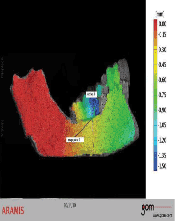
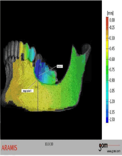
| The displacement field of the posterior mandible is obtained during denture loading. The highest displacement values are noticed just below the restoration (RPD). Line section is within the right and left buccal cortical laminae of the posterior mandible and is placed using software (Figure 1 and 3). Both sides of RPD model (right and left) have size of displacement values from 0 to 1.50 mm. Overall mean displacement for the last stage of the posterior mandible is about 0.45 mm. It is noticed that the right side of the RPD model indicates anterior increase and posterior decrease of the displacement field. The left side of the RPD model shows the oposite spreading of the displacement field direction. Bone displacement registred just below the attack (offensive) point of the applied load is 0.45 mm. The mentioned displacement value is the part of the section line and its marked as a stage point 0. This point lies in the direction of applied load. Buccal cortical laminae below and around the abutment tooth experienced displacement of 0.45-0.55 mm. |
| The second experimental model-FPD model |
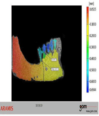
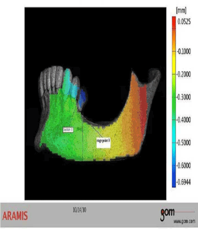
| With respect to the same limitations implemented in the previous experiment, the displacement field is obtained during loading of 300 N (last stage). The load is directed on the left and right distally extending cantilever units, simultaneously. The vertical (sagittal) line in the (Figures 2 and 4) joins the points of reference placed on the observed object, mandible and cantilever, and it changes its length during loading. The scale in (Figures 2 and 4) enables registering of quantative changes in the posterior mandible, and it is presented in milimeters. The range of the displacement values is from 0.05 to 0.69 mm. Displacement values below and around the distal (posterior) abutment tooth (0.20- 0.30 mm, right and left, respectively), were lower than in the case of RPD model (0.45-0.55 mm). Generally, mean displacement value for the last stage of the posterior mandible is 0.20 mm. Both sides of the FPD model indicate anterior increase of the displacement value within spreading of displacement field direction. |
| Discussion |
| In this research Digital Image Correlation technique was used to calculate and analyze displacement generated by static non-impact loading on the surface of experimental models with different structural designs. The study has indicated that Digital Image Correlation method (DIC) can be used to measure displacement on the posterior mandible bone surface and replacements during loading. DIC have not been widely used in dental research so far, although it has several advantages over measurements by others digital methods. A significant advantage of the applied technique compared to laser scanning systems, such as ESPI, is that the movement of the object has no influence on the measurements. Laser measurements require an absolute rigor of specimens, and unavoidable small movements of a person can be expected during photogrammetric measurements. Therefore, the relative displacement of the tracked replacement compared to an immobile reference area on the adjacent teeth provides reliable data acquisition. |
| On the other hand, strain gauges have traditionally been used to determine strains on surfaces of bone and bone simulants [11-13] but they are limited to detecting strains in a small region [14,15] Additionally, a strain gauge averages the strains measured over the gauge length, possibly leading to lower readings than the actual values, and sensitivity of measurement to temperature changes is a cause of concern [16]. An attractive feature of the digital image correlation method is that, instead of an average displacement/strain value generated on the small surface where the strain gauge is positioned, the full displacement/ strain field showing local details can be obtained for the whole surface of the model under observation. DIC is much less sensitive to ambient vibrations, can detect rigid body motion and simultaneously measure 3D displacements in a high dynamic range (submicrons to millimeters) of measuring capacity [15-17] Unlike single camera systems that have difficulty measuring displacement on complex surfaces geometries [18], the two camera system allows accurate measurements on flat surfaces and slightly less accurate measurement of the investigated curves. A current limitation of the applied optical measurement technique is the camera system’s field of view. Right and left side were assesed separatly. It means that the experiment included only the posterior part (segment) of the restored mandible models viewed from sagittal (lateral) aspect where lateral sides of the restored mandible were excluded. To obtain the most accurate information on crevices and curves of observed models, alternative surface pattern applications have to be assessed. |
| This investigation is an in vitro study that uses cadaveric specimen-mandible. As all other cadaveric analyzes the experiment may suffer from the donor-related variability of the investigated bone properties, the absence of soft tissue in the anatomic part investigated and impossibility of positioning mandible as in real situation. Nevertheless, this type of study, analyzed with DIC, could help us to understand the nature of stress distribution through the hierarchical structure of bone. It must be considered that its difficult to simulate the viscoelastic properties of periodontal ligament and residual ridge mucosa, only analysing in the mouth can reflect the true conditions. Modeling of soft tissue has not been done in this research, and also viscoelastic properties of the periodontal ligament (PDL) and mucosa layer were not included when obtaining results. Knowing their dimensions and physical characteristics it was considered that their biomechanical behavior might influence only the intensity of displacement, but not change the direction of displacement field [19,20]. After all, it was the intention of this study to analyze and present DIC as a possible method in the field of dental biomechanics. That is the reasons why displacement values were presented qualitatively as gradient of different colors, in order to give full insight into the biomechanical behavior of the analyzed structures. Study presented biomechanical view of two possible therapeutic solutions for the case of the mandible shortened dental arch. Treatment modalities employed in the study presented different biomechanical behaviour for possible comparison. The focus of analyse was placed on displacement in the upper part of the posterior mandible bone (pars alveolaris) and the distal abutments. According to the obtained displacement values the software generated section line positioned in the bone structure of the posterior mandible. Although, not very much in the same location for both models, section lines are characterised by reference points in the bone that exibit greatest displacement during loading. Thus, proper comparison of displacement values between two experimental models was enabled. |
| Differences are found between two experimental models (RPD and FPD). The displacement Y is presented in all four figures, and it has vertical direction. Higher values of displacement are registred in the RPD model in comparing to FPD model. Higher displacements are probably the reason of different kind of fixation within two types of therapy options. In the RPD model all remaining teeth were splinted in the full cast restoration in combination with attachment retained RPD. This type of RPD allows movement of the free-end saddles toward the edentulous ridge when loaded and consequently higher displacement in the posterior mandible bone. The movement resulted in transfer a majority of the applied load to the edentulous ridge, which led to much less displacement beneath the anterior retainers (see Figure 3). Similar analyses were performed using finite element method (FEM) and concluded that superioinferior denture displacement caused by vertical direction loading was larger in the posterior segments than in the anterior segments of the posterior mandible [21,22]. |
| The difference in the obtained values may be attributed to the effect of splinting remaining teeth in the experiment. The improvement in stress distribution to the supporting structures with fixed splinting was demonstrated for both mandibular and maxillary RPDs [23,24]. |
| In the second experimental model all remaining teeth were splinted in the full-arch reconstruction with cantilever FPD. The rigid construction of the cantilever FPD showed smaller displacement values in the posterior supporting marginal bone compared to the values found in the first experiment (RPD model). Vertical loading of cantilevered distally units can cause only limited stress transfer to distant portions of the posterior mandible (and abutments surrounding) [25]. Splinting of the remaining teeth allowed uniform distribution of load to the supporting alveolar bone and remaining teeth [26]. |
| It seems that when displacement pattern for both models were compared, the FPD loading did not induce overall significantlly stronger displacement values of the posterior mandible. However, the FPD therapy influenced the displacement magnitude on the marginal bone of adjacent anterior abutments when loaded on a distally extended unit. This may be explained that when connecting two or more teeth rigidly, loading is distributed and transmitted to both or each single abutments (and retainers) [27]. The effect of connecting teeth seems to be limited locally, because the direction of the displacements is within the upper part (marginal bone) of the mandible in both cases (RPD and FPD therapy). Beside abovementioned, cantilever FPD may not provide the best biomechanical solution on periodontally compromised abutment teeth due to their possible overloading during masticatory functions. In such cases, properly designed and adjusted RPD with fixed splinting of remaining teeth might be considered as therapeutic option. |
| Conclusion |
| Dimensional alterations in the posterior segment of the human cadaver mandible restored with different therapy modalities were assessed using digital image correlation method. The optical method used in this study visualized displacement distribution in the mandible models during occlusal loading. Within the limitations of this study, the following conclusions were derived: |
| - Displacements within residual ridge bone are mostly influenced by the teeth and denture vertical movements in both models (RPD and FPD); |
| - It was concluded that the mechanical behaviour of the cantilever FPD was found to be more favourable than that of the alternative RPD in terms of displacements; |
| - Findings provide that FPD therapy induce lesser displacement in the marginal bone of the posterior (distal) abutments; |
| - RPD therapy indicates higher displacement of the residual ridge. This fact is a consequence of greater vertical movement of the removable partial denture than the cantilever. |
| Acknowledgements |
| The authors would like to thank the Innovation Center of Faculty of Mechanical Engineering University of Belgrade-Serbia, Faculty of Mechanical Engineering- University of Belgrade- Serbia andSchool of Medicine, University of Belgrade- Serbia. |
| References |
|
Figures at a glance
 |
 |
 |
 |
| Figure 1 | Figure 2 | Figure 3 | Figure 4 |
Relevant Topics
Recommended Journals
Article Tools
Article Usage
- Total views: 14106
- [From(publication date):
specialissue-2011 - Dec 23, 2025] - Breakdown by view type
- HTML page views : 9473
- PDF downloads : 4633
