10 Years Clinical Follow-Up of the First Hemicap® Resurfacing Mini Prosthesis in the Knee of a Former Active 55 Year Old Man with a Full Thickness Osteochondrale Lesion
Received: 04-Jan-2017 / Accepted Date: 03-Feb-2017 / Published Date: 10-Feb-2017
Abstract
The treatment of osteochondral lesions remains a clinical challenge. A “treatment-gap” has been identified, where middle-aged patients with focal knee-lesions are considered too old for biological treatment, and yet too young for uni- or total arthroplasty (UKA/ TKA). In order to try to fill this gap, a novel metal mini-prosthesis – Hemicap® - was invented and clinically tested, and was introduced in Denmark (DK) in 2006. The aim of this case report is to present this first “Danish patient” at his 10-year clinical follow-up.
Keywords: Osteochondral; Arthroplasty; Hemicap procedure
79128Introduction
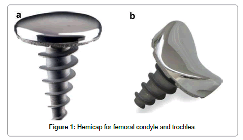
The HemiCAP® resurfacing implant consists of two components: a fixation component and a modular articular component, connected with a Morse Taper (HemiCAP® Focal Femoral Condyle Resurfacing Prosthesis, Arthrosurface Inc, Franklin, MA, USA). The fixation component is a titanium cancellous screw with full-length cannulation. The cobalt chrome articular component is available in 15 or 20 mm diameter and comes in 16 different offset configurations which 15 correspond to the superior/inferior and medial/lateral radius of curvatures at the implant site. The trochlear implant is 15 or 20 mm in diameter with a single concave shape fitting the trochleal 17 sulcus (Figure 1). A polyethylene inlay is available for the patella and is recommended to be used 18 in combination with the trochlear implant. [1-11].
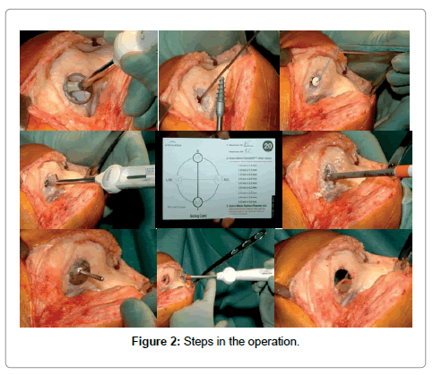
Surgical procedure The procedure was initiated with a standard arthroscopy to identify cartilage status and treat any concomitant intra articular pathology. The defect was exposed using a small para-patellar incision. The cartilage lesion was sized and a special centralized drill guide was used to place a k-wire perpendicular and central to the articular cartilage surface. Over the k-wire, the reaming for the fixation screw was performed and the screw was implanted into the bone. Mapping instruments were used to measure the surface curvature, and a matching surface reamer prepared the inlay implant bed. Sizing trials were used to confirm an accurate fit to the surrounding cartilage. The resurfacing implant was press- fit onto the fixation screw and was seated slightly recessed – (0.5 mm) - to the surrounding articular cartilage surface (Figure 2). A standardized rehabilitation protocol with free range of motion was allowed immediately postoperatively.
Case Presentation
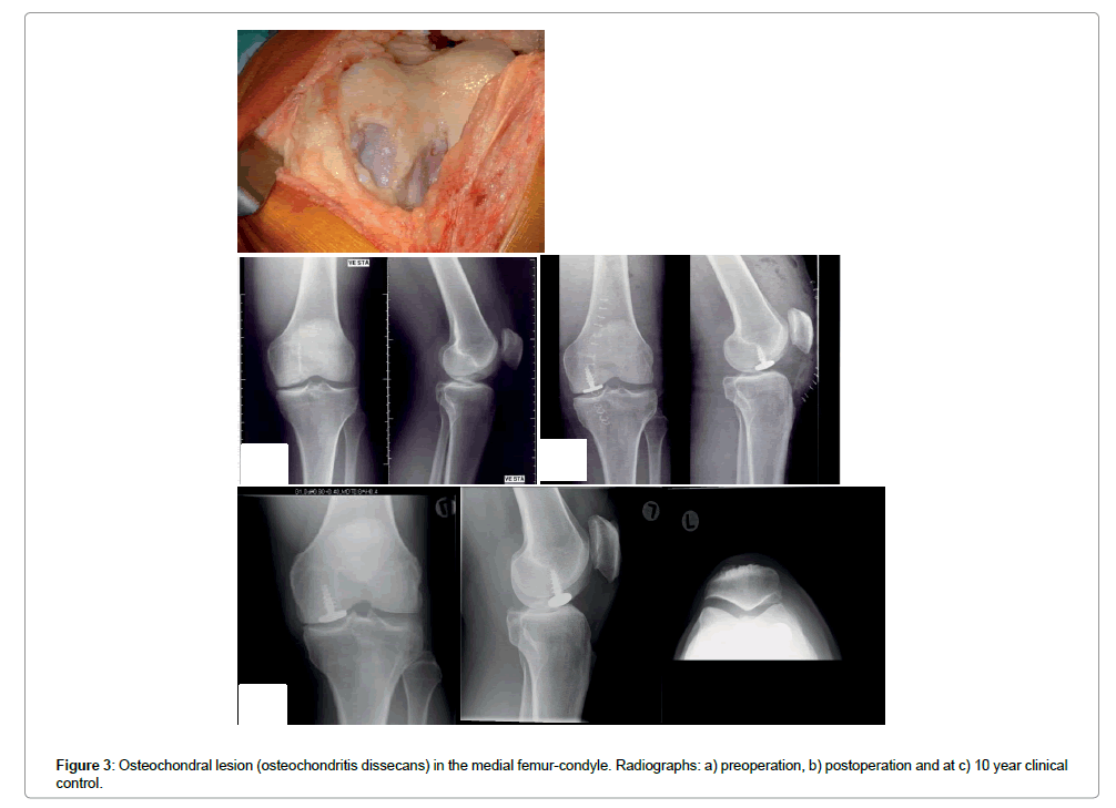
55 year-old man a construction worker with his own small subcontracting company having problems working due to increasing knee pain. He had been using strong painkillers (Paracetamol, NSAIDs and periodically morphine) to be able to continue to work, and had two arthroscopies and micro- fractures done without clinical effect on his large osteochondritis dissecans in the medial condyle (Figure 3). Radiographs and MR and arthroscopy showed nice, healthy cartilage in the other compartments. He had been unable to work for the last 6 months prior to operation because of pain and impaired function, and was now at risk of long-term unemployment.
He was chosen to be the first person in Denmark to undergo the Hemicap procedure. He followed our postoperative protocol, and was pain free at his 6 week appointment, not on any medications and had returned to fulltime work. The Patient continued to work his construction job for another 9 years and to this day remains active and pain free.
He was followed up in clinic and with radiographs at 3 months, 1, 2, 5 and 10 years. VAS preoperative at 7 fell to 1 – 2 at the present and AKSS rising from 45 postoperative and at 10 years 90 – 95. KLgrade was preoperative 0 – 1 in all compartments, and now at 10-years, 2 in medial compartment and 1 in the others. There are no signs of any loosening of the Hemicap® (Table 1).
| Prep | 3 months | 1 year | 2 years | 5 years | 10 years | |
|---|---|---|---|---|---|---|
| Pain score - VAS | 8 | 4 | 2 | 1 | 1 | 2 |
| AKSSfunc. | 45 | 80 | 95 | 95 | 90 | 90 |
| AKSSobj. | 50 | 85 | 90 | 90 | 90 | 90 |
| KL grade | ||||||
| Medil | 1 | 1 | 1 | 1 | 1 | 2 |
| Lateal | 0 | 0 | 0 | 1 | 1 | 1 |
Table 1: Clinical and radiographic outcomes.
Conclusion
The 10-year follow-up result after treatment with this new metal inlay mini-prosthesis -Hemicap® - in a challenging “older” patient with focal full-thickness (osteo) chondral lesion ( osteochondritis dissecans ) and a history of failed previous cartilage surgery demonstrated excellent long-term pain and subjective/objective outcome improvements. The patient went from being on the verge of losing his livelihood with all the financial and emotional risks that come with the unemployment to returning to full time work with minimal to no limitations [1,2,12,13]. In the US alone, 3.6 million people fall into this “treatment gap” and may significant benefit from having this procedure done [14].
Conflict of Interest
There is no conflict of interests.
References
- Becher C, Kalbe C, Thermann H (2011) Minimum 5-year results of focal 68 articular prosthetic resurfacing for the treatment of full thickness articular cartilage 69 defects in the knee. Arch Orthop Trauma Surg131: 1135-1143.
- Bollars P, Bosquet M, Vandekerckhove B, Hardeman F, Bellemans J (2012) 72 Prosthetic inlay resurfacing for the treatment of focal, full thickness cartilage defects 73 of the femoral condyle: A bridge between biologicals and conventional arthroplasty. Knee Surg Sports TraumatolArthrosc 20: 1753-1759.
- Cicuttini F, Ding C, Wluka A, Davis S, Ebeling PR, et al. (2005) Association with cartilage defects with loss of knee cartilage in healthy, middle-age adults: A prospective study. Arthritis Rheum 52: 2033-2039.
- Dhollander AAM, Almquist KF, Moens K,Vandekerckhove PJ, Verdonk R, et al. (2015) The use of a prosthetic inlay resurfacing as a salvage procedure for a failed cartilage repair. Knee Surg Sports TraumatolArthrosc 23: 2208-2212.
- Gobbi A, Karnatzikos G, Kumar A (2014) Longterm results after microfracture treatment for full-thickness knee chondral lesions in athletes. Knee Surg Sports TraumatolArthtosc 22: 1986-1996.
- Kreuz PC, Steinwachs MR, Erggelet C, Krause SJ, Konrad G, et al. (2006) Results after microfracture of full-thickness chondral defects in different 102 compartments in the knee. Osteoarthritis Cartilage 14: 1119-1125.
- Laursen JO, Lind M (2015) Treatment of full-thickness femoral cartilage lesions using condyle resurfacing prosthesis. Knee Surg Sports TraumatolArthrosc.
- Laursen JO (2016) Treatment of full-thickness cartilage lesions and early OA using large condyle resurfacing prosthesis: UniCAP. Knee Surg Sports TraumatolArthrosc 24: 1695 - 1671.
- Laursen JO (2016) High mid?term revision rate after treatment of large, full?112 thickness cartilage lesions and OA in the patellofemoral joint using a large inlay resurfacing prosthesis: HemiCAP?Wave®. Knee Surg Sports TraumatolArthrosc.
- Mithoefer K, Williams RJ, Warren RF, Potter HG, Spock CR, et al. (2005) Themicrofracture technique for the treatment of articular cartilage lesions in the knee: a prospective cohort study. J Bone Joint Surg (Am) 87: 1911-1920.
- Li CS, Karlsson J, Winemaker M, Sancheti P, Bhandari M (2014) Orthopedic surgeons feel that there is a treatment gap in management of early OA: international survey. Knee Surg Sports TraumatolArthrosc 22: 363-378.
- Heir S, Nerhus TK, Rotterud JH, Loken S, Ekeland A, et al. ( 92 2010 ) Focal cartilage defects in the knee impair quality of life as much as severe osteoarthritis: a comparison of knee injury and osteoarthritis outcome score in 4 patient categories scheduled for knee surgery. Am J Sports Med 38:Â 231- 237.
- Kellgren JH, Lawrence JS (1957) Radiological assessment of osteo-arthrosis. Ann 98 Rheum Dis 16: 494-502.
- London NJ, Miller LE, Block JE (2011) Clinical and economic consequences of 121 the treatment gap in knee osteoarthritis management. Med Hypotheses 76: 887-892.
Citation: Laursen JO (2017) 10 Years Clinical Follow-Up of the First Hemicap® Resurfacing Mini Prosthesis in the Knee of a Former Active 55 Year Old Man with a Full Thickness Osteochondrale Lesion. J Med Imp Surg 2: 112.
Copyright: © 2017 Laursen JO. This is an open-access article distributed under the terms of the Creative Commons Attribution License, which permits unrestricted use, distribution, and reproduction in any medium, provided the original author and source are credited.
Select your language of interest to view the total content in your interested language
Share This Article
Recommended Journals
Open Access Journals
Article Usage
- Total views: 5135
- [From(publication date): 0-2017 - Jul 12, 2025]
- Breakdown by view type
- HTML page views: 4208
- PDF downloads: 927


