A Brief Review on Analysis of Cytokines' Role in Research
Received: 05-May-2023 / Manuscript No. jcb-23-99672 / Editor assigned: 08-May-2023 / PreQC No. jcb-23-99672 (PQ) / Reviewed: 22-May-2023 / QC No. jcb-23-99672 / Revised: 24-May-2023 / Manuscript No. jcb-23-99672 (R) / Published Date: 31-May-2023 DOI: 10.4172/2576-3881.1000441
Abstract
About 20 years ago, several organisations looking for genes with sequence homology to the cytokine family IL-1 found the IL-36 cytokines. One of these investigations, written up in 2001 by Debets and colleagues, showed that human keratinocytes expressed IL-1 and IL-1, now known as IL-36 and the IL-36R antagonist (IL-36Ra), respectively. This was especially true after stimulation with IL-1 and TNF. Three pro-inflammatory cytokines from the IL-36 family—IL-36, IL-36, and IL-36—as well as a receptor antagonist—IL-36Ra—bind to and signal through a heterodimeric receptor made up of IL-36R and the IL-1R accessory protein (IL-1RAcP). A result that directly connects dysregulated IL-36 pathway activation to inflammatory skin disorders is the development of generalised pustular psoriasis in people with inactivating mutations in the gene for IL-36Ra, a severe type of psoriasis. This review’s focus is on the cellular origins of IL-36 cytokines, the effects of IL-36 signalling on different cell types, and the relationship between IL-36 and a range of inflammatory skin conditions, such as different types of psoriasis, hidradenitis suppurativa, atopic dermatitis, and allergic contact dermatitis.
Keywords
Heterodimeric; Glioblastoma; Central nervous system; Chemotherapy drugs
Introduction
The most frequent malignant primary tumour of the central nervous system (CNS) in adults, glioblastoma (GBM), is on the rise all over the world. GBM has a high mortality rate and high recurrence rate, which contribute to its aggressive behaviour. Patients with GBM typically experience an accelerated neurologic decline and a decline in quality of life. The overall survival still hovers around 15 months, and the 5-year survival rate is as low as 5% despite standard of care therapy that includes surgical resection followed by concomitant radiation and temozolomide. Numerous tumour forms, including melanoma, non-small cell lung cancer, and non-Hodgkin lymphoma, have been successfully treated with immunotherapy. For patients with CNS tumours, including GBM, immunotherapy has been widely studied; nonetheless, monotherapy immunotherapy was ineffective for this type of tumour. Numerous molecular traits of GBM, including low mutational burden, poor T-cell infiltration, significant tumor-elicited T-cell dysfunction, and the presence of tumor-associated macrophages (TAMs), which suppress lymphocyte-induced inflammation and phagocytes, could be to blame for immunotherapy’s failure [1]. In fact, one of the difficulties in treating GBM is that it is immunogenic. Chemotherapy drugs and peripheral immune cells frequently cannot cross the blood-brain barrier (BBB), and infiltrating lymphocytes are uncommon in GBM. Therefore, the most prevalent type of cell in the tumour is the resident immune cells in the CNS, or microglia. In the presence of damage-associated molecular patterns (DAMPs), these cells become reactive and polarise to a classic activated state (M1) capable of engulfing debris and secreting crucial pro-inflammatory cytokines, such as interleukin (IL)-1b, tumour necrosis factor (TNF)-a, and chemoattractant mediators. IL-4, IL-10, IL-13, and tumour growth factor (TGF)-b are examples of anti-inflammatory cytokines that can be released by microglia when they are polarised to the alternate state (M2), on the other hand [2]. The M1/M2 cell ratio Worldwide, the usage of engineered nanoparticles (NPs) is expanding quickly in the creation of novel materials that offer beneficial qualities for treatments and consumer goods. It is crucial to prevent these new NP/nanomaterials (NM) from having qualities that have a negative impact on humans’ acute or chronic health or that have an unfavourable impact on the variety of species in the environment given their expanding use.
Numerous studies have concentrated on NP hazard characterization as well as exposure evaluation to determine which NPs may pose a risk to human health in work settings, during pharmacological therapy, or in the general population/consumer population. It’s crucial to understand the molecular starting processes ultimately responsible for the consequences seen from NP exposure in order to more accurately forecast NP toxicity and build adverse outcome pathways (AOPs) for various kinds of NPs.
Family of the IL-36
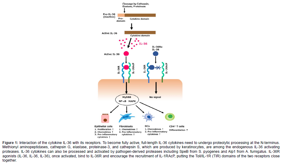
Interleukin 1 (IL-1) family cytokines, which include the recently identified IL-36, IL-37, and IL-38 proteins, have important roles as inflammation-initiators and are typically among the first cytokines released in response to infection or damage. IL-1 family cytokines have the power to initiate intricate cascades of further cytokine production from a variety of cell types, including resident tissue macrophages and dendritic cells as well as keratinocytes and endothelial cells lining local blood vessels. IL-36, IL-36, and IL-36 are all encoded by separate genes, and there is growing evidence that these cytokines are important in the inflammation of the skin, notably in psoriasis. IL-36, IL-36, and IL-36 are all produced as biologically inactive leaderless cytokines. As with other IL-1 family members including IL-1 and IL-18, proteolytic processing of IL-36 cytokines is necessary to activate their proinflammatory action. According to research by Sims and colleagues, removing a modest number of residues from the N termini of IL-36, IL-36, and IL-36 significantly boosts each protein’s biological activity by a factor of more than 10,000. Inhibitors of IL-36 proteolytic activation may have significant potential for the treatment of inflammatory skin disorders because IL-36 cytokines appear to play a significant role as initiators of inflammation in the skin barrier. However, it is currently unknown which proteases cause the IL-36 cytokines to become active [3].
The IL-36R signaling
The cytokines of the Interleukin-36 (IL-36) family are extensively expressed in the skin lesions of psoriasis and are essential to the disease’s aetiology. It is well established that IL-36 antagonist (IL-36Ra) reduces the pathophysiology of psoriasis by blocking IL-36/IL-36R signalling. It is unknown, though, if IL-36R possesses a unique naturally soluble receptor. Here, we discovered a naturally occurring soluble IL-36R (sIL-36R) that inhibits IL-36/IL-36R signalling and demonstrate that sIL-36R reduces inflammation in psoriasis. By DNA sequencing, it was discovered that one IL-36R variant produced the extracellular domain of IL-36R (sIL-36R) in keratinocytes. A competitive experiment was done to verify that sIL-36R would operate as a ruse to annoy IL-36R signalling. There are 11 distinct ligands that make up the IL-1 family of cytokines: IL-1 (also known as IL-1F1), IL-1 (also known as IL- 1F2), IL-1Ra (also known as IL-1F3), IL-18 (also known as IL-1F4), IL-1F5 to IL-1F10, and IL-1F11 (also known as IL-33). When they bind to the type I IL-1 receptor (IL-1RI) and activate the common coreceptor IL-1 receptor accessory protein (IL-1RAcP), IL-1 and IL-1 are known to cause proinflammatory effects, whereas IL-1Ra functions as a competitive inhibitor of IL-1 binding to IL-1RI and thus has an anti-inflammatory effect. Numerous investigations revealed that IL- 18 is a proinflammatory cytokine that is unmistakably more than an IFN- inducer, while IL-33 was characterised as an immunoregulatory [4]. (Figure 1)
Figure 1: Interaction of the cytokine IL-36 with its receptors. To become fully active, full-length IL-36 cytokines need to undergo proteolytic processing at the N-terminus. Methionyl aminopeptidases, cathepsin G, elastase, proteinase-3, and cathepsin S, which are produced by keratinocytes, are among the endogenous IL-36 activating proteases. IL-36 cytokines can also be processed and activated by pathogen-derived proteases including SpeB from S. pyogenes and Alp1 from A. fumigatus. IL-36R agonists (IL-36, IL-36, IL-36), once activated, bind to IL-36R and encourage the recruitment of IL-1RAcP, putting the Toll/IL-1R (TIR) domains of the two receptors close together.
Expression and action of IL-36R
An essential part of the adaptive immune system, CD4+ T helper (Th) cells can develop into multiple regulatory and effector lineages that have an impact on cancer, infectious diseases, inflammatory disorders, and autoimmune diseases. Foxp3-expressing regulatory Th cells (Treg) are potently immunosuppressive and aid in maintaining immune homeostasis. Treg can grow intrathymically or in the periphery.2 Contrarily, effector Th cells (Teff) can be divided into a number of broad categories (Th1, Th2, Th9, Th17, Th22, and TFH) based on the dominating signature cytokines they generate and the expression of related master transcription factors.4 It’s interesting to note that particular cytokines and other variables control whether naive Th cells differentiate into the Treg or Teff lineages [5]. (Figure 2)
Figure 2: Effects of IL-36 pathway activation on cells. Following stimulation by TNF, IL-17A, IL-22, and IL-1, keratinocytes release IL-36 cytokines. CatS, a protease found in neutrophil extracellular traps (NETs), or pathogen-associated proteases can all be used to proteolytically convert full-length IL-36 cytokines into their active versions. While IL-36 activation of myeloid cells results in overexpression of costimulatory molecules and secretion of IL-1, IL-6, and IL-23, which promote Th17 development, activated IL- 36 promotes plasmacytoid dendritic cells to release IFN. TNF, IL-17A, and IL-22 are produced by Th17 and Th22 cells, and these molecules boost keratinocytes' production of IL-36.
Immune cells, both innate and adaptive
The goal of adenovirus-based vaccines is to elicit a protective immune response against the transgenic product by altering the adenoviral genome to produce nonreplicating virion particles able to contain a desired transgene. This adenovirus-based vaccine technology is employed in licenced vaccines against the COVID-19 disease (Ad26, Ad5, and ChAdOx1-based vector vaccines) and the disease brought on by Ebola virus infection (Ad26 in combination with a Modified Vaccinia Ankara component) 1, 2, 3. Fundamental research has been done to comprehend the structure, tropism, and host response to adenoviruses. These studies have also shed light on the mechanism of action of adenovirus-based vectors. The most recent research on viral cellular entry and innate immune responses is discussed here, with special emphasis on adenovirus-based vectors approved for use as preventative vaccinations in humans. Also highlighted is how innate sensing mechanisms and cell entrance affect adaptive immune responses [6].
Materials and Methods
In a case-control study, we included 53 stroke-free normotensives as controls (CS), 59 stroke-free hypertensives (HPT), and 63 cases with stroke and hypertension (HPT-S). Using commercially available ELISA kits from Biobase Biotech, Shanghai, China, sociodemographic information and blood samples were obtained for the estimation of Interleukin-10 (IL-10), IL-6, IL-8, IL-1, and monocyte chemoattractant protein-1 (MCP-1). For the purpose of predicting stroke in hypertensives, the Receiver Operator Characteristics (ROC) analysis was utilised to determine the diagnostic accuracy of cytokines. A combination IL-10 and MCP-1 bioscore model was created to forecast stroke in hypertensives. The likelihood of IL-10 and MCP-1 in predicting stroke among hypertensives was evaluated using the multiple logistic regression analysis. R was used to perform the statistical analysis [7].
Radiographic and clinical evaluate
Preoperative evaluations included evaluations of comorbidities, neurologic symptoms including motor impairments, clinical characteristics, and Karnofsky Performance Status (KPS). After the surgery, with a follow-up of about a thousand days, the overall survival and progression free survival (PFS) were examined [8].
Results and Discussion
Conditions for cell culture and exposure
A SV-40 hybrid (Ad12SV40) transformed human bronchial epithelial cell line known as BEAS-2B was cultured in LHC-9 media in collagen-coated flasks (PureColTM) at 37 °C with 5% CO2, with medium replacement every other day. Following seeding, the cells (passages 8–50) were cultivated for 24 hours in LHC-9 media in 35 mm 6-well culture plates (300 000 cells per well) or on 100 mm culture dishes. Before being exposed to Si10 and Si50, the cells were starved for 24 h in serum-free DMEM/F12. After a 20-hour exposure, cytokine release (CXCL8, IL-6, IL-1, and IL-1) and TGF- release (after a 4-hour exposure) were examined. After 2 hours of exposure, Western blotting was used to analyse the phosphorylation of p38, JNK, and NF-B. In 35 mm (six-well) and 5 mL in the culture dishes, respectively, the total exposure volume was 1 mL.
Medicine
Physiological saline (SAL, 0.9% NaCl) was used to dissolve nicotine hydrogen tartrate salt (0.5 mg/kg, Merck®), and the pH was raised to 7.4. The solutions were made just before use and administered intraperitoneally (i.p.) at a volume of 1 mL per 100 g of body weight. The identical amount of saline is given to the control animals [10].
Analysis of the cytokines IL-6, CXCL8, IL-1, and IL-1
IL-6 and IL-8 structural and functional properties and their use as biomarkers for the early identification of oral cancer. Several POC biosensing platforms have been developed to find IL-6 and IL-8 in early oral cancer detection. Optical and electrochemical biosensors are designed, made, and have transduction mechanisms for POC detection of IL-6 and IL-8. Challenges associated with implementing these biosensors in clinical settings and employing them to analyse real samples. The cell culture media were collected 20 hours after exposure and centrifuged at 300 x g and 10,000 x g to remove floating SiNPs. Sandwich ELISA was used in accordance with the manufacturer’s instructions to measure the levels of IL-6, CXCL8, IL-1, and IL-1 protein. A plate reader (TECAN Sunrise) with specialised software was used to measure and quantify absorbance [11].
Melanoma tumour immune cell composition
A high concentration and combination of plasma EV (pEV) from several cellular origins are thought to be present in human plasma. However, there are no precise subpopulations of pEV that have been verified or approved. This is because distinguishing and identifying vesicles is technically challenging. This job is further complicated by the high heterogeneity of pEV in terms of size, surface marker composition, and cellular and subcellular origin. Nevertheless, it was hypothesised that platelets secrete a sizable amount of pEV, up to 107/ml plasma, as shown by flow cytometry. Notably, these pEV were discovered to affect target cells in both homeostasis and disease in a variety of ways. For patients whose data are accessible in TCGA, we used RNA-seq data analysis using TIMER deconvolution to estimate immune cell subsets. For each important immune cell subset, TIMER contains an input matrix of reference gene expression signatures that is utilised to estimate the relative proportions of each cell type of interest.
Conclusion
The IL-36 cytokines have gained prominence as important contributors to skin inflammation since their discovery about 20 years ago. The presence of IL36RN mutations in DITRA variants of GPP serves as the best example of this. Additionally, IL-36R targeting with neutralising antibodies has demonstrated efficacy in Phase I and Phase II clinical studies regardless of IL36RN mutational status, offering hope for GPP patients. Despite the fact that the clinical goals of the initial clinical trials assessing IL-36R blockage in PPP were not met, there were some indications of a quicker response in treated groups and better responses in patients with more advanced disease.
Acknowledgement
None
References
- Carrier Y, Ma HL, Ramon HE, Napierata L, Small C, et al. (2011) Inter-regulation of Th17 cytokines and the IL-36 cytokines in vitro and in vivo: implications in psoriasis pathogenesis. J Invest Dermatol 131: 2428-2437.
- Miura S, Garcet S, Salud-Gnilo C, Gonzalez J, Xuan Li, et al. (2021) IL-36 and IL-17A Cooperatively Induce a Psoriasis-Like Gene Expression Response in Human Keratinocytes. J Invest Dermatol 141:2086-2090.
- Debets R, Jackie C Timans, Homey B, Zurawski S, Theodore R Sana, et al. (2001) Two novel IL-1 family members, IL-1 delta and IL-1 epsilon, function as an antagonist and agonist of NF-kappa B activation through the orphan IL-1 receptor-related protein 2. J Immunol 167:1440-1446.
- Towne JE, Kirsten E Garka, Blair R Renshaw, Duke Virca G, John ES (2004) Interleukin (IL)-1F6, IL-1F8, and IL-1F9 signal through IL-1Rrp2 and IL-1RAcP to activate the pathway leading to NF-kappaB and MAPKs. J Biol Chem 279:13677-13688.
- Boutet MA, Bart G, Pen MA hoat M, Amiaud J, Brulin B, et al. (2016) Distinct expression of interleukin (IL)-36alpha, beta and gamma, their antagonist IL-36Ra and IL-38 in psoriasis, rheumatoid arthritis and Crohn's disease. Clin Exp Immunol 184:159-173.
- Ovesen SK, Schulze-Osthoff K, Iversen L, Johansen C (2021) IkB is a Key Regulator of Tumour Necrosis Factor-a and Interleukin-17A-mediated Induction of Interleukin-36g in Human Keratinocytes. Acta Dermato-Venereologica 101:386.
- Vigne S, Palmer G, Lamacchia C, Martin P, Talabot A, et al. (2011) IL-36R ligands are potent regulators of dendritic and T cells. Blood 118: 5813-5823.
- Henry CM, Sullivan GP, Clancy DM, Afonina IS, Kulms D, et al. (2016) Neutrophil-Derived Proteases Escalate Inflammation through Activation of IL-36 Family Cytokines. Cell Rep 14:708-722.
- Shao S, Tsoi LC, Swindell WR, Chen J, Uppala R, et al. (2021) IRAK2 Has a Critical Role in Promoting Feed-Forward Amplification of Epidermal Inflammatory Responses. J Invest Dermatol.
- Han Y, Mora J, Huard A, da Silva P, Wiechmann S, et al. (2019) IL-38 Ameliorates Skin Inflammation and Limits IL-17 Production from gammadelta T Cells. Cell Rep 27:835-846.
- Telfer NR, Chalmers RJ, Whale K, Colman G (1992) The role of streptococcal infection in the initiation of guttate psoriasis. Arch Dermatol 128:39-42.
Indexed at, Google Scholar, Crossref
Indexed at, Google Scholar, Crossref
Indexed at, Google Scholar, Crossref
Indexed at, Google Scholar, Crossref
Indexed at, Google Scholar, Crossref
Indexed at, Google Scholar, Crossref
Indexed at, Google Scholar, Crossref
Indexed at, Google Scholar, Crossref
Indexed at, Google Scholar, Crossref
Indexed at, Google Scholar, Crossref
Citation: Carrie QH (2023) A Brief Review on Analysis of Cytokines’ Role in Research. J Cytokine Biol 8: 439. DOI: 10.4172/2576-3881.1000441
Copyright: © 2023 Carrie QH. This is an open-access article distributed under the terms of the Creative Commons Attribution License, which permits unrestricted use, distribution, and reproduction in any medium, provided the original author and source are credited.
Select your language of interest to view the total content in your interested language
Share This Article
Recommended Journals
Open Access Journals
Article Tools
Article Usage
- Total views: 2343
- [From(publication date): 0-2023 - Dec 19, 2025]
- Breakdown by view type
- HTML page views: 1952
- PDF downloads: 391


