A Case Report On Radiation Dose Reduction in Computed Tomography
Received: 13-Dec-2021 / Accepted Date: 27-Dec-2021 / Published Date: 03-Jan-2022
Abstract
Computed tomography (CT) is an imaging method that utilizes special x-ray equipment to make itemized pictures, or scans, of areas inside the body. It is at times called computerized tomography or computerized axial tomography (CAT).
In spite of all inclusive agreement that Computed tomography (CT) predominantly helps patients when utilized for fitting signs, concerns have been raised in regards to the expected danger of illness enlistment from CT because of the dramatically expanded utilization of CT in medication. Keeping radiation portion as low as actually reachable, steady with the indicative assignment, stays the main system for diminishing this expected normal risk. This article sums up the overall specialized methodologies that are usually utilized for radiation portion the board in CT. Portion the board techniques for paediatric CT, heart CT, double energy CT, CT perfusion and interventional CT are explicitly examined, and future points of view on CT portion decrease are introduced.
Keywords: Radiology; CT scan; Radiation; Disease; X-ray
Introduction
A few radiation portion decrease methodologies are utilized. CTA of the aorta ought to be performed provided that the clinical sign is fitting and the output ought to be restricted to the space of interest. ECG-gating builds the radiation portion and ought to be restricted distinctly to the assessment of aortic root and rising aorta, for example, in the pre-TAVR assessment. Assuming ECG gating is done, imminent ECG setting off ought to be utilized, in light of the fact that it diminishes radiation portion by 60% and there is no compelling reason to get information all through the heart cycle. In the event that review ECG gating is utilized, ECG-based cylinder current balance is utilized, i.e., greatest chamber current is applied in just a single specific heart stage and the cylinder current is diminished to 30% for the other cardiovascular stages [1-3]. This decreases radiation portion by 40%– half the cylinder current ought to be kept to the base conceivable, which is dictated by body habitus. A large portion of the advanced scanners likewise have programmed tube current balance, where the cylinder current is changed by the body district, with lower dosages used for more slender regions and higher dosages for thicker regions. The cylinder voltage ought to likewise be kept to the most un-workable for a specific patient size. In youngsters, 80 kVp is utilized, though for grown-ups 100 kVp is utilized on the off chance that the weight list (BMI) <30 and 120 kVp is utilized if the BMI >30. The utilization of the double source, high-pitch scanner for CTA generously diminishes radiation portion in contrast with standard helical CT [4].
More up to date remaking calculations, for example, iterative recreation, decline commotion in the checked picture, consequently empowering the utilization of lower radiation portions strategies, including low cylinder current and cylinder voltages Additionally, the utilization of virtual noncontract pictures from double energy scanners takes out the requirement for genuine noncontract pictures in multiphasic concentrates like TEVAR convention and can bring about radiation portion reserve funds up to 60% [5].
Computed Tomography Report
Computed Tomography (CT) is the pillar of cutting edge kids’ lung imaging. Present day CT equipment offers significant benefits over past ages of innovation. A few frameworks additionally permit examining at amazingly high paces, with the whole chest filtered in under a large portion of a second. This permits even un-co-usable small kids to be filtered without requiring profound sedation or general sedation, with the guide of delicate immobilization hardware. Time-settled, or 4D (CT cine fluoroscopy) as of late become an essentially material imaging apparatus, and is especially valuable in the evaluation of dynamic huge aviation routes sickness, permitting non-obtrusive appraisal of tracheobronchomalacia [6]. IV differentiation organization is useful for some signs for youth thoracic CT, especially as youngsters come up short on the mediastina fat planes that permit simpler qualification between mediastina structures adults.
Radiation portion
Radiation portion in CT is a significant wellspring of worry for the clinical local area and the overall population. A multicentre concentrate on exhibited that radiation portion from heart CT fluctuated from 5 to 30 mSv with a normal portion of 12 mSv. With the expanded utilization of cardiovascular imaging, there have been endeavours to build up radiation portion decrease strategies [7].
While there are numerous strategies to decrease radiation, the decision of ECG-gating strategy is generally significant in deciding radiation portion decrease. As referenced above, forthcoming ECGgating just requires light during a particular timespan heart cycle; hence, radiation portion can be diminished to 1–4 mSv utilizing this strategy. In a meta-examination of 20 investigations contrasting forthcoming and review ECG-setting off, the pooled successful portion was 3.5 mSv with tentatively set off checks contrasted with 12 mSv utilizing review filtering. These outcomes were affirmed by the PROTECTION III preliminary in which a radiation level of 3.5 mSv was accomplished with forthcoming checking instead of 11.2 mSv with review ECGgating. Thusly, planned ECG-setting off is liked in the decrease of radiation portion, especially in patients with low, consistent pulses. Concentrates additionally exhibited the capacity to arrive at radiation dosages lower than 1 mSv utilizing ECG-set off high-pitch examining with DSCT, Once more, be that as it may, this strategy is especially helpful in patients with low to direct, normal pulses [8-10].
Radiation portion can be additionally diminished by utilizing iterative remaking. With iterative remaking, an adjustment circle is acquainted with the reproduction cycle bringing about further developed goal in high difference locales and commotion decrease in low differentiation regions, which are requirements for radiation portion decrease. This turns out to be especially helpful when joined with other portion decrease methods to accomplish impressive portion decreases. In one review, the mix of low kV, high pitch examining with iterative reproduction brought about a radiation portion as low as 0.06 mSv. As well as accomplishing lower radiation portion levels, the utilization of iterative remaking was found to further develop picture quality and symptomatic precision.
Radiation exposure
The development in accessibility and utilization of CT during the most recent quite a few years has saved many lives. Regardless of the multitude of advances in innovation prompting method improvement and radiation portion decrease, CT actually represents a huge radiation trouble, especially in the paediatric populace. There is a proceeded with need to improve current CT procedures and assess practical imaging choices to guarantee that openness to ionizing radiation is kept as low as sensible to accomplish exact determinations. Present day CT scanners can change portion openness by understanding age, weight, or potentially body part thickness.
Case Presentation
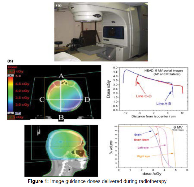
All dose reduction strategies are predicated on the assumption that the CT scanner’s radiation dose levels and image quality fall within manufacturer specifications and other general quality criteria. This can be accomplished through a quality control program that is designed and overseen by a qualified medical physicist (Figure 1).
Fixed tube current (technique charts)
Unlike traditional radiographic imaging, a CT image never looks “over-exposed” in the sense of being too dark or too light; the normalized nature of CT data (i.e., CT numbers represent a fixed amount of attenuation relative to water) ensures that the image always appears properly exposed. As a consequence, CT users are not technically compelled to decrease the tube-current-time product (mAs) for small patients, which may result in excess radiation dose for these patients. It is, however, a fundamental responsibility of the CT operator to take patient size into account when selecting the parameters that affect radiation dose, the most basic of which is the mAs 25, 34.
As with radiographic and fluoroscopic imaging, the operator should be provided with appropriate guidelines for as selection as a function of patient size. These are often referred to as technique charts. In CT, the tube current exposure time and tube potential can all be altered to give the appropriate exposure to the patient. However, users most commonly standardize the tube potential (kV) and gantry rotation time (s) for a given clinical application. The fastest rotation time should typically be used to minimize motion blurring and artifact, and the lowest kV consistent with the patient size should be selected to maximize image contrast 35-40. Hence tube current is the primary parameter that is adapted to patient size.
Conclusion
Radiation portion decrease procedures took into account radiation portion decreases of half or more in 78% of CAC CT studies. Notwithstanding, hazard renaming was affected in 3% (portion decrease of 75%) up to 21% (portion decrease of 60%) of people, contingent upon the procurement strategy. Explicit portion diminished conventions, including either tube current decrease or IR or unearthly forming with tin filtration that showed low renaming rates may possibly be utilized in CAC examining and in future populace based evaluating for CVD hazard delineation. Tube current decrease with IR is most seriously explored in current writing as a strategy for portion decrease in CAC imaging. In opposition to tin-channel, tube current decrease is relevant on all kind of CT scanners with restricted effect on hazard definition. Future exploration in portion decrease strategies of CAC imaging should zero in on bigger patient investigations assessing CVD hazard renaming rates, Agatston score dissemination and the reproducibility of the portion diminished convention.
References
- Gilbert Whittemore (1988) Multiple Exposures: Chronicles of the Radiation Age. Catherine Caufield 83: 697-698.
- Martland HS, Humphries RE (1973) Oestrogenic sarcoma in dialpainters using luminous paint. Arch Pathol 7:406-417.
- Stannard JN. Baalman RW (1988) Radioactivity and health: a history. Vol 1: Laboratory research. Columbus, OH: Battelle Press, USA.
- National Research Council (1988) Committee on the Biological Effects of Ionizing Radiations. Health risks of radon and other internally deposited alpha-emitters BEIR IV. Washington, DC: National Academy Press, USA.
- Henshaw PS, Hawkins JW, Meyer HL (1944) Incidence of leukaemia in physicians. J Natl Cancer Inst 4:339-346.
- Ulrich H (1946) the incidence of leukaemia in radiologists. N Engl J Med 234:45-46.
- Folley JH, Borges W, Yamawaki T (1952) Incidence of  leukaemia in survivors of the atomic bomb in Hiroshima and Nagasaki, Japan. Am J Med 13:311-321.
- Leukaemia and aplastic anaemia (1957) in patients irradiated for ankylosing spondylitis. London, UK: Medical Research Council.
- Wagoner JK, Archer VE, Carroll BE (1964) Cancer mortality patterns among US uranium miners and millers, 1950 through 1962. J Natl Cancer Inst 32:787-801.
Citation: Susana L (2021) A Case Report On Radiation Dose Reduction in Computed Tomography. OMICS J Radiol 10: 358.
Copyright: © 2021 Susana L. This is an open-access article distributed under the terms of the Creative Commons Attribution License, which permits unrestricted use, distribution, and reproduction in any medium, provided the original author and source are credited.
Select your language of interest to view the total content in your interested language
Share This Article
Open Access Journals
Article Usage
- Total views: 2370
- [From(publication date): 0-2021 - Dec 08, 2025]
- Breakdown by view type
- HTML page views: 1785
- PDF downloads: 585

