A New Minimal Invasive Technique in the Treatment of Resistant Plantar Fasciitis by Percutaneous Partial Plantar Fasciotomy, Drilling of the Calcaneus and Resection of Calcaneal Spur
Received: 02-Jan-2018 / Accepted Date: 15-Jan-2018 / Published Date: 22-Jan-2018 DOI: 10.4172/2329-910X.1000256
Abstract
Background: Plantar fasciitis is a common cause of plantar heel pain that may cause significant discomfort and disability. Many surgical techniques were involved in treatment like open plantar fasciotomy, endoscopic techniques, and percutaneous fasciotomy. This study was conducted to evaluate the results of surgical treatment of resistant plantar fasciitis by a new minimal invasive technique of combined percutaneous partial plantar fasciotomy, drilling of the calcaneus and resection of the calcaneal spur.
Methods: Between June 2013 and July 2015, twenty-five patients presented by resistant plantar fasciitis had undergone a minimal invasive surgery by percutaneuos partial plantar fasciotomy, drilling of the calcaneus and resection of the calcaneal spur.
Results: Heel pain was relieved within an average of 4 weeks (range: 3-8 weeks). Postoperatively twenty two 88% patients were rated as having excellent results, three patients 12% were rated as having good results and no patients 0% were rated as having a poor result without any improvement.
Conclusion: This technique compares favorably with other reported open surgical procedures and other minimal invasive and high-cost techniques. It is a relatively safe, short procedure, with a rapid learning curve and it is not associated with serious complications.
Keywords: Resistant plantar fasciitis; Minimal invasive surgery; Percutaneous partial plantar fasciotomy; Drilling; Resection of calcaneal spur
Introduction
Plantar fasciitis is a common cause of inferior heel pain in adults and accounting for approximately 11% to 15% of all foot problems [1]. Multiple treatment modalities are employed for treatment chronic plantar fasciitis including nonsteroidal anti-inflammatory drugs (NSAID), massages, activity modifications, orthoses, local infiltration of steroids and shock wave therapy [2]. After the failure of conservative measures for at least six months more aggressive surgical techniques have been reported such as open plantar fasciotomy with heel spur resection, endoscopy and minimally invasive procedures have become popular [3]. The percutaneous plantar fasciotomy procedure was first described by Harvey Pelzer [4]. The technique of percutaneous plantar fasciotomy is a simple procedure with rare complications, low costeffective method compared with more invasive and high-cost techniques [5]. The aim of this study to evaluate the results of a new minimal invasive surgery in treatment resistant plantar fasciitis by a percutaneous partial plantar fasciotomy, drilling of the calcaneus and resection of the calcaneal spur and compare it with other techniques.
Patients and methods: Between June 2013 and July 2015, twenty-five patients presented by resistant plantar fasciitis had undergone a minimal invasive surgery by percutaneuos partial plantar fasciotomy, drilling of the calcaneus and resection of the calcaneal spur. All patients were selected from outpatient clinic at Mansoura university hospital after a failure of conservative measures for at least six months including nonsteroidal anti-inflammatory drugs (NSAID), massages, activity modifications, orthoses, local infiltration of steroids and shock wave therapy. Symptoms were present for an average of 11 months (range: 6-23 months). All patients subjected to full clinical examination of the foot to locate the site of pain and to exclude other causes of heel pain like infection and tumor. Radiological examination included plain X-ray (Figure 1). It was performed to exclude infection, stress fracture, tumor and degenerative disease of mid-tarsal joints. All patients had a significant calcaneal spurs. This study included 25 patients with 32 heel pain. Of 25 patients, only 7 were bilateral. Out of twenty five patients 20 patients (80%) were female and 5 patients (20%) were male. The mean age of patients was 42 years (range: 35-59 years).
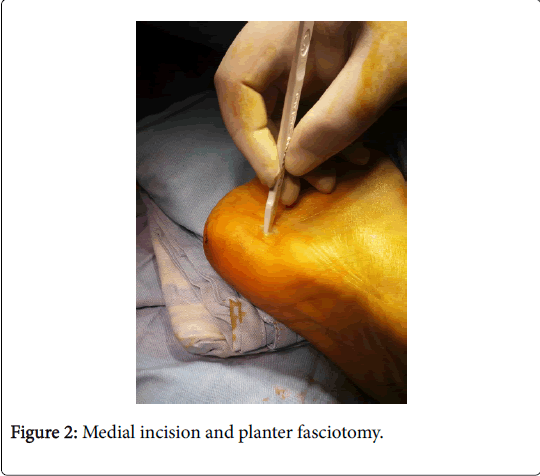
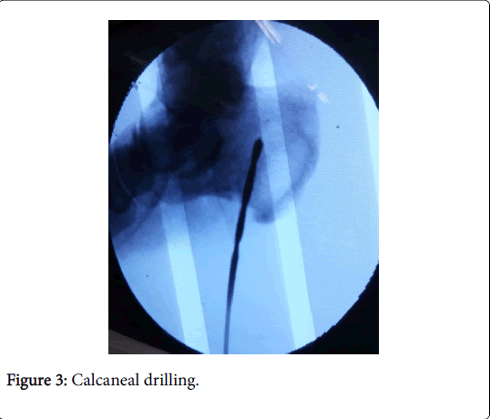
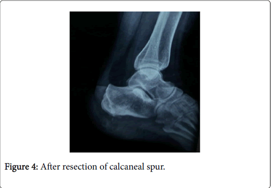
Surgical technique: The operation was performed under a local anesthesia which is achieved by blocking the tibial and saphenous nerves. After the preparation and draping of the foot and toes, a percutaneous partial plantar fasciotomy was done through a stab medial horizontal incision on the heel of 1 cm length located at the point of intersection of 2 lines under image with 2 k-wires (Figure 2): the first line is parallel to the inferior surface of calcaneal spur while the other line is perpendicular to the first line at the base of bone spur. The foot and toes positioned in a maximum dorsiflexion to place tension on plantar fascia then the plantar fascia is palpated and identified by a mosquito and separated from the heel fat bad then by a surgical blade superficial to the fascia and after estimating the distance required to be released by palpation about 50% were only resected till reaching the underlying muscle. Care was taken to leave at least 50% of the plantar fascia intact as the fascial band about 14 mm. After a percutaneous partial plantar fasciotomy a sequential drilling of the calcaneous was done around the calcaneal spur through the same medial incision (Figure 3). It was done by a drill bit 2.5 mm and drill holes were done under C-arm. Resection of the calcaneal spur was done by surgical burr inserted on a power drill and through the same skin incision under image intensifier and the spur was resected from its tip to the base of the calcaneal spur (Figure 4).
The post-operative follow-up: The patients discharged on the same day of the operation in just a crepe bandage. Each patient was instructed for partial weight bearing for 2 weeks after the surgery and ambulates afterward in a silicon heel for about 4 weeks. The patients underwent follow-up program for evaluating the clinical results in terms of pain, activity level and patient satisfaction.
The modified criteria of the Roles and Maudsley score (RM score) was applied as a rating scale for the patients. It was defined as
Eexcellent: No pain, with satisfactory treatment outcome, with unlimited painless walking.
Good: Substantially decreased symptoms, with satisfactory treatment outcome, with more than one hour painless walking.
Acceptable: Somewhat decreased symptoms with more tolerable pain level than before treatment, and slightly satisfied with the treatment outcome.
Poor: Identical or worse symptoms and with no satisfactory outcome. Treatment was considered successful when the patient had an excellent or good score. The visual analog pain scale was used to determine the effectiveness and patient satisfaction with the procedure [6]. Successful result was considered if the patient reported a percentage decrease in the VAS score larger than 60% from baseline. Comparisons for the patients preoperatively and postoperatively were performed with two-sided Chi-square tests. In all analyses, statistically significant is considered if p value was less than 0.05.
Results
The minimum follow-up time after surgery was two years. It ranged from 24 to 32 months, with an average of 27 months. There were no intraoperative complications. Also, postoperatively there was neither infection nor tender scar. All patients had overall clinical improvements and returned to unrestricted activity and their preoperative occupations. The results showed more improvement as regards to the preoperative pain level of 7.9 (± 1.2) with a range of 6-10 by utilizing the visual analog pain scale. The pain level at final followup was 1.9 (± 2.7) with a range of 0-6 with a more significant difference. Postoperatively, twenty two 88% patients were rated as having excellent results; three patients 12% were rated as having good results. No patients were rated as a poor result without any improvement. Heel pain was improved within an average of 4 weeks after the surgery (range: 3-8 weeks). After 4 months there was no pain during follow-up. All patients with satisfied results returned to their former occupations or activities and all of them were satisfied with the incision scar. There was no recurrence of pain during follow-up period. No patients developed a lateral arch pain and flat foot at the final follow-up.
Discussion
The plantar fasciitis is thought to be a consequence of inflammation at the insertion of the plantar fascia on the medial tubercle of the calcaneus. Patients complain of a sharp pain localized at the inferior aspect of the os calcis. This pain is usually more intense upon the first steps in the morning or after a period of rest. By the general public this condition is sometimes referred to as heel spurs. Plantar fasciitis is frequently associated with the presence of a heel spur (exostosis). However, many asymptomatic patients have bony heel spurs, whereas many patients with plantar fasciitis do not have a calcaneal spur [7]. The possible risk factors include occupations requiring prolonged standing, obesity, weight bearing, and heel spurs [8]. The treatment of chronic plantar fascitis is a difficult problem, but most patients eventually improve with conservative therapy. About 5% of the patients need surgical treatment after a failure of conservative treatment [5]. Many surgical procedures have been described for treatment of chronic planter fasciitis, it included open surgery, endoscopic techniques, and percutaneous fasciotomy. Open surgical interventions recommended a surgical removal or release of the plantar fascia, and removal of heel spurs. The open surgery techniques were associated with frequent complications such as persistent pain, infections, nerve disturbance, prolonged recovery time and tender skin scars [9]. On the other hand the endoscopic techniques for plantar fascia release which is a very common procedure reported high rate of success. However, the postoperative painful portals and nerve entrapment are possible complications [10-13]. In the recent articles, the procedure of percutaneous partial plantar fasciotomy is an alternative to open surgical methods with many advantages and fewer complications [14].
In this study, i used a new minimal invasive technique of combined percutaneous partial plantar fasciotomy, drilling of the calcaneus and resection of calcaneal spur. Drilling of the calcaneus plays a role in relieving the heel pain. The idea of pain relief with drilling due to decreasing both intraosseous pressure and calcaneal bone marrow oedema. The technique of sequential drilling of the body of os calcis was described by Hassab and El Sherriff to decompress the calcaneus and they achieved 62 improvements of 68 cases [15]. In my study; the resection of calcaneal spurs have been done by a new technique by using a surgical burr on a power drill through the same incision under image and the spur was resected totally. Excision of the spur was done as it is reported as a risk factor and also heel spur removal is directed at releasing the plantar fascia. Griffith reported in his study; the spur was excised through U-shaped incision around the heel and he reported six good outcomes in 6 patients. Also, Du Vries reported in his study; open fascial release was done and calcaneal spur excision through a medial horizontal incision about 6 cm. and reported 37 good outcomes from 37 patients [15]. Many articles reported the success of open surgical techniques for plantar fascial release but a number of complications have been reported like flat foot, infections, wound dehiscence, arch pain, painful neuromas and prolonged recovery [16,17]. In the study conducted by Davies et al. they reported in their study of 43 patients with 47 painful heels who underwent partial plantar fasciotomy and nerve decompression. The satisfaction was 49% after an average of 31 months follow-up [18]. Berlin described a technique of a transverse puncture incision over the origin of the fascial pain, severed the medial attachment and then performed a partial fasciotomy laterally. He reported 89% good to excellent relief in 82 patients undergoing percutaneous plantar fasciotomy [19]. Benton- Weil et al. reported in their study 83% patient satisfaction in 51 patients. Percutaneous plantar fascial release procedure was done, but there was no indication on the extent of plantar fascia released [5].
In my study this minimally invasive procedure is performed under a local anesthesia with a small skin incision which is one stab medial horizontal incision, approximately 1 cm in length, the incision is an ideal one as it lies in line with the relaxed skin tension lines and is on a non weight bearing area, minimizing scarring and yielded good healing with a minimal risk of infection. In the current study; there were no complications reported after partial plantar fasciotomy like: lateral arch pain, instability and flat foot, as the partial release was done by preserving the release of the plantar fascia less than 50% of its width. So, the complications of lateral column instability and increased fore foot and lateral column stress is kept to a minimum.
This technique was effective in resolving the chronic heel pain. This pain improved quicker within an average of 4 weeks after the surgery (range: 3-8 weeks). The patients were able to wear normal shoes, and return to their normal activities by 4-12 weeks postoperatively. No cases with pain recurrence were reported during follow-up time. As regards to the final results; Excellent and good outcomes were obtained in 22 patients (88%), 3 patients (12%), respectively. No patients were rated as poor results. All patients returned to their former occupations and activities. All patients were satisfied with the incision scar especially the female patients.
Conclusion
In this new minimal invasive technique, the combination of the percutaneous partial planter fasciotomy, drilling of calcaneus and resection of the calcaneal spur in resistant planter fasciitis improved significantly the results. It is a relatively safe, short procedure, with a rapid learning curve and it is not associated with serious complications.
Conflict of Interest
There are no conflicts of interest.
Funding
Nil.
Ethical approval
The study was done under the local ethical committee.
Informed consent
Informed consent was obtained from all individual participants included in the study.
References
- League AC (2008) Current concept reviews: Planter fasciitis. Foot Ankle Surg 29: 358-366.
- Martin JE, Hosch JC, Goforth WP, Murff RT, Lynch DM, et al. (2001) Mechanical treatment of plantar fascitis. A prospective study. J Am Podiatr Med Assoc 91: 55–62.
- Vohra PK, Japour CJ (2009) Ultrasound guided planter fascia release technique a retrospective study of 66 feet. J Am Podiatr Med Assoc 99: 183-190.
- Pilzer HP, Berlin MJ (1983) Percutaneous plantar fasciotomy: A new approach. Current Podiatry 32: 36.
- Benton WW, Borrel AH, Weil LS Jr, Weil LS Sr (1998) Percutaneous plantar fasciotomy: A minimally invasive procedure for recalcitrant plantar fascitis. J Foot Ankle Surg 37: 269-272.
- Roles NC, Maudsley RH (1972) Radial tunnel syndrome: Resistant tennis elbow as a nerve entrapment. J Bone Joint Surg Br 54: 499-508.
- Singh D, Angel J, Bentley G, Trevino SG (1997) Fortnightly review: Plantar fasciitis. BMJ 315: 172-17Â5.
- Riddle DL, Pulisic M, Pidcoe P, Johnson RE (2003) Risk factors for Plantar fasciitis: A matched case Âcontrol study. J Bone Joint Surg Am 85: 872-87Â7.
- Neufeld SK, Cerrato R (2008) Plantar fasciitis: Evaluation and treatment. J Am Acad Orthop Surg 16: 338–346.
- Barrett SL, Day AV (1991) Endoscopic plantar fasciotomy for chronic plantar fasciitis/heel spur syndrome: Surgical technique–early clinical results. J Foot Surg 30: 568-570.
- Barrett SL, Day SV (1993) Endoscopic plantar fasciotomy: Two portal endoscopic surgical techniques–clinical results of 65 procedures. J Foot Ankle Surg 2: 248-256.
- Barrett SL, Day SV, Brown MG (2011) Endoscopic plantar fasciotomy: Preliminary study with cadaveric specimens. J Foot Surg 30: 170-172.
- O’Malley MJ, Page A, Cook R (2000) Endoscopic plantar fasciotomy for chronic heel pain. Foot Ankle 21: 505-510.
- Thomas JL, Christensen JC, Kravitz SR, Mendicino RW, Schuberth JM, et al. (2010) American college of foot and ankle surgeons heel pain committee. The diagnosis and treatment of heel pain: A clinical practice guideline-revision 2010. J Foot Ankle Surg 49: 1–19.
- Bazaz R, Ferkel RD (2007) Results of endoscopic plantar fascia release. Foot Ankle Int 28: 549-556.
- Ali S, Gilpert N, Galloway T (1995) Planter fasciitis: A review of the literature. British J Podiatric Med Surg 7: 61-62.
- Sammarco GJ, Idusuyi OB (1998) Stress fracture of the base of the third metatarsal after an endoscopic plantar fasciotomy: A case report. Foot Ankle Int 19: 157-159.
- Davies MS, Weiss GA, Saxby TS (1999) Plantar fasciitis: How successful is surgical intervention? Foot Ankle Int 20: 803-807.
Citation: Maaty MT, Khalek MA (2018) A New Minimal Invasive Technique in the Treatment of Resistant Plantar Fasciitis by Percutaneous Partial Plantar Fasciotomy, Drilling of the Calcaneus and Resection of Calcaneal Spur. Clin Res Foot Ankle 6: 256. DOI: 10.4172/2329-910X.1000256
Copyright: ©2018 Maaty MT, et al. This is an open-access article distributed under the terms of the Creative Commons Attribution License, which permits unrestricted use, distribution, and reproduction in any medium, provided the original author and source are credited.
Select your language of interest to view the total content in your interested language
Share This Article
Recommended Journals
Open Access Journals
Article Tools
Article Usage
- Total views: 7648
- [From(publication date): 0-2018 - Dec 23, 2025]
- Breakdown by view type
- HTML page views: 6616
- PDF downloads: 1032


