Case Report Open Access
A Rare Case of Gastrointestinal Obstruction: Bouveret Syndrome
Ruben Gonzalo Gonzalez*, Marian de Andrés Fuertes, Sara Regaño Diez, José Luis Conty Serrano, Enrique José San Emeterio González, Guillermo Soler Dorda and Francisco Javier Gil PiedraDepartment of General Surgery, Hospital Laredo, Av de los Derechos Humanos, s/n, 39770 Laredo, Cantabria, Spain
- *Corresponding Author:
- Ruben Gonzalo Gonzalez
Department of General Surgery, Hospital Laredo
Av de los Derechos Humanos, s/n
39770 Laredo, Cantabria, Spain
Tel: 620 108 834
E-mail: rubsoygg@hotmail.com
Received date: March 23, 2015; Accepted date: April 07, 2015; Published date: April 15, 2015
Citation: Gonzalez RG, Fuertes MDA, Diez SR, Serrano JLC, González EJSE, et al. (2015) A Rare Case of Gastrointestinal Obstruction: Bouveret Syndrome. J Gastrointest Dig Syst 5:277. doi:10.4172/2161-069X.1000277
Copyright: © 2015 Gonzalez RG, et al. This is an open-access article distributed under the terms of the Creative Commons Attribution License, which permits unrestricted use, distribution and reproduction in any medium, provided the original author and source are credited.
Visit for more related articles at Journal of Gastrointestinal & Digestive System
Abstract
Bouveret Syndrome (BS) is a rare type of gallstone ileus in which a gallstone enters the intestinal tract via a cholecystoenteric fistula and is lodged in the duodenum or the stomach. It should be considered in any patient who presents with pneumobilia without recent endoscopic retrograde cholangiopancreatography (ERCP) or biliary surgery. Morbidity and mortality rates have decreased in recent years, but still remain high, at 25%, and may be related to the advanced age of the typical patient and their resulting comorbidities, as well as to diagnostic delay. Endoscopy is preferred as the first therapeutic option, but is frequently unsuccessful, and surgery is often required. We present a case report of a patient with symptoms of gastric outlet obstruction, with interesting radiological findings and successful surgical treatment after failure of endoscopic techniques.
Keywords
Biliary diseases; Bilioenteric fistula; Bouveret´s syndrome; Gastrointestinal obstruction
Introduction
Gallstone ileus is a rare complication of cholelithiasis which presents with the clinical picture of intestinal obstruction. Bouveret syndrome (BS) is the most infrequent variety of gallstone ileus, in which the obstruction has a pyloric-duodenal location caused by the impaction of a large gallstone in the duodenum after passage through a cholecystoduodenal or choledochogastroduodenal fistula. This syndrome was first described in 1896 by Leon Bouveret, who published two clinical cases of patients with gastric outlet (pyloric) obstruction secondary to the presence of a gallstone [1]. Since then, a little over 300 cases have been described in the literature [2]. To achieve a diagnosis, both radiological and endoscopic techniques are required. Endoscopic treatment should always be attempted, but it usually fails, thus making surgical treatment necessary.
The diagnostic and therapeutic management of this rare cause of gastric obstruction is discussed below.
Case Report
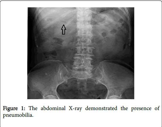
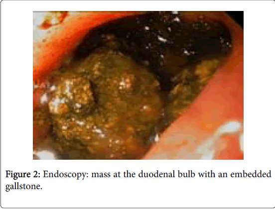
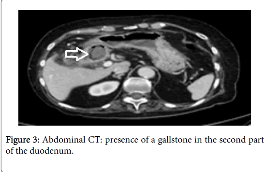
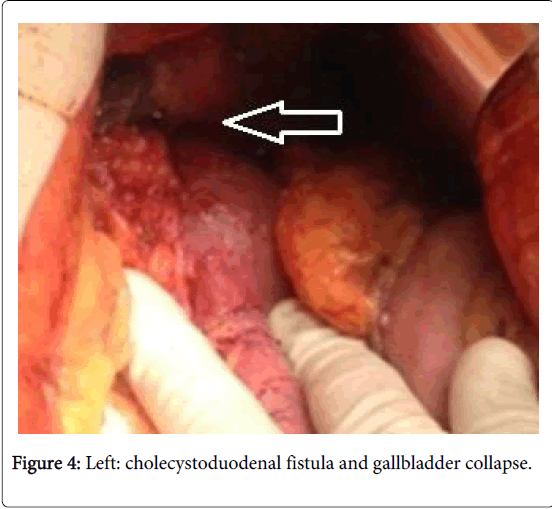
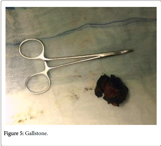
The subject was an 81-year old woman with a history of well-controlled hypertension, cardiac arrhythmia due to atrial fibrillation, and pulmonary thromboembolism treated with anticoagulants, as well as a previous hysterectomy. She presented at the Emergency Department with a three-day history of epigastric pain, nausea, and postprandial vomiting, as well as a 24-hour history of haematemesis. Neither fever nor jaundice was present. The abdomen was distended and tympanic. Palpation was painful in the right epigastrium and hypochondrium. No masses or enlarged organs were palpated, and Murphy’s sign was negative. Amongst routine investigations, only the c-reactive protein was abnormal: 8.70 mg/dL. A plain abdominal X-ray demonstrated the presence of pneumobilia (Figure 1). A gastroscopy was performed due to haematemesis. It showed the presence of a mass at the duodenal bulb with an embedded gallstone (Figure 2). Given that Bouveret syndrome was suspected, an abdominal CT scan was carried out, which demonstrated the presence of a cholecystoduodenal fistula and showed a 3 cm diameter gallstone at the duodenal bulb (Figures 3 and 4). Endoscopic extraction of the gallstone failed, and the patient underwent emergercy surgery. The surgical procedure revealed the presence of a collapsed, thickened-wall gallbladder and a cholecystoduodenal fistula. A gastrotomy was performed, a brown 4x3 cm gallstone was extracted after mobilization from the duodenum towards the stomach, and the fistulous hole was explored in detail (Figure 5). The gallbladder and the fistula were left intact. The postoperative outcome was satisfactory, and she is currently asymptomatic.
Discussion
Biliodigestive fistulas are rare, occurring in fewer than 1% of cholelithiasis patients. Most are cholecystoduodenal (60%), but cholecystocolonic (17%), cholecystogastric (5%), and cholecystoduodenal fistulas can also be found. Risk factors include a long history of cholelithiasis, recurrent episodes of acute cholecystitis, large stones (>2.5 cm), female gender, and advanced age. Most patients have small lithiases, which they eliminate through their faeces without causing any obstruction. Only 5-6% of patients develop gallstone ileus, the most common location of the obstruction being the terminal ileum (50-90% of cases), followed by the proximal ileum-jejunum (20-40%). The pyloric-duodenal location (true BS) is the least common (<5% of cases) [3,4].
SB’s clinical picture corresponds to that of a high intestinal obstruction, and usually presents non-specific symptoms: epigastric pain, nausea, vomiting, and/or ileus. Only one-third of patients present with jaundice and/or alteration of hepatic enzymes [5]. In our case, the patient presented a non-specific clinical picture, without any clear signs of intestinal obstruction.
Diagnosis can be radiological or endoscopic. Typical findings from simple abdominal X-ray are known as Rigler's triad: pneumobilia (Figure 1), intestinal obstruction, and ectopic gallstone [6]. CT-scan is regarded as the best diagnostic technique, and identifies Rigler's triad in 75% of cases [7]. High digestive endoscopy is the cornerstone of specific diagnosis, as it allows lithiasis to be visualized directly and other diagnoses to be ruled out [4] (Figure 2). In our case, due to the finding of haematemesis, an early gastroscopy was indicated. This demonstrated the presence of a mass effect at the level of the duodenal bulb. Subsequently the abdominal CT-scan confirmed the diagnosis of Bouveret syndrome (Figures 3 and 4).
The ultimate goal of treatment should be to remove the intestinal obstruction caused by the lithiasis. Given the advanced age and the extensive comorbidity with which these patients usually present, endoscopic extraction should always be attempted, since this is a minimally invasive procedure with a low incidence of complication [8]. However, endoscopic strategies usually fail; therefore, surgical treatment is required to remove the lithiasis. In our case, the endoscopic extraction of the gallstones was attempted twice, but without success, due to their large size.
The surgical approach can be performed either through laparotomy or laparoscopy. During the procedure, the whole small bowel should be examined, since in 16% of cases additional gallstones can be found at other locations. If possible, the lithiasis should be mobilized towards the stomach and removed through gastrotomy. If this is not feasible, one should try to mobilize towards the jejunum and remove it through an enterotomy. If both manoeuvres fail, a duodenotomy is carried out as a last resort. Performing cholecystectomy and fistula closure in the same surgical procedure is associated with a morbidity and mortality of 35%, vs. 12% in those patients in whom only lithiasis is removed. Additionally, if we consider that a cholecystoduodenal fistula can work as a biliodigestive anastomosis, and that the fistulous tract usually closes spontaneously (especially if there is no residual lithiasis), cholecystectomy and fistula closure can be assessed subsequently in each case, as a possible further step [9,10]. In our case, the patient was operated on urgently through median laparotomy, and the gallstone was removed through gastrotomy, after mobilization from the duodenum (Figure 5).
In conclusion, although BS is a rare complication of cholelithiasis, its diagnosis should always be considered when dealing with a patient with high intestinal obstruction and pneumobilia without recent ERCP or biliary surgery. Therefore, endoscopic treatment should always be considered as the first option. However, endoscopic treatment often fails due to the large size of the lithiasis; in such cases, surgical treatment should be performed, removing the gallstone and assessing in a further step whether cholecystectomy and fistula closure are necessary or not.
References
- Bouveret L (1896) Stenose du pylore adherent a vesicule. Rev Med (Paris) 16: 1-16.
- Mavroeidis VK, Matthioudakis DI, Economou NK, Karanikas ID (2013) Bouveret syndrome-the rarest variant of gallstone ileus: a case report and literature review. Case Rep Surg 2013: 839370.
- Rojas J, Caban P, Hernandez JA, Daz C, Vidal A (2005) Syndrome de Bouveret. Caso clÃnico y revision de la literatura. Rev Chil Cirug 57: 508-510.
- Cappell MS, Davis M (2006) Characterization of Bouveret's syndrome: a comprehensive review of 128 cases. Am J Gastroenterol 101: 2139-2146.
- Nickel F, Müller-Eschner MM, Chu J, von Tengg-Kobligk H, Müller-Stich BP (2013) Bouveret's syndrome: presentation of two cases with review of the literature and development of a surgical treatment strategy. BMC Surg 13: 33.
- Rigler LG, Borman CN, Noble JF (1941) Gallstone obstruction: pathogenesis and roentgen manifestations. JAMA 117: 1753-1759.
- Singh AK, Shirkhoda A, Lal N, Sagar P (2003) Bouveret's syndrome: appearance on CT and upper gastrointestinal radiography before and after stone obturation. AJR Am J Roentgenol 181: 828-830.
- Wittenburg H, Mössner J, Caca K (2005) Endoscopic treatment of duodenal obstruction due to a gallstone ("Bouveret's syndrome"). Ann Hepatol 4: 132-134.
- Masannat YA, Caplin S, Brown T (2006) A rare complication of a common disease: Bouveret syndrome, a case report. World J Gastroenterol 12: 2620-2621.
- Buchs NC, Azagury D, Chilcott M, Nguyen-Tang T, Dumonceau JM, et al. (2007) Bouveret's syndrome: management and strategy of a rare cause of gastric outlet obstruction. Digestion 75: 17-19.
Relevant Topics
- Constipation
- Digestive Enzymes
- Endoscopy
- Epigastric Pain
- Gall Bladder
- Gastric Cancer
- Gastrointestinal Bleeding
- Gastrointestinal Hormones
- Gastrointestinal Infections
- Gastrointestinal Inflammation
- Gastrointestinal Pathology
- Gastrointestinal Pharmacology
- Gastrointestinal Radiology
- Gastrointestinal Surgery
- Gastrointestinal Tuberculosis
- GIST Sarcoma
- Intestinal Blockage
- Pancreas
- Salivary Glands
- Stomach Bloating
- Stomach Cramps
- Stomach Disorders
- Stomach Ulcer
Recommended Journals
Article Tools
Article Usage
- Total views: 15622
- [From(publication date):
April-2015 - Aug 04, 2025] - Breakdown by view type
- HTML page views : 11033
- PDF downloads : 4589

