A Rare Case of Metastasis of Lung Carcinoid to Breast
Received: 14-Dec-2017 / Accepted Date: 05-Jan-2018 / Published Date: 12-Jan-2018 DOI: 10.4172/2167-7964.1000286
Abstract
Metastases to the breast from non-mammary primary tumors are uncommon. We present a rare, but typical, case of metastasis to the breast from lung carcinoid tumor, 20 years after the primary cancer was treated.
Keywords: Metastasis to the breast; Mammography; Ultrasound; Lung carcinoid
Introduction
Secondary tumors deposits in the breast are uncommon and account for 0.5-2% of all breast malignancies. Usually metastatic cancer to the breast emanates from a contralateral breast cancer but virtually any cancer can give metastatic deposits to the breast.
The first case of metastatic lesion to the breast has been reported by Trevithick in 1903 and it was about a case of reticulum cell sarcoma, metastatic to the breast [1].
The most common extramammary cancers that give metastatic deposits to breast are malignant melanoma, lung carcinoma, carcinoid tumors from a variety of primary sites, prostate carcinoma in men, ovarian carcinoma, gastric carcinoma, renal cell carcinoma, various types of sarcoma (especially rhabdomyosarcoma in younger females), colorectal carcinoma, carcinoma of the urinary bladder, lymphoma, various malignant tumors of the neck and head, medulloblastoma, neuroblastoma, malignant mesothelioma, endometrial carcinoma [2].
Metastatic tumors to the breast have a variable gross appearance, depending on the type of metastasis. In most cases (85%) the metastasis is represented as a solitary, unilateral lesion, which is well demarcated from the surrounding breast parenchyma. Less frequently (10%) the metastases are seen as multiple lesions and even less (5%) a diffuse involvement of the breast is present [3]. There may be metastatic disease to the ipsilateral axillary lymph nodes and this factor does not imply that the malignancy is a primary breast tumor, as metastatic deposits can involve simultaneously the breast and the axillary lymph nodes [4].
On mammography metastatic lesions may manifest as a single or multiple well defined masses, usually at the subcutaneous fat. Because they do not stimulate despoplastic response, spiculations are very rare. Microcalcifications are rare and when present they raise the suspicion of an association with ovarian carcinoma or medullary carcinoma of the thyroid. Skin thickening can be seen in cases with extensive lymphatic involvement [5]. However, the mammographic characteristics of metastases to the breast are not specific.
On ultrasound the metastatic lesions are usually round or ovoid masses with well circumscribed, maybe lobulated margins. They are hypoechoic because of their high cellularity and show increased vascularity. Additionally, metastatic deposits have normal or enhanced through-transmission. These lesions usually do not demonstrate the classic thick echogenic halos. Instead, they can incite inflammatory response and for this reason they may appear to be encompassed within an ill-defined area of increased echogenity that represents peritumoral inflammatory edema [6]. On ultrasound also the sonographic characteristics of the metastatic lesions are not specific and the differential diagnosis must be done from high-grade NOS lesions or certain special-type carcinomas such as medullary, colloid or invasive papillary carcinoma.
Usually at the time of the diagnosis of a metastasis to the breast metastatic deposits to other sides (e.g. bones, lungs) are present and this makes the diagnosis of metastatic disease to the breast unquestionable. However, in a small percentage of patients (25-40%) the breast metastatic deposit may be the initial presenting finding [7].
The differential diagnosis from a primary breast tumor is usually difficult because the metastatic lesions can mimic any usual or unusual type of primary breast tumors. However, the differential diagnosis is critical for the appropriate patient management.
Any information from the patient’s medical history (prior malignancies), as well as from the physical examination (unexplained masses occurring elsewhere) and from laboratory examination are of great importance.
The diagnosis should be given by needle. The histological and cytological appearance of the metastatic tumor is related to the primary tumor and this fact stresses even more the significance of the patient’s medical history which must be given to the pathologists.
In cases that the differential diagnosis is difficult, there are same ancillary findings and tumor markers that may be helpful in assessment.
The assessment of Estrogen (ER) and Progesterone Receptors (PrR) is a helpful finding as ER, PrR positivity is more indicative for primary breast cancer. Nevertheless there are primary breast tumors that are negative for these receptors while there are metastatic deposits that are positive for estrogen and progesterone receptors (e.g. metastatic deposits from ovarian and prostatic cancers).
The primary invasive breast malignancies tend to occur within areas of surrounding benign or atypical proliferative changes. On the other, hand metastatic deposits tend to occur in areas of breast that are devoid of surrounding benign or atypical proliferative changes. They usually occur in the subcutaneous fat.
The primary invasive breast malignancies tend to distort and expand the surround breast structures while metastatic deposits tend to displace them.
The primary invasive breast malignancies prone to incite desmoplastic response in contrast to the metastatic deposits that prone to incite inflammatory response.
Finally, the primary breast malignancies often have intraductal components while the metastatic deposits usually have no intraductal components.
Clinical Presentation
A 67 year-old woman came for her annual screening mammography. There was not any palpable finding and she had negative family history of breast cancer. From the individual history it was known that the patient underwent a lung surgery (right lower lobe lobectomy) 20 years ago, because of a lung carcinoid measuring 3 cm with invasive trend to the surrounding tissue and without atypia (pT2N0).
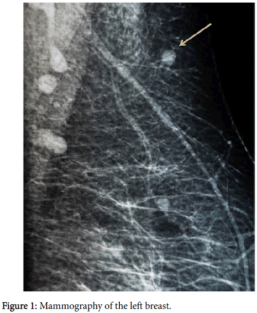
The mammography of the left breast reveals a well circumscribed lesion at the upper half of the breast, near the axilla and just beneath the skin in the subcutaneous fat (Figure 1).
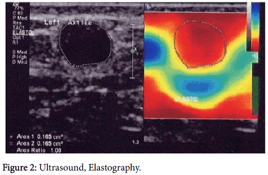
At ultrasonography, a round, very hypoechoid lesion was found at the left axilla, measuring 5.2 × 4.7 mm. The lesion appeared with well demarcated margins shown a hyperechoid halo, hypervascularity and the elastographic measurements were indicative for a hard lesion (Figure 2).
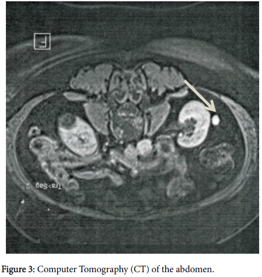
The lesion was classified as BIRADS 4A and a core biopsy was decided. The histologic diagnosis showed a tumor with neuroendocrinological differentiation (ER-ve, PrR-ve, E-cadherin-ve). This diagnosis established the metastatic disease. The Computer Tomography of the lungs reveals no metastatic deposits to the lungs but the Computer Tomography and Magnetic Tomography of the abdomen showed a metastatic lesion at the right kidney (Figure 3).
The bone scintigraphy reveals multiple secondary deposits at frontal bones, at thoracic vertebrae and at costs.
The laboratory examinations showed a slightly elevation to the chromogranin A(CglA) levels (5.40 nmol/l).
After this diagnostic workup a systematic chemotherapy was decided (cisplastin/etoposide) with 4-6 cycles depending on the response.
Conclusion
This case indicates that metastatic disease to the breast, although rare, exists and may be originate from any malignancy. Having this in mind, all women with known previous malignancies should be examined thoroughly when they undergo their routine annual mammography. On the other hand, when we radiologists, discover breast tumors with uncommon histological characteristics, during routine mammographic examinations, we must suggest physical examination of whole body and full radiological and laboratory examinations in order to discover the primary malignancy which will guide to the appropriate therapy.
References
- Hajdu SI, Urban JA (1972) Cancers metastatic to the breast. Cancer 29: 1691-1696.
- Wozniak TC, Naunheim KS (1998) Bronchial carscinoid tumor metastatic to the breast. Ann Thorac Surg 65: 1148-1149.
- Dillon D, Guidi AJ, Schnitt SJ (2014) Pathology of invasive breast cancer. In: Harris JR, Lippman ME, Morrow M, Osborne CK. Diseases of the breast. (5th edn) pp: 401-402.
- Toombs BD, Kalisher L (1977) Metastatic diseases to the breast clinical, pathologic and radiographic features. AJR Am J Roentgenol 129: 673-676.
- Bohman LG, Basett LW, Gold RH, Voet R (1982) Breast metastases from extramammary malignancies. Radiology 144: 309-312.
- Derchi LE, Rizzatto G, Giuseppetti GM, Dini G, Garaventa A (1985) Metastatic tumors in the breast: sonographic findings. J Ultrasound Med 4: 69-74.
- Thomas Stavros (2004) Malignant solid breast nodules: specific types. pp: 667-670.
Citation: Soilemezi E (2018) A Rare Case of Metastasis of Lung Carcinoid to Breast. OMICS J Radiol 7: 286. DOI: 10.4172/2167-7964.1000286
Copyright: ©2018 Soilemezi E. This is an open-access article distributed under the terms of the Creative Commons Attribution License, which permits unrestricted use, distribution, and reproduction in any medium, provided the original author and source are credited.
Select your language of interest to view the total content in your interested language
Share This Article
Open Access Journals
Article Tools
Article Usage
- Total views: 4689
- [From(publication date): 0-2018 - Dec 08, 2025]
- Breakdown by view type
- HTML page views: 3784
- PDF downloads: 905


