Review Article Open Access
A Tunisian Case of Dyssegmental Dysplasia: Silverman-handmarker Type
Yacoubi Mohamed Tahar1,2, Nadia Ben Jamaa1, Achour Radhouane3*, Ksibi Imen4, Mokni Moncef2, Delesoide Anne Lise5
1Research unit, Faculty of medicine of Sousse, Tunisia
2Department of pathology, University hospital of Sousse, Tunisia
3Emergency Department of Maternity and Neonatology Center, Faculty of Medicine of Tunis, Tunis, Tunisia
4Neonatology Department of Maternity and Neonatology Center, Faculty of Medicine of Tunis, Tunis, Tunisia
5Department of Biology of development, Hospital Robert DEBRE of Paris, France
- Corresponding Author:
- Achour Radhouane
Emergency Department of Maternity and Neonatology Center
Faculty of Medicine of Tunis, Tunis, Tunisia
Tel: 21698549398
E-mail: Radhouane.a@live.com
Received March 30, 2016; Accepted April 25, 2016; Published April 30, 2016
Citation: Tahar YM, Jamaa NB, Radhouane A, Imen K, Moncef M, et al. (2016) A Tunisian Case of Dyssegmental Dysplasia: Silvermanhandmarker Type. J Preg Child Health 3:249. doi:10.4172/2376-127X.1000249
Copyright: © 2016 Tahar YM, et al. This is an open-access article distributed under the terms of the Creative Commons Attribution License, which permits unrestricted use, distribution, and reproduction in any medium, provided the original author and source are credited.
Visit for more related articles at Journal of Pregnancy and Child Health
Abstract
Objective: Dyssegmental dysplasia, Silverman-Handmaker Type, is an autosomal recessive lethal disorder, originally considered as a Kniest-like skeletal dysplasia with camptomelia. It is linked to functional null mutations of the perlecan gene (HSPG2) located on chromosome 36.1-35. We report the first case diagnosed in our department. Study: A 31 year-old mother, prim pare and non-consanguineous with her husband, given a 15-16 weeks-aged foetus with dwarfism and dyssegmental dysplasia detected prenatally by ultrasonography. A medical termination of the pregnancy was indicated. Radiographs and autopsy were performed. External examination showed a facial dysmorphy, thick neck, pterygia and micromelia with curved limbs, chest narrowing and an abducted hallux in the right foot. No anomaly in visceral examination was found. The radiographs showed short limbs with Kniest like large metaphyses, bowing of long bones and pleomorphic vertebral bodies with irregular segmentation and absent ossification of pelvic bones. The cranial vault was normal. Histologically, the resting cartilage showed scattered, large puddle-like spaces. The physeal growth zones were disorganized. Conclusion: Prenatal diagnosis is useful to detect this type of skeletal anomaly in order to indicate medical termination of the pregnancy earlier. Fetopathological examination and skeletal radiographs are mandatory in order to establish a precise diagnosis allowing a suitable genetic counselling.
Keywords
Dyssegmental dysplasia; Silverman-Handmaker dysplasia; Dyssegmental dwarfism; Mutation of the perlecan gene
Introduction
The dyssegmental dysplasia, Silverman-Handmaker type (DDSH) is known as an anisospondilic camptomicromelic dwarfism.
The DDSH is a rare entity; initially described by HANDMAKER and coll. in 1983, since then some thirty cases have been reported [1-4].
It consists in a lethal form of micromelic dwarfism characterized by disorganization of the vertebral bodies, camptomelia and micromelia.
In this study, we report the first case diagnosed in our department of a male fetus with clinical and radiographic evidence of a lethal form of dyssegmental dysplasia compatible with the Silverman-Handmaker type diagnosed prenatally.
Case report
A 31-years-old mother, prim pare, having no consanguinity with her husband, performed an ultrasonography at 22 weeks of gestation showing short limbs with angulations, a thick neck and low-set ears. A medical termination of the pregnancy was indicated. Radiographic and autopsy were performed.
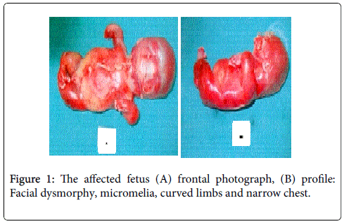
The external examination showed a male foetus anatomically aged of 15-16 gestational weeks (hypotrophic) with a facial dysmorphy associating flat face low-set ears and retrognathia, short and thick neck, pterygia and micromelia with curved limbs, chest narrowing and an abducted hallux in the right foot. No anomaly in visceral examination (Figure 1).
The radiographs showed short limbs with Kniest like large metaphyses, bowing of long bones and pleomorphic vertebral bodies with irregular segmentation and absent ossification of pelvic bones. The cranial vault was normal (Figure 2).
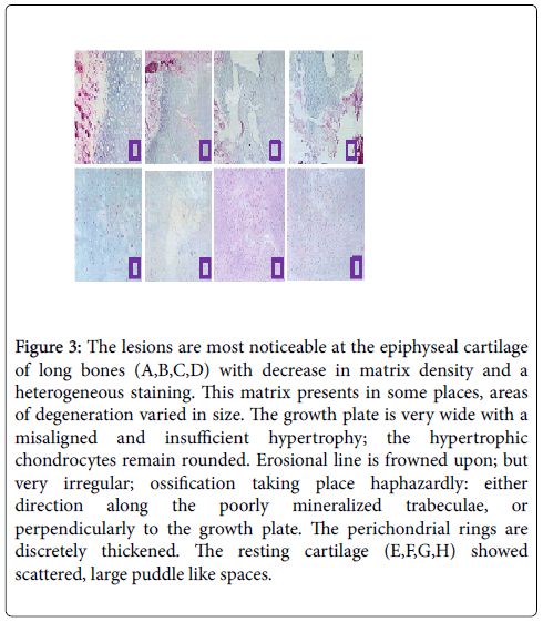
Histologically, the lesions are most noticeable at the epiphyseal cartilage of long bones with decrease in matrix density and a heterogeneous staining. This matrix presents in some places, areas of degeneration varied in size. In which, the chondrocytes have a fusiform aspect. The growth plate is very wide with a misaligned and insufficient hypertrophy; the hypertrophic chondrocytes remain rounded.
Erosional line is frowned upon; however, it appears very irregular; ossification taking place haphazardly: either direction along the poorly mineralized trabeculae, or perpendicularly to the growth plate. The perichondrial rings are discretely thickened. The resting cartilage showed scattered, large puddle-like spaces. The primary bone and physeal bone density appear to substantially normal and not increased (Figure 3).
Figure 3: The lesions are most noticeable at the epiphyseal cartilage of long bones (A,B,C,D) with decrease in matrix density and a heterogeneous staining. This matrix presents in some places, areas of degeneration varied in size. The growth plate is very wide with a misaligned and insufficient hypertrophy; the hypertrophic chondrocytes remain rounded. Erosional line is frowned upon; but very irregular; ossification taking place haphazardly: either direction along the poorly mineralized trabeculae, or perpendicularly to the growth plate. The perichondrial rings are discretely thickened. The resting cartilage (E,F,G,H) showed scattered, large puddle like spaces.
Discussion
The dyssegmental dysplasias are lethal forms of fetal and neonatal short-limbed dwarfism in which vertebral segmentation defects and short, thick, bowed long bones are the most important radiographic features. There are two types.
"Dyssegmental dysplasia, Silverman-Handmaker type" (DDSH) is characterized by stillbirth or death within the first few days of life and by distinct and more severe radiographic changes than the second type (Rolland Desbuquois) [1].
The antenatal diagnosis of short-limbed dwarfism is usually made in one of two clinical situations: the patient is referred for ultrasound secondary to a family history of dwarfism, or routine exam reveals a femur measuring less than the 5th percentile for gestational age.
Aleck [3], Rieubland [4] and other authors [1,2,5] described the typical findings of dyssegmental dysplasia which includes micromelic dwarfism, cephalocele, mid-facial defects, small orbits, flat face, micrognathia, cleft palate, short neck and reduced joint mobility. Cardiac defects and hydronephrosis were also reported.
Radiological findings associate disorganization of vertebral bodies, elongated scapulae, abnormal size, shape and ossification of the acromion, the coracoid process, and the body of the scapulae [6-8].
The histological examination usually notice short endochondral growth plate, calcospherites, mucoid degeneration of resting cartilage; haphazard arrangement of collagen fibers on scanning electron microscopy of bone was described [2,3].
Prabhu et al. described arachnoid cyst and venous angioma in CT scan and MRI of the brain. These lesions were not reported previously with DDSH. Ophthalmological examination demonstrated subluxation of the lenses and prominent choroidal vasculature in the fundus [2].
Dyssegmental dysplasia, Silverman-Handmaker type (OMIM 224410) is an autosomal recessive form of lethal dwarfism. It is caused by mutations in the perlecan gene HSPG2) [4]. Patients of Hispanic origin are well represented in the Silverman-Handmaker variety.
Perlecan is a large heparan sulfate proteoglycan present in all basement membranes and in some other tissues such as cartilage, and is implicated in cell growth and differentiation [6]. The cartilage matrix from these patients stained poorly with antibody specific for perlecan, but there was staining of intracellular inclusion bodies. Biochemically, truncated perlecan was not secreted by the patient fibroblasts, but was degraded to smaller fragments within the cells [5,6].
In our study, it is unfortunate that it was not possible to undertake molecular or perlecan investigations in the fetus but the diagnosis was radio logically and histologically confirmed.
The differential diagnosis includes fibrochondrogenesis, chondrodysplasia punctata and Weissenbacher-Zweymuller syndrome. Fibrochondrogenesis is characterized by limb and vertebral deformities including shortened dumbbell-shaped metaphyses and pear-shaped vertebral bodies. The short limbs noted in fibrochondrogenesis are in a proximal or rhizomelic pattern. This is in contrast to dyssegmental dysplasia which is characterized by micromelia or disproportionate shortening of the entire extremity.
Chondrodysplasia punctata is typified by vertebral bodies with coronal clefts, metaphyseal splaying and stippled epiphyses. The ultrasound diagnosis is reported elsewhere in this issue, and the rhizomelic, potentially lethal variety has been diagnosed using radiography.
In conclusion, in the absence of molecular and biochemical tests, prenatal ultrasound is the only tool available for prenatal diagnosis. So that the medical termination of pregnancy is indicated; radiographs and fetopathological study are performed to confirm the diagnosis of DDSH allowing a suitable genetic counselling.
Acknowledgements
This work was supported by the Ministry of public Health in Tunisia and the Research Unit of fetopathology UR 03/08/21.
References
- Gonzalvo GO, Sanchez MB (1990) Evidence of heterogeneity in dyssegmental dysplasia. An EspPediatr 33: 213-223.
- Prabhu VG, Kozma C, Leftridge CA, Helmbrecht GD, France ML (1998) Dyssegmental dysplasia Silverman-Handmaker type in a consanguineous Druze Lebanese family: long term survival and documentation of the natural history. Am J Med Genet 75: 164-170.
- Aleck KA, Grix A, Clericuzio C, Kaplan P, Adomian GE, (1987) Dyssegmentaldysplasias: clinical, radiographic, and morphologic evidence of heterogeneity. Am J Med Genet 27: 295-312.
- Rieubland C, Jacquemont S, Mittaz L, Osterheld MC, Vial Y, et al (2010) Phenotypic and molecular characterization of a novel case of dyssegmental dysplasia, Silverman-Handmaker type. Eur J Med Genet 53: 294-298.
- Arikawa-Hirasawa E, Wilcox WR, Le AH, Silverman N, Govindraj P, et al. (2001) Dyssegmental dysplasia, Silverman-Handmaker type, is caused by functional null mutations of the perlecan gene. Nat Genet 27: 431-434.
- Hirasawa AE, Wilcox WR, Yamada Y (2001) Dyssegmental dysplasia, Silverman-Handmaker type: unexpected role of perlecan in cartilage development. Am J Med Genet 106: 254-257.
- Handmaker SD, Robinson LD, CampbellJA, Chinwah O, GorlinRJ (1977) Dyssegmental dwarfism: a new syndrome of lethal dwarfism. Birth Defects Orig XIII: 79-90.
- Fasanelli S, Kozlowski K, Reiter S, Sillence D (1985) Dyssegmental dysplasia report of two cases with a review of the literature. Skeletal Radiol 14: 173-177.
Relevant Topics
Recommended Journals
Article Tools
Article Usage
- Total views: 10540
- [From(publication date):
April-2016 - Aug 15, 2025] - Breakdown by view type
- HTML page views : 9675
- PDF downloads : 865


