A typical Depiction of Renal Nephrocalcinosis Reflecting the Superiority of Ultrasound to Computed Tomography
Received: 23-Jan-2022 / Manuscript No. roa-22-55845 / Editor assigned: 03-Mar-2022 / PreQC No. roa-22-55845(PQ) / Reviewed: 17-Mar-2022 / QC No. roa-22- 55845 / Revised: 22-Mar-2022 / Manuscript No. roa-22-55845(R) / Published Date: 29-Mar-2022 DOI: 10.4172/2167-7964.1000369
Abstract
Nephrocalcinosis is defined by calcium phosphate or calcium oxalate deposits in the kidney parenchyma, particularly in tubular epithelial cells and interstitial tissue. It should be differentiated from nephrolithiasis where calcium salts deposits are located in the kidney and urinary tract. We report a case of a 5 years old child with history of hypoparathyroidism of recent discovery , and the clear upper hand that ultrasound had in the assessment of medullary nephrocalcinosis in patients with metabolic disorders.
Keywords
Nephrocalcinosis; Ultrasound; Children; Imaging modalities
Image Article
A renal calcifying disorder generally includes two entities: nephrocalcinosis and nephrolithiasis. In addition to their difference in location, nephrolithiasis may occur in healthy individuals while nephrocalcinosis suggests an underlying genetic or metabolicendocrine disorder, such as hypoparathyroidism often detected as an incidental finding as reflected in our case, nephrocalcinosis may be classified according to the radiological type: medullary, cortical or diffuse. [1]
Unlike nephrolithiasis, nephrocalcinosis is asymptomatic, and may progress to renal insufficiency. Early diagnosis is important because therapeutic interventions may stabilize, slow or, rarely, reverse disease.
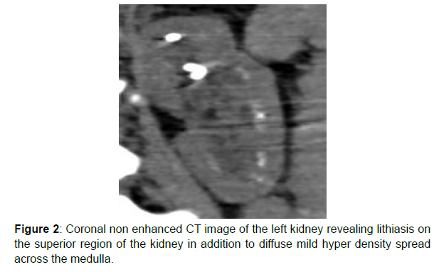
Therefore surveillance in high-risk patients is a crucial component of management. Diagnostic imaging for nephrocalcinosis includes radiographs, computed tomography (CT), and ultrasonography (US). Radiographs are insensitive due to poor delineation of renal anatomy and confounding by bowel gas. CT offers superior contrast resolution and definition of anatomic structures and is the gold standard for nephrolithiasis ultrasound however offers a higher sensitivity in term of early diagnosis of mild and moderate nephrocalcinosis compared to the CT [2] (Figures 1 and 2).
References
- Monet-Didailler C, Chateil JF, Allard L, Godron-Dubrasquet A, Harambat J (2021) Nephrocalcinosis in children. Nephrol Ther. 17: 58-66.
- Boyce AM, Shawker TH, Hill SC, Choyke PL, Hill MC, et al. (2013) Ultrasound is superior to computed tomography for assessment of medullary nephrocalcinosis in hypoparathyroidism. J Clin Endocrinol Metab 98: 989-994.
Indexed at, Google Scholar, Crossref
Citation: Najwa A, Zaynab IH, Ismail HM, Najlae L, Latifa C, et al. (2022) A typical Depiction of Renal Nephrocalcinosis Reflecting the Superiority of Ultrasound to Computed Tomography. OMICS J Radiol 11: 368. DOI: 10.4172/2167-7964.1000369
Copyright: © 2022 Najwa A, et al. This is an open-access article distributed under the terms of the Creative Commons Attribution License, which permits unrestricted use, distribution, and reproduction in any medium, provided the original author and source are credited.
Select your language of interest to view the total content in your interested language
Share This Article
Open Access Journals
Article Tools
Article Usage
- Total views: 4481
- [From(publication date): 0-2022 - Dec 09, 2025]
- Breakdown by view type
- HTML page views: 3766
- PDF downloads: 715


