Make the best use of Scientific Research and information from our 700+ peer reviewed, Open Access Journals that operates with the help of 50,000+ Editorial Board Members and esteemed reviewers and 1000+ Scientific associations in Medical, Clinical, Pharmaceutical, Engineering, Technology and Management Fields.
Meet Inspiring Speakers and Experts at our 3000+ Global Conferenceseries Events with over 600+ Conferences, 1200+ Symposiums and 1200+ Workshops on Medical, Pharma, Engineering, Science, Technology and Business
Review Article Open Access
Application of Blood Flow Restriction in Resistance Exercise Assessed by Intramuscular Metabolic Stress
| Okita K1* and Takada S2 | |
| 1Department of Sport Education, Hokusho University, Japan | |
| 2Department of Cardiovascular Medicine, Hokkaido University Graduate School of Medicine, Japan | |
| Corresponding Author : | Koichi Okita Department of Sport Education Hokusho University, 23 Bunkyodai Ebetsu 069-8511, Hokkaido, Japan Tel: +81-11-386-8011 Fax: +81-11-387-1542 E-mail: okitak@hokusho-u.ac.jp |
| Received July 22, 2013; Accepted November 12, 2013; Published November 14, 2013 | |
| Citation: Okita K, Takada S (2013) Application of Blood Flow Restriction in Resistance Exercise Assessed by Intramuscular Metabolic Stress. J Nov Physiother 3:187. doi:10.4172/2165-7025.1000187 | |
| Copyright: © 2013 Okita K, et al. This is an open-access article distributed under the terms of the Creative Commons Attribution License, which permits unrestricted use, distribution, and reproduction in any medium, provided the original author and source are credited. | |
Visit for more related articles at Journal of Novel Physiotherapies
| Keywords |
| Resistance training; Skeletal muscle; Energetic metabolism; Blood flow restriction; Magnetic resonance spectroscopy |
| Introduction |
| Skeletal muscle bulk and strength are now becoming important therapeutic targets in exercise therapy [1,2]. In order to get more muscle bulk and strength, we have to perform high-intensity resistance training with mechanical load greater than 65% of one repetition maximum (1 RM) [3-5]. However, such intensive loads could not be often applied for weak subjects. |
| In recent years, several lines of studies have provided the compelling data showing that low-intensity resistance training with blood flow restriction (BFR) leads to muscle hypertrophy and strength increase [6-18], and results in adaptations equal to those of high-intensity resistance training [17,18]. The researchers suggested that the supplementation of low-intensity resistance exercise with BFR might provide additional stress and enhanced recruitment in the skeletal muscle fibers, but the exact details were not clarified. |
| Metabolic stresses such as depletion of phosphocreatine, an increase in inorganic phosphate, a decrease in intramuscular pH, and lactate accumulation have been also suggested to be potent stimuli for obtaining training effects [19-21]. It was speculated that BFR might advance the metabolic stress and also the recruitment in the skeletal muscle even during low-intensity resistance exercise. Lately, we elucidated the intramuscular energetic metabolism during low-intensity resistance exercise with BFR by using 31P-magnetic resonance spectroscopy (31P-MRS), which could evaluate the metabolic by-products, pH and muscle fiber recruitment in exercising muscle [22]. This mini review presents our important findings concerning application of BFR in resistance exercise and discusses the future evolution of this training manner. |
| Exercise with Blood Flow Restriction |
| Actually, there is a large variety of methods in previous papers demonstrating dramatic muscle hypertrophy and strength increase with exercise with BFR and might be also no standard protocol. They have employed various exercise intensities ranging from 20 to 50% 1 RM and pressure ranging from 100 to 300 mmHg [6-18]. There are generally the two major concepts in the resistance training with BFR. One is to increase the training effects more than those obtained by usual resistance training procedures [8,17] especially in physically active people and athletes. The other is to obtain the favorable effects of resistance training without using a conventional high-intensity exercise load especially in inactive, female, senior persons and/or diseased persons [7,15]. We followed the latter concept and designed the investigation. We employed 20% 1 RM as low-intensity resistance exercise and 65% 1 RM as high-intensity resistance exercise, in accord with the majority of previous studies [6,7,10,15,18] and the recommendation of the American College of Sports Medicine [3]. We also precisely controlled the exercise conditions (such as load and repetition) to exactly compare the effects between different protocols [12-14,22]. |
| Methods |
| Measurement of intramuscular metabolic stress during resistance exercise |
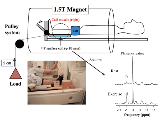
| Details of the measurements were described in our previous studies [12-14,22]. We employed young, healthy subjects without orthopedic or cardiovascular diseases. Subjects performed unilateral plantar flexion exercises in a 55-cm bore, 1.5-tesla superconducting magnet (MagnetomVision VB33G; Siemens, Erlangen, Germany). The experimental exercises were set for 2 min with 30 repetitions per 1 min, lifting the weight 5 cm above ground. Exercise protocols were as follows: low-intensity resistance exercise using 20% 1 RM, 20% 1 RM with BFR and high-intensity resistance exercise using 65% 1 RM. In 20% 1 RM with BFR, an 18.5-cm-wide pressure cuff was placed around the thigh of the right leg. The air pressure was inflated for 10 seconds before the exercise protocol, and promptly released after the exercise was finished. BFR was carried out using 130% of the subject’s resting systolic blood pressure with a pneumatic rapid inflator, following the report by Takano et al. [15]. Intramuscular metabolism was measured with an 80-mm surface coil placed under the muscle belly of the right gastrocnemius. The data were obtained at rest and every 30 sec during exercise (Figure 1). Intramuscular phosphocreatine and pH were estimated by well-established manners [23,24]. |
| Results |
| Intramuscular metabolism during exercise with blood flow restriction |
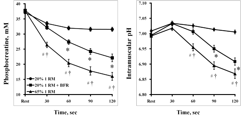
| Intramuscular phosphocreatine and pH was significantly decreased in 20% 1 RM with BFR and H, but not in 20% 1 RM without BFR. Changes of phosphocreatine and pH in 20% 1 RM with BFR were significantly greater than those in 20% 1 RM without BFR. However, those in 20% 1 RM with BFR were significantly lower than those in 65% 1 RM without BFR (Figure 2). |
| By applying BFR, additional changes of intramuscular metabolites and pH were obtained even during low-intensity resistance exercise. However, these changes did not necessarily reach the level of those during high-intensity resistance exercise. There was a wide range of individual response to this exercise. Contrary to the speculations by previous studies [8], the results have suggested that the metabolic stress in skeletal muscle during low-intensity resistance exercise with BFR is not generally equivalent to that in high-intensity resistance exercise [22]. It is inconceivable that everyone would get the same favorable training effects from a uniform procedure of resistance exercise with BFR. |
| Discussion and Perspectives |
| The metabolic stress in skeletal muscle during low-intensity (20% 1 RM) resistance exercise was significantly increased by applying BFR, but did not necessarily reach the level of that during usual highintensity (65% 1 RM) resistance exercise without BFR. This new method of resistance training needs to be examined for optimization of the protocol to reach equivalence with the high-intensity resistance training. |
| Optimization of a protocol for resistance training with blood flow restriction |
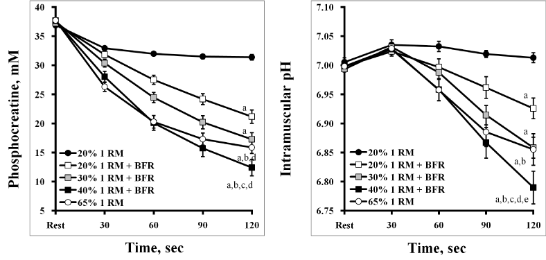
| In a resistance exercise with BFR, skeletal muscle stress could be varied by exercise load, number of repetition, duration, and cuff pressure according to subject’s tolerance [13]. Although our data showed that the muscular metabolic stress could not generally reach the level of those during high-intensity resistance exercise, it is possible that an increase (30 ~ 40% 1 RM) of exercise intensity might greatly enhance intramuscular metabolic stress during BFR exercise (Figure 3) [12]. |
| The cuff pressure for BFR seems to be another important factor of determining training stress. We employed the pressure of 130% of the subject’s resting systolic blood pressure following the report by Takano et al. [15]. In the previous study [22], we showed that the combination of low BFR pressure (below systolic pressure) at a load of 20% 1 RM in the BFR protocol created significantly lower metabolic stress than that during the moderate BFR pressure protocol (above systolic pressure). The applied BFR pressure would need to be higher than the blood pressure level during exercise. We also showed a little additional effect of raising cuff pressure on muscular metabolic stress. However, the level of BFR pressure might occasionally need to be selected according to the exercise intensity and subject’s status. |
| Third important point is concerning the total exercise volume (intensity×repetition). Basically, muscle metabolic stress is dependent on work rate, intensity×frequency (repetition rate). However, in BFR exercise muscular by-products progressively accumulated because of interruption of recovery even in relaxation period (metabolic freeze effect) [25,26]. Therefore, effective muscle stress might be achieved by increasing exercise volume, simply total repetitions without changing intensity and/or frequency. Thus, less intensity with increasing repetitions might become effective. |
| Implications in 31P-MRS measurements during BFR exercise |
| 31P-MRS can measure the high-energy metabolites, pH and also fast-twitch (FT) fiber recruitment in exercising muscle. It has been known that the metabolic stress parameters, such as lactate and H+ in blood level, are correlated to the elevated post exercise growth hormone (GH) concentration [27,28]. In addition, the metabolic stress might stimulate some other hormonal release and cytokine production, including insulin-like growth factor 1 and interleukin-6 as well as GH [6,13,15]. Those growth factors and cytokines have been suggested to regulate muscle growth/hypertrophy [29,30]. FT fiber recruitment might be also an important factor for successful muscle hypertrophy and strength gain by resistance training [3,4]. Therefore, 31P-MRS is an extremely useful tool for examining the effects of various modes of exercises. Recently, we confirmed that enhanced metabolic stress, defined as phosphocreatine depletion and intramuscular pH decrease, proportionally contributed to training effects, muscle hypertrophy and strength gain [14]. |
| Summary |
| In this mini review, we showed the usefulness and effectiveness of resistance exercise with blood flow restriction by using a novel technique with 31P-MRS. We hope that this unique procedure will be widely applied in physical therapy. |
| Grants |
| This study was supported in part by Grant-in-Aid for Scientific Research from Japan Society for the Promotion of Science (JSPS), KAKENHI (23500784) and a grant of MEXT Supported Program for the Strategic Research Foundation at Private Universities, 2011-2013. |
References |
|
Figures at a glance
 |
 |
 |
| Figure 1 | Figure 2 | Figure 3 |
Post your comment
Relevant Topics
- Electrical stimulation
- High Intensity Exercise
- Muscle Movements
- Musculoskeletal Physical Therapy
- Musculoskeletal Physiotherapy
- Neurophysiotherapy
- Neuroplasticity
- Neuropsychiatric drugs
- Physical Activity
- Physical Fitness
- Physical Medicine
- Physical Therapy
- Precision Rehabilitation
- Scapular Mobilization
- Sleep Disorders
- Sports and Physical Activity
- Sports Physical Therapy
Recommended Journals
Article Tools
Article Usage
- Total views: 15509
- [From(publication date):
December-2013 - Aug 29, 2025] - Breakdown by view type
- HTML page views : 10898
- PDF downloads : 4611
