Research Article Open Access
Application of Glycerin Poultice to the Semitendinous and Semimembranous above the Popliteal Fossa Improves Motor Disturbance in Parkinson Disease
| Yuzuru Yasuda* | |
| Department of Neurology, Yasuda Clinic, 62-7, Takenokaido-cho, Takehana, Yamashina-ku, Kyoto 607-8080, Japan | |
| Corresponding Author : | Yuzuru Yasuda Department of Neurology Yasuda Clinic, 62-7, Takenokaido-cho Takehana, Yamashina-ku Kyoto 607-8080, Japan Tel: +81 75 583 2288 Fax: +81 75 583 2228 E-mail: yuzuru-yasuda@mra.biglobe.ne.jp |
| Received August 18, 2012; Accepted September 18, 2012; Published September 21, 2012 | |
| Citation: Yasuda Y (2012) Application of Glycerin Poultice to the Semitendinous and Semimembranous above the Popliteal Fossa Improves Motor Disturbance in Parkinson Disease. J Nov Physiother S1:005. doi:10.4172/2165-7025.S1-005 | |
| Copyright: © 2012 Yasuda Y. This is an open-access article distributed under the terms of the Creative Commons Attribution License, which permits unrestricted use, distribution, and reproduction in any medium, provided the original author and source are credited. | |
Visit for more related articles at Journal of Novel Physiotherapies
Abstract
It has been reported that application of glycerin poultice to the flexor digotorum profundus in the middle phalanx of the little finger improves motor disturbance of Parkinson disease. I investigated whether the application of glycerin poultice to the semitendinous and semimembranous above the popliteal fossa (GSSP) improved motor disturbance in Parkinson disease. Step length and diameter of the pupil in 20 patients with Parkinson disease and 20 control subjects was evaluated before and after application of GSSP. Motor disturbance in patients with Parkinson disease was evaluated before and after application of GSSP. The application of GSSP increased step length and constricted the pupil in all subjects, which means that application of GSSP caused the decreased tension of the semitendinous and semimembranous and lowered sympathetic nerve activity. With application of GSSP, motor disturbance in patients with Parkinson disease was improved, which is caused by reversing the mechanism of the muscle mechanosensitive reflex.
| Keywords |
| Parkinson disease; Glycerin poultice; Semitendinous and semimembranous above the popliteal fossa, Sympathetic nerve activity; Muscle mechanosensitive reflex |
| Introduction |
| It has been reported that patients with Parkinson disease (PD) show improvement of motor disturbance with application of glycerin poultice to the flexor digitorum profundus in the middle phalanx of the little finger (GFML) on both sides [1]. GFML is as follows; glycerin poultice was made by means of gauze (3×7 mm) immersed by 50% glycerin and the poultice was attached precisely to the flexor digotorum profundus in the middle phalanx of the little finger on both sides. In all subjects, application of GFML increased dorsiflexion of the little finger suggesting that the tension of the flexor digitorum profundus was decrease, and application of GFML constricted the pupil suggesting that sympathetic nerve activity was lowered. The rationale of application of GFML is to reverse the mechanism of the muscle mechanosensitive reflex. Because, muscle mechanosensitive reflex is evoked by an increase of muscle tension, which in turn causes sympathetic nerve activity, namely, an increase of mean blood pressure, heart rate, and breathing [2]. Static contraction of the muscle and tendon stretch has the same effect concerning the muscle mechanosensitive reflex [3]. The precise mechanism of the mechanoreceptors response contributing to sympathetic nerve activation remains unknown. Recently it has been reported that muscle mechanosensitive receptors evoking the reflex cardiovascular responses locate close to the myotendinous junction [4]. |
| Thereafter, I thought that application of glycerin poultice to the tendon of the lower extremity may improve motor disturbance in PD as well as application of GFML, and researched which muscles have the principal mechanosensitive receptor. And I found that the tendon of the semitendinous and semimembranous above the popliteal fossa in the most patients with PD is very stiff and investigated whether application of glycerin poultice to the semitendinous and semimembranous above the popliteal fossa (GSSP) will improve motor disturbance in PD. |
| Patients and Methods |
| Step length |
| 20 patients with clinically diagnosed PD (8 males and 12 females, 69.1 ± 8.0 years of age (mean ± SD)), and 20 controls (7 males and 13 females, 66.1 ± 10.1 years of age) participated in this study. Patients with PD did not show an on-off phenomenon or orthostatic hypotension; the duration of PD was 5.2 ± 2.9 years. The Hoehn and Yahr score and the Unified Parkinson’s Disease Rating Scale motor examination (full score, 108) [5] was 2.4 ± 0.5, 32.5 ± 6.5, respectively. Anti-parkinsonian drugs were not interrupted during the course of the study. All control subjects had no previous history of neurological disease and were neurologically and physically normal. All subjects were right handed and right footed according to Waterloo Footedness Questionnaire-Revised, and Waterloo Handedness Questionnaire- Revised [6,7] and did not have severe cervical and lumbar spondylosis. All participants gave their consent to participate in this study. The Research Council of Yasuda Clinic approved the study protocol. |
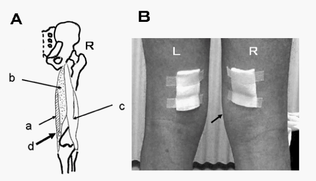
| Glycerin poultice was formulated and applied as follows: gauze (4×5 cm) was immersed in 50% glycerin and the poultice was attached precisely to the semitendinous and semimembranous with the lower margin of the poultice being one cm above the medial epicondyle of femur. (Figure 1A and 1B). |
| The step length (the distance from left ankle to right one) was photographed by digital movie camera, (SANYO DMX-HD1; shutter speed, 1/60 second). The distance between the camera and the subjects was 3m and the axis of the lens was situated 80 cm from the floor, therefore the step length was adequately evaluated. Two markers at a distance of 95 cm which were not clearly visible by the subjects, were situated on the floor. To avoid paradoxical movement, any visible lines were not present on the floor. To overcome placebo effects, the subject was not given previous notice about any effects with application of GSSP and was requested to walk naturally. Right step length (the distance by which the right foot moved forward in front of the left one) and left step length (vice versa) were photographed four times, respectively, to gain the mean step length. The same maneuver was carried out ten minutes after GSSP was applied on both sides. The step length was measured on the screen of a personal computer and the true value was calculated by the ratio to the distance of two markers (Figure 2). |
| Diameter of the pupil |
| To avoid accommodation reflex, the subject was requested with an easy manner to look at a mark which is 4 m away. The subject’s left pupil was photographed at a close distance with SANYO DMX-HD1 digital camera and the indicator, with a length of 20 mm, was situated on the lower eye lid. 10 minutes after GSSP was applied on both sides, the left pupil was also photographed. The diameter of the left pupil was measured on the screen of a personal computer and the true value was calculated by the ratio to the indicator. |
| Motor disturbance in PD |
| The motor disturbance was measured according to UPDRS motor examination [5] in the patients with PD before GSSP was applied to both sides. Ten minutes after GSSP was applied bilaterally, the score was also measured. |
| Statistical analysis |
| Statistical analysis was performed with the ANOVA in the groups and conditions (Student t-test as a post hoc test) and with paired Student t-test in two conditions. Significance was assumed at a p<0.05. |
| Results |
| Increase of the step length |
| In patients with PD, step length was 32.7 ± 10.8 cm and 36.7 ± 9.5 cm before and after application of GSSP (p<0.001), respectively. In controls, step length was 41.7 ± 6.4 cm and 45.5 ± 6.0 cm before and after application of GSSP (p<0.001), respectively. |
| Step length of patients with PD was less than that of controls before and after application of GSSP (p<0.01, p<0.01, respectively) (Table 1). |
| Constriction of the pupil |
| The diameters of the left pupil was 3.28 ± 0.65 mm and 2.91 ± 0.67 mm before and after application of GSSP, respectively (p<0.001) and that of the control was 3.27 ± 0.59 mm and 2.87 ± 0.51 mm, respectively (p<0.001), The pupil was constricted in all subjects with application of GSSP. |
| Improvement of motor disturbance in PD |
| UPDRS motor examination [5] showed improvement of all categories (Table 2) with application of GSSP. Total score of UPDRS before application of GSSP was 32.65 ± 5.96, and that after application of GSSP was 15.05 ± 4.59 (p<0.001). Scores regarding tremor at rest (/20), rigidity (/20), leg agility (/8) before application of GSSP were 1.65 ± 2.10, 7.40 ± 0.88, and 2.85 ± 0.74, respectively, and those after application with GSSP were 0.60 ± 0.88, 3.75 ± 1.01, and 1.35 ± 0.81, respectively (p=0.001, p<0.001, p<0.001, respectively). |
| Discussion |
| With application of GSSP, step length increased in all subjects, which indicates that the tension of both thigh flexors decreased. Although precise mechanism of action of glycerin is not clear, it has been reported that topical glycerin application improves skin moisture [8] and plasticity [9]. With application of GSSP, glycerin immersed the tendon of semitendinous and semimembranous and softened them through the skin, which is the cause of decreased tension of the thigh flexors. Moreover, with application of GSSP, the pupil was constricted in all subjects. Application of GSSP produced two significant findings; decreased tension of semitendinous and semimembranous, and constriction of the pupil. |
| The mechanism of the effect with application of GSSP is concerned with muscle mechanosensitive reflex. The muscle mechanosensitive reflex is evoked by an increase of muscle tension, which in turn causes sympathetic nerve activity [2-4] and application of GSSP decreased the tension of the semitendinous and semimembranous, which in turn reduced sympathetic nerve activity because pupil was constricted with application of GSSP in all subjects of this study [10]. The rationale of application of GSSP is to reverse the mechanism of the muscle mechanosensitive reflex, which corresponds to decreased activity of muscle mechanosensitive receptor close to the myotendoinous junction [4] of the semitendinous and semimembranous. |
| UPDRS motor examination [5] showed improvement of all categories with application of GSSP. Especially, scores regarding tremor, rigidity and movements of hands were excellent as well as with application of GFML. All scores regarding movement of legs with application of GSSP were more improved than those of GFML. Especially, leg ability was excellent with application of GSSP. The improvement of tremor in patients with PD is due to beta-blocking effect with application of GSSP because beta-blocker is effective in the treatment of tremor by supposedly affecting the muscle spindle by blocking the adrenoreceptor [11]. And the improvement of rigidity of patients with PD is concerned with complex sympathetic modulation of the muscle spindle afferent sensitivity [12,13] which is also derived from lowered sympathetic nerve activity with application of GSSP as well as GFML. |
| As application of GSSP causes sympathetic modulation of the muscle spindle afferent sensitivity as well as GFML, it is beneficial especially to the patients with leg muscle tonus being high, for example, spastic hemiparesis, spastic paraparesis and so on. In fact, many patients with spastic hemiparesis showed marked improvement of gait disturbance with application of GSSP to the side of hemiparesis. Moreover, the patient with transverse myelitis who shows spastic paraparesis showed marked improvement of gait disturbance with application of GSSP to both sides (data not shown). In this study, patients with PD showed improvement of leg movement, especially leg ability, and normal controls also showed increase of step length, which suggests that application of GSSP is effective for prevention of falls of the aged. |
| As application of GSSP lowers sympathetic nerve activity by reversing the mechanism of muscle mechanosensitive reflex as well as that of GFML, the principal meschanoseisitive receptors would exist in or near the tendon of the semitendinous and semimembranous above the popliteal fossa, and of the flexor digitorum profundus in the middle phalanx of the little finger in human. Moreover, another principal mechanosensitive receptor may exist in human. The future direction for research is to investigate whether application of glycerin poultice to the principal mechanosensitive receptors will become a useful tool for the relief of the disorder whose mechanism is connected with increased sympathetic nerve activity. |
References
- Yasuda Y (2009) A novel method improving motor disturbance in Parkinson disease: application of glycerin poultice to the flexor digitorum profundus in the middle phalanx of the little finger. NeuroRehabilitation 24: 219-223.
- Mitchell JH, Kaufman MP, Iwamoto GA (1983) The exercise pressor reflex: its cardiovascular effects, afferent mechanisms, and central pathways. Annu Rev Physiol 45: 229-242.
- Hayes SG, Kindig AE, Kaufman MP (2005) Comparison between the effect of static contraction and tendon stretch on the discharge of group III and IV muscle afferents. J Appl Physiol 99: 1891-1896.
- Nakamoto T, Matsukawa K (2007) Muscle mechanosensitive receptors close to the myotendinous junction of the Achilles tendon elicit a pressor reflex. J Appl Physiol 102: 2112-2120.
- Fahn S, Elton R (1987) Members of the UPDRS Development Committee. Unified Parkinson├ó┬?┬?s disease rating scale: Recent developments in Parkinson├ó┬?┬?s disease. Macmillan Health Care Information, Florham Park, NJ, 153-163.
- Elias LJ, Bryden MP, Bulman-Fleming MB (1998) Footedness is a better predictor than is handedness of emotional lateralization. Neuropsychologia 36: 37-43.
- Kang Y, Harris LJ (2000) Handedness and footedness in Korean college students. Brain Cogn 43: 268-274.
- Bisset DL, McBride JF (1984) Skin conditioning with glycerol, J Soc Cosmet Chem 35: 345-350.
- Overgaard Olsen L, Jemec GB (1993) The influence of water, glycerin, paraffin oil and ethanol on skin mechanics. Acta Derm Venereol 73: 404-406.
- Yu Y, Koss MC (2002) alpha (1A)-adrenoceptors mediate sympathetically evoked pupillary dilation in rats. J Pharmacol Exp Ther 300: 521-525.
- Rincon F, Louis ED (2005) Benefits and risks of pharmacological and surgical treatments for essential tremor: disease mechanisms and current management. Expert Opin Drug Saf 4: 899-913.
- Hammar I, Jankowska E (2003) Modulatory effects of alpha1-,alpha2-, and beta -receptor agonists on feline spinal interneurons with monosynaptic input from group I muscle afferents. J Neurosci 23: 332-338.
- Roatta S, Windhorst U, Ljubisavljevic M, Johansson H, Passatore M (2002) Sympathetic modulation of muscle spindle afferent sensitivity to stretch in rabbit jaw closing muscles. J Physiol 540: 237-248.
Tables and Figures at a glance
| Table 1 | Table 2 |
Figures at a glance
 |
 |
| Figure 1 | Figure 2 |
Relevant Topics
- Electrical stimulation
- High Intensity Exercise
- Muscle Movements
- Musculoskeletal Physical Therapy
- Musculoskeletal Physiotherapy
- Neurophysiotherapy
- Neuroplasticity
- Neuropsychiatric drugs
- Physical Activity
- Physical Fitness
- Physical Medicine
- Physical Therapy
- Precision Rehabilitation
- Scapular Mobilization
- Sleep Disorders
- Sports and Physical Activity
- Sports Physical Therapy
Recommended Journals
Article Tools
Article Usage
- Total views: 14742
- [From(publication date):
specialissue-2012 - Aug 28, 2025] - Breakdown by view type
- HTML page views : 10169
- PDF downloads : 4573
