Assessment of Dentists Knowledge towards Cone Beam Computed Tomography in Public and University Teaching Hospitals in Khartoum State
Received: 18-Oct-2017 / Accepted Date: 17-Nov-2017 / Published Date: 22-Nov-2017 DOI: 10.4172/2167-7964.1000280
Abstract
Purpose: Cone beam computed tomography (CBCT) is an excellent three-dimensional (3D) imaging technique. It was introduced in 1996. In developing countries including Sudan this technique has been recently introduced.
The aim of this study was to assess the current knowledge of dentists in University teaching hospitals and public hospitals in Khartoum State with regards to the usage and application of CBCT.
Material and method: After obtaining permission and ethical clearance from the responsible authorities, this study on CBCT was conducted among dentists in University and public hospitals by using a self-administered questionnaire to assess the knowledge on CBCT among dentists. The questionnaire was distributed among 250 dentists in Universities teaching hospitals and public hospitals in Khartoum state. It includes allocated information comprised demographic data (age, gender, years of employment, educational degree, duration of experience and working area) in addition to fifteen questions concerning) regarding the CBCT technology.
Result: 220 Dentist responded to the questionnaire. 2.7% have been found to have a high level of knowledge, 18.6% had an average level of knowledge, 54.1% low level and the remaining 20.9% had very low level of knowledge. A significant difference was noted among the category of gender (p=0.00). Based on educational level, the highest score of knowledge was found among specialist (p=0.004).
Conclusion: This study concludes that the knowledge of the dentists either general practitioners or specialists about cone beam computed tomography as a new imagining modality is inadequate.
Keywords: Knowledge; Cone beam computed tomography; Dentists
Introduction
Cone beam computed tomography (CBCT), is a three-dimensional (3D) dental and maxillofacial imaging modality that has been developed in recent years [1].
Cone beam computed tomography is a medical imaging technique consisting of X-ray computed tomography where the X-rays are divergent, forming a cone with cone beam CT, an X-ray beam in the shape of a cone is moved around the patient to produce large number of images in the form of views or slices [2,3].
CBCT produces high quality images and it has the ability to give sub-millimeter resolution in terms of images with high diagnostic quality [4].
Cone beam technology was first presented in the European market in 1996 by QR S.R.L. (NewTom 9000) and into the US market in 2001. CT scanner has the ability to produce three-dimensional (3-D) images of dental structures, soft tissues, nerve paths and bone in the craniofacial region in a single scan. Therefore, images obtained with cone beam CT allow for more precise treatment planning [2,5].
The continuous technological advance of these devices and software, together with a reduction of cost for the clinician and the patient, has made CBCT progressively more reachable. Additionally, the many obvious advantages of CBCT over conventional Computed Tomography (CT) and conventional panoramic and intraoral imaging have resulted in substantial increase in the applications of CBCT in dentomaxillofacial imaging [6,7].
Whereas oral health professionals have long depended on 2-D imaging for diagnosis and treatment planning, this technology typically requires multiple exposures andwith them, multiple doses of damaging radiation. Nowadays, with a properly prescribed 3-D scan, practitioners have gained the ability to collect much more data-often with a single scan and potentially with a lower effective patient dose [8,9].
CBCT, as with any technology, has disadvantages. One major disadvantage is it can only demonstrate limited contrast resolution, mainly due to relatively high scatter radiation during image acquisition and inherent flat panel detector related artifacts. CBCT is not sufficient for soft tissue evaluation. The risk of adult patient fatal malignancy related to CBCT is between 1/100,000 and 1/350,000 andwhen using the technology for children, the risk could be twice as much [2].
The patient’s history and clinical examination must justify the use of CBCT by demonstrating that the benefits to the patient outweigh the potential risks. CBCT should not be routinely used for detection of caries, periodontal bone loss andperiapical pathosis or for routine orthodontic diagnosis [2].
Clinicians should use CBCT only when the need for imaging cannot be answered adequately by lower dose conventional dental radiography or alternate imaging modalities [3].
The European Academy of Dental and Maxillofacial Radiology has issued guidelines for the use of this technology in European countries. Nevertheless, in many other countries including Sudan, such instruction is lacking (9).
In view of this and the importance of dentist’s knowledge towards new technologies, this survey was designed with an aim to assess the current knowledge among dentists in Khartoum state towards the usage and application of CBCT.
Material and Methods
The present study was carried out among medicals, specialists and house officers working in University teaching hospitals and public hospitals in Khartoum state to assess their knowledge on CBCT.
The study protocol was approved by the Ethical Committee of faculty of Dentistry, University of Science and Technology, Omdurman.
A total of 250 dentists were invited to participate in this study, but only 220 dentists responded to the questionnaire which was comprised of demographic data (age, sex, years of employment, educational degree) and fifteen other questions regarding the technology of CBCT.
The questionnaire was designed to be informative with the provision of four options from which the study participants were asked to choose one answer.
Question 1 was about a single CBCT examination.
Questions 2, 4, 6, 9, 12 and 13 assessed the indication for the use of CBCT.
Questions 3 and 14 were about the advantages of CBCT over CT and intraoral radiograph.
Questions 5, 7 and 8 evaluated the use of CBCT. Question 19 assessed the application of CBCT in orthodontics.
Question 11 determined the uses of CBCT in implant.
Question 15 considered the prescription of CBCT.
The questionnaire was adopted from the study done among dentists in Iran [10]. (The questionnaire is available from the corresponding author).
Pretesting of questionnaire was done on 10 randomly selected dentists after whom it was finalized based on the result of pretest.
The questionnaire was personally handed over to 250 Participants who worked at public and University hospitals in Khartoum state.
A brief discussion and clarification about the questionnaire sections was done during the meeting and before attempting to answer the questions.
The distribution of the questionnaires among the hospitals lasted for a time period of two weeks and a reminder was given to the participants after a week for the completion of the questionnaire.
The study participants included medicals, specialists and house officers. There was no obligation for answering the questionnaire and dentists were warranted that the results of this study will be used only for educational purposes and will not be used for assessing the dentists. Prior consent was obtained from the participants and their confidentiality was maintained.
In the current survey, analysis of data was done by the SPSS software version 22. Comparison between two mean was undertaken by using Mann-Whitney test while comparison between more than two groups was accomplished by using Kruskal-Wallis test. A P value less than 0.05 were considered as significant.
Results
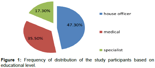
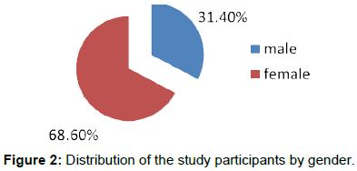
Out of the 250 questionnaires that were distributed among dentists 220 were answered. Generally, a notable response rate was observed (88%). The participants comprised 104 house officers (47%), 78 medical officers (35%) and38 specialists (17%), including 151 females (69%) and 69 males (31%) (Figures 1 and 2).
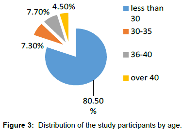
There were 69 men (31.4%) and 151 women (68.6%) accounting for a sex ratio of 2.2 and aged between 23 and 56 years old with an average of 39.5 ± 7.73 years old (Figure 3).
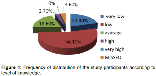
The grading scales for assessing the level of knowledge were as follows; 0-20 was considered as very low; 21-40 was considered as low; 41-60 was considered as average; 61-80 was considered as high and 81- 100 was considered as very high. 20.9% had a very low average of knowledge, 54.1% had a low level of knowledge, 18.6% had an average level of knowledge and 2.7% had a high level of knowledge (Figure 4).
Dentists̓ relative frequency of distribution for the answers of the fifteen questions related to CBCT are illustrated in Table 1.
| Questions | Correct | Incorrect |
|---|---|---|
| Question 1: Prescribing CBCT | 66 (30%) | 143 (65%) |
| Question 2: Justifiability for indications of CBCT | 27 (12.3%) | 182 (82.17%) |
| Question 3: Comparing CT and CBCT | 65 (29.5%) | 144(65.5%) |
| Question 4: Most common indications of CBCT . | 111 (50.5%) | 97 (41.1%) |
| Question 5: CBCT and TMJ | 45 (20.5%) | 158 (71.8%) |
| Question 6: Contraindication of CBCT | 107 (48.6%) | 99 (45%) |
| Question 7: Indication of CBCT in root fractures | 138 (62.7%) | 67 (30.5%) |
| Question 8: CBCT and articular disc | 68 (30.9%) | 135 (61.4%) |
| Question 9: Indication of CBCT for implant surgery in edentulous patients | 31 (14.1%) | 170 (77.3%) |
| Question 10: Contraindication of CBCT in orthodontics | 51 (23.2%) | 150 (68.2%) |
| Question 11: Contraindications of CBCT in implant surgery | 58 (26.4%) | 142 (64.5%) |
| Question 12: Contra indication of CBCT | 40 (18.2%) | 162 (73.6%) |
| Question 13: Comparing CBCT with Orthopantomogram (OPG) | 78 (35.5%) | 121 (55%) |
| Question 14: Comparing CBCT with intraoral radiographies | 96 (43.6%) | 108 (49.1%) |
| Question 15: Order for prescription | 86 (39.1%) | 115 (52.3%) |
Table 1: Dentists̓ relative frequency of distribution for the answers of the fifteen questions related to CBCT
The statistical analysis did not show any significant correlation between the level of knowledge and age (p=0.2), years of employment (p=0.1) and working area in either University or public hospitals (p=0.1) (Tables 2-4)
| Age | Number of Dentists | Mean | Standard deviation |
|---|---|---|---|
| Less than 30 | 177 | 32.8 | 14.1 |
| 35-30 | 16 | 36.2 | 13.7 |
| 40-36 | 17 | 33.3 | 13.5 |
| Over 40 | 10 | 42 | 13.3 |
Table 2: Comparisons of knowledge scores by age.
| Years of employment | Number | Mean | Standard deviation |
|---|---|---|---|
| Less than 5years | 163 | 32.3 | 14.2 |
| 5-10 Years | 36 | 36.7 | 12.03 |
| 10-15 Years | 11 | 36 | 13.7 |
| Over 15 years | 10 | 39.3 | 18.1 |
| p value = 0.1 | |||
Table 3: Comparison of knowledge scores according to number of years for employment.
| Working area | Number | Mean | Standard deviation |
|---|---|---|---|
| University teaching Hospital | 143 | 34.6 | 14.2 |
| Public hospital | 65 | 30.4 | 13.7 |
| Both | 12 | 36.6 | 13.4 |
Table 4: Comparison of knowledge scores by working area.
However, upon comparison of the level of knowledge in with regards to to gender, a significant difference was seen between males and females (p=0.001) (Table 5).
| Gender | Number | Mean | Standard deviation |
|---|---|---|---|
| Male | 69 | 34.8 | 13.6 |
| Female | 151 | 32.9 | 14.3 |
Table 5: Comparison of knowledge scores by gender.
Additionally, a significant difference in knowledge was found among dentists based on their educational degree (house officers, medical officers and specialists) (p=0.01) (Table 6).
| Educational degree | Number | Mean | Standard deviation |
|---|---|---|---|
| House officer | 104 | 31.2 | 13.4 |
| Medical | 78 | 34.04 | 14.6 |
| Specialist | 38 | 39.07 | 13.6 |
Table 6: Comparison of knowledge scores by educational degree.
Discussion
The selection of a proper diagnostic technique plays a pivotal role in the treatment of disease. An appropriate diagnostic method can provide essential information, in addition to the minimizing of cost and harm to patients. In dental radiography, efforts have been made to reduce patient exposure to radiation. Cone-beam computed tomography (CBCT) and digital radiography were developed to attain this goal [11].
In Sudan, no published study was done considering the knowledge about CBCT imaging among dentists and this may be due to the ignorance of the new techniques existence and the few number of the CBCT machines. Therefore, it is necessary to shed light and to assess the knowledge in regards to CBCT to initiate continuing dental training on that subject.
The present study was conducted to evaluate Sudanese dentists’ knowledge regarding CBCT.
Two hundred and twenty dentists including house officers, medical officers and specialists contributed in this cross-sectional study.
The present study found that the majority of respondents had a low level of knowledge about CBCT and only few respondents had a high level of knowledge regarding this topic. A possible clarification for this finding can be the unavailability of CBCT at the work place with a consequent difficulty in the acquirement of knowledge on a given system without practical experience. Therefore, the lack of CBCT units in the dental office setup may represent a significant factor contributing to dentists’ unfamiliarity with this technology with CBCT education being chiefly limited to textbooks.
This is in line with the study carried out by Kamburoĝlu et al., among Turkish dental students which underscored the problems with acquiring knowledge on a given system without practical experience [12].
In the current study no relationship has been found between age and knowledge about CBCT, a finding that could possibly be owing to its recent recognition as an imaging modality andthe lack of CBCT units at their work areas also the lack of practical experience and unfamiliarity with image characteristics in image acquisition. In addition to the advanced level of software knowledge regarding understanding and interpreting of CBCT images [13].
The results of the present study reveal a significant difference between genders with regards to CBCT knowledge. Males had greater knowledge compared to females and this can be explained by the fact that most of the training courses in CBCT are held outside Sudan and the majority of females have certain obligations that prevent them from attending such courses to improve their level of knowledge.
Concerning the number of years of employment, there was no significant difference in the knowledge of individuals with different numbers of years of employment. This can also be attributed to the absence of CBCT in their working areas together with the lack of a sufficient theoretical knowledge and practical experience through continuous education programs.
Moreover, specialists demonstrated a higher level of knowledge about CBCT than house officers and medical officers. This difference may be due to the characteristics of the specialist’s job as it involves various modalities of three dimensional imaging compared with a general dentist. Given the fact that the advantages of CBCT are clear over other methods of imaging, it should not be limited to specialty branches and comprehensive training must return the real and logic role of this modality.
This is in accordance with the study done by Mahdizadeh et al. which revealed that specialists had greater knowledge about CBCT compared to other conventional intraoral radiographies [11].
Regarding the knowledge of dentists on the basis of the field of practice either private or public hospitals, there was no significant difference found. This finding reflects the generalizability in the lack of knowledge towards the CBCT technology and the need for improvement of the level of education regarding this new technology, additionally effort should be spent to increase the availability of CBCT machines in hospitals to encourage dentists to raise their knowledge in regards to it.
One of the limitations of the present study is the type of questionnaire used (self-reporting questionnaire) which could be a possible source of bias. Another limitation is the use of convenient sampling technique which could compromise the generalizability of the current results. However, the consensus achieved from this study on the general need of the dental practitioners to have a formal and structured training in CBCT cannot be overlooked.
Conclusion
The results of this study indicate that there is a gap in knowledge of CBCT applications among Sudanese dentists as it is a new innovation in the field of dental radiology, with a consequent restriction in the ability to explore this imaging modality to the fullest.
Introduction of training in CBCT at undergraduate as well as postgraduate level will assist dentists in using this technique in an efficient way to upgrade the accuracy and reliability of oral and maxillofacial diagnosis, treatment planning and outcomes.
Acknowledgement
We wish to thank the dentists who participared in this study, without their participation this work would have been impossible. Special thanks to Dr. Sakina Abbasher and Azza Tajelsir for helping us in revising and editing the English language.
Competing Interests
The authors declare that they have no competing interests.
Ethics Approval and Consent to Participate
The study protocol was approved by the Ethical Committee of faculty of Dentistry, University of Science and Technology, Omdurman.
References
- Parashar V, Whaites E, Monsour P, Chaudhry J, Geist JR (2012) Cone beam computed tomography in dental education: A survey of US, UK, and Australian dental schools. J Dent Edu 76: 1443-1447.
- Scarfe WC, Farman AG, Sukovic P (2006) Clinical applications of cone-beam computed tomography in dental practice. J Can Dent Assoc 72: 75-80.
- Hatcher DC (2010) Operational principles for cone-beam computed tomography. J Am Dent Assoc 141: 3S-6S.
- Qirresh E, Rabi H, Rabi T (2016) Current status of awareness, knowledge and attitude of dentists in palestine towards cone beam computed tomography: A survey. Oral Biol Dent 4: 1-4.
- Kiljunen T, Kaasalainen T, Suomalainen A, Kortesniemi M (2015) Dental cone beam CT: A review. Phys Med 31: 844-860.
- Boeddinghaus R, Whyte A (2008) Current concepts in maxillofacial imaging. Eur J Radiol 66: 396-418.
- Kapila S, Conley RS, Harrell WE (2011) The current status of cone beam computed tomography imaging in orthodontics. Dentomaxillofac Radiol 40: 24-34.
- Rugani P, Kirnbauer B, Arnetzl GV, Jakse N (2009) Cone beam computerized tomography: Basics for digital planning in oral surgery and implantology. Int J Comput Dent 12: 131-145.
- Horner K, Islam M, Flygare L, Tsiklakis K, Whaites E (2009) Basic principles for use of dental cone beam computed tomography: Consensus guidelines of the European Academy of Dental and Maxillofacial Radiology. Dentomaxillo-Facial Radiol 38: 187-195.
- Tofangchiha M, Arianfar F, Bakhshi M, Khorasani M (2015) The assessment of dentists’ knowledge regarding indications of cone beam computed tomography in Qazvin, Iran. Biotech Health Sci 2: e25815.
- Mehdizadeh M, Booshehri SG, Kazemzadeh F, Soltani P, Motamedi MRK (2015) Level of knowledge of dental practitioners in Isfahan, Iran about cone-beam computed tomography and digital radiography. Imag Sci Dent 45: 133-135.
- Kamburoğlu1 K, Kurşun Ş, Akarslan ZZ (2011) Dental students’ knowledge and attitudes towards cone beam computed tomography in Turkey. Dentomaxillofac Radiol 40: 439-443.
- Lavanya R, Babu DG, Waghray S, Chaitanya NC, Mamatha B, et al. (2016) A Questionnaire Cross-Sectional Study on Application of CBCT in Dental Postgraduate Students. Pol J Radiol 81: 181-189.
Citation: Abdelmonaim Y, Fayez A, Abid R, Abdelraziq K, Mohammed R, et al. (2017) Assessment of Dentists Knowledge towards Cone Beam Computed Tomography in Public and University Teaching Hospitals in Khartoum State. OMICS J Radiol 6: 280. DOI: 10.4172/2167-7964.1000280
Copyright: ©2017 Abdelmonaim Y, et al. This is an open-access article distributed under the terms of the Creative Commons Attribution License, which permits unrestricted use, distribution, and reproduction in any medium, provided the original author and source are credited.
Select your language of interest to view the total content in your interested language
Share This Article
Open Access Journals
Article Tools
Article Usage
- Total views: 5128
- [From(publication date): 0-2017 - Dec 10, 2025]
- Breakdown by view type
- HTML page views: 4185
- PDF downloads: 943


