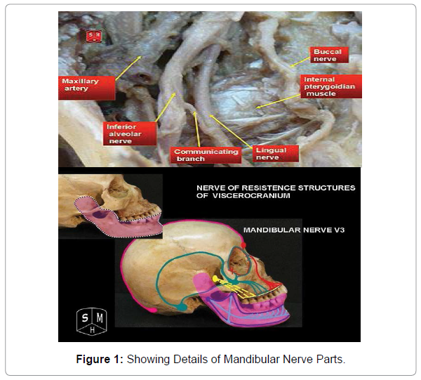
Brief clarification Image of Mandibular Nerve
Received: 13-Oct-2021 / Accepted Date: 20-Oct-2021 / Published Date: 27-Oct-2021 DOI: 10.4172/2167-7964.1000346
Image Article
The mandibular nerve supplies the teeth and gums of the mandible, the skin of the transient area, part of the auricle, the lower lip, and the lower part of the face the mandibular nerve likewise supplies the muscles of rumination and the mucous layer of the foremost 66% of the tongue [1].
The enormous tangible root rises up out of the parallel piece of the trigeminal ganglion and ways out the cranial pit through the foramen ovale. Portio minor, the little engine base of the trigeminal nerve, passes under the trigeminal ganglion and through the foramen ovale to join with the tactile root right external the skull.
The mandibular nerve quickly passes between tensor veli palatine, which is average, and parallel pterygoid, which is sidelong, and emits a meningeal branch (nerves spinosus) and the nerve to average pterygoid from its average side. The nerve then, at that point, separates into a little front and enormous back trunk [2].
The front division radiates branches to three significant muscles of rumination and a buccal branch which is tactile to the cheek. The back division emits three primary tangible branches, the auriculotemporal, lingual and second rate alveolar nerves and engine filaments to supply mylohyoid and the foremost tummy of the digastric muscle.
Clinical image of mandibular nerve
The peripheral mandibular nerve might be harmed during a medical procedure in the neck district, particularly during extraction of the submandibular salivary organ or during neck analyzations because of absence of exact information on varieties in the course, branches and relations. A physical issue to this nerve during a surgery can misshape the demeanour of the grin just as other looks. The minimal mandibular part of the facial nerve is found shallow to the facial corridor and (front) facial vein. Subsequently the facial corridor can be utilized as a significant milestone in finding the negligible mandibular nerve during careful procedures [2]. Harm can cause loss of motion of the three muscles it supplies, which can make a hilter kilter grin due absence of compression of the depressor labia inferiors muscle. This might be adjusted with resection of the muscle, which will in general be effective.
In the brainstem, the mandibular branch emerges from 3 cores (mesencephalon, head tangible and spinal) which lead to its enormous tactile root. The engine core of the trigeminal nerve, situated in the stuffing of the upper space of the pons, leads to the engine base of the nerve,
The mandibular nerve supplies both engine and tangible data, which implies it’s connected to development and faculties. One of its most fundamental capacities is controlling the developments of the muscles that permit you to bite [3]. These incorporate the masseter, the parallel and average pterygoids, and the temporalis muscle
Branches of mandibular nerve
Masseteric nerve (motor)
Deep temporal nerves, anterior and posterior (motor)
Buccal nerve (a sensory nerve)
Lateral pterygoid nerve (motor)
The trigeminal nerve has three branches coming to all through the face and oral hole. The upper branch is liable for sensations for the scalp, temple and front of the head. The centre branch is liable for sensations in the cheek, upper jaw, top lip, top teeth and gums, and side of the nose.
References
- Ava Yoon, Vinay Puttanniah (2016) Mandibular Nerve Entrapment, in Peripheral Nerve Entrapments 217-228.
- Amir Haschemi (1981) Partial anastomosis between the lingual and mandibular nerves for restoration of sensibility in J Craniomaxillofac Surg 9:225-227.
- Rajwant Kaur (2021) Pseudo paralysis of Marginal Mandibular Nerve Branch by Big Submandibular Gland Sailolith: A Case Report Otolaryngology 6.
Citation: Malakuv T (2021) Brief clarification Image of Mandibular Nerve. OMICS J Radiol 10: 346. DOI: 10.4172/2167-7964.1000346
Copyright: © 2021 Malakuv T. This is an open-access article distributed under the terms of the Creative Commons Attribution License, which permits unrestricted use, distribution, and reproduction in any medium, provided the original author and source are credited.
Select your language of interest to view the total content in your interested language
Share This Article
Open Access Journals
Article Tools
Article Usage
- Total views: 2646
- [From(publication date): 0-2021 - Dec 17, 2025]
- Breakdown by view type
- HTML page views: 1915
- PDF downloads: 731

