Research Article Open Access
Chronic Ankle Instability: Results with Brostrom-Evans Procedure
| Albiñana Cuningham Juan, Macklin Vadell Alberto and Peratta Marcela* | |
| Cerviño Av. 4679, 2nd floor, 1426 Buenos Aires, Argentina | |
| Corresponding Author : | Marcela Peratta Cerviño Av. 4679, 2nd floor 1426 Buenos Aires, Argentina E-mail: marcelaperatta@gmail.com |
| Received March 12, 2013; Accepted April 12, 2013; Published April 15, 2013 | |
| Citation: Juan AC, Alberto MV, Marcela P (2013) Chronic Ankle Instability: Results with Brostrom-Evans Procedure. J Nov Physiother S3:007. doi:10.4172/2165-7025.S3-007 | |
| Copyright: © 2013 Juan AC, et al. This is an open-access article distributed under the terms of the Creative Commons Attribution License, which permits unrestricted use, distribution, and reproduction in any medium, provided the original author and source are credited. | |
Visit for more related articles at Journal of Novel Physiotherapies
Abstract
Ankle inversion injuries account for 40% of all sport injuries and up to 20-24% of these will have residual pain, instability, or a combination of both. There are two types of chronic ankle instability: functional and mechanical. In this study, we will retrospectively analyze the Brostrom-Evans technique used to treat the mechanical chronic instabilities. We have reviewed ten procedures in nine recreational athletes, 6 male and 4 female, one of which was bilateral. In two cases, a calcaneal osteotomy was associated to correct the hindfoot varus. The Brostrom-Evans technique solves the chronic instability problem by sacrificing the normal inversion movement of the ankle. We would recommend modifying the hindfoot varus and then performing a Brostrom-Evans plasty in those cases in which a calcaneus varus is associated to a chronic instability.
| Keywords |
| Calcaneal osteotomy; Brostrom-Evans technique; Hindfoot varus; Chronic instability |
| Introduction |
| Persistent pain, instability or a combination of both can be observed after an acute ankle sprain. |
| Instability can be anatomically classified as tibiotalar, subtalar or rotatory (tibio and subtalar). |
| In 1965, Freeman divided ankle instability into organic and functional instability [1]. |
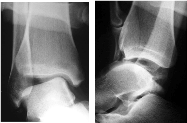
| An insufficient lateral collateral ligament complex associated with an impaired or torn peroneus brevis characterizes organic lateral ankle instability. Stress x-rays such as anterior drawer test or stress varus test are positive in these cases (Figure 1). |
| Patients with functional ankle instability have subjective complaints about their ankle giving way, but they lack radiographic and clinical evidence of instability [2]. |
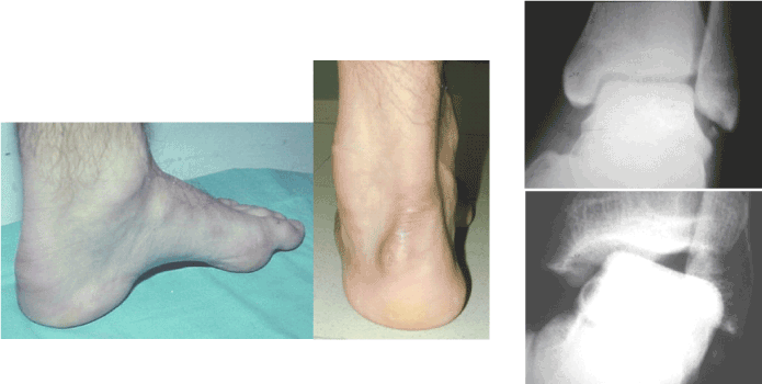
| In both types of instability predisposing factors can coexist such as: ossubperoneum, adduct cavus foot, calcaneus varus, genu varus and tibialintrarotation (Figure 2). |
| Initial treatment must begin with functional rehabilitation and proprioceptive training. |
| Patients, who are recalcitrant to conservative measures, may be candidates for operative intervention. |
| Many operative techniques have been described to reconstruct the lateral collateral ligament complex of the ankle. |
| Our work group has used the Brostrom technique modified by Gould for the isolated tibiotalar instabilities and the Chrisman-Snook technique for the severe instabilities with some degree of subtalar involvement [3,4]. Recently we have used the Brostrom associated to the Evans procedure, considering it provides an increase in stability by passive retention compared to the Brostrom-Gould technique and less morbidity than the Chrisman-Snook technique [5,6]. |
| The purpose of this paper is to describe this technique and present the initial results of our series. |
| Materials and Methods |
| From February 2002 to January 2004, ten patients, 6 male and 4 female, with an average age of 40.5 years (22 to 58) underwent surgical procedure in eleven ankles. For this study we have reviewed 10 ankles in 9 patients. |
| Out of the evaluated group, one patient was operated bilaterally and another patient had a calcaneal osteotomy associated to the Brostrom-Evans [7] technique to correct the hindfoot varus. |
| Surgical technique |
| The patient is placed in a lateral decubitus position, a sciatic block or epidural anesthesia is administered and a tourniquet is placed at the most proximal end of the thigh. |
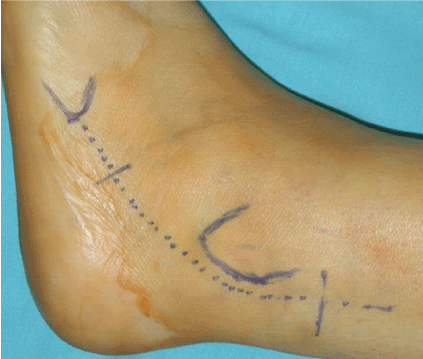
| Using a longitudinal approach, a skin incision is made 5 cm (Figure 3) proximal to the fibular malleolus to the base of the 5th metatarsal. Subcutaneous tissue is carefully dissected protecting the sural nerve. |
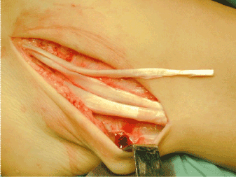
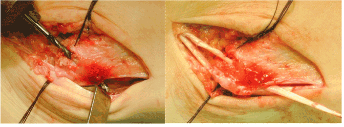
| The inferior extensor retinaculum and the tibiotalar joint capsule are individualized. The peroneal sheath is incised, and the anterior part of the hemisection of the peroneus brevis is harvested preserving the distal insertion at the base of the 5th metatarsal (Figure 4). The articular capsule is dissected exposing the fibrous remnant of the anterior talofibular ligament. The peroneal tendons are retracted posteriorly in order to inspect the calcaneofibular ligament that is generally elongated or avulsed. Both ligaments are repaired without completing the suture, if necessary, we employ one or two suture anchors to fixate the ligaments. |
| With a 4.5 mm diameter drill, a trans-osseous tunnel is performed from anterior to posterior and from distal to proximal, at a midpoint between the anterior talofibular ligament and the calcaneofibular ligament, exiting at about 2.5 cm from the distal end of the peroneal malleolus, to pass the hemitendon of the peroneus brevis (Figure 5). |
| Suture of the calcaneofibular ligament is performed, followed by the suture of the anterior talofibular ligament. We suture the hemitendon at the entrance and at the exit of the bone tunnel (Figure 6). |
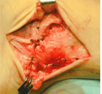
| Finally we closed the articular capsule and extensor retinaculum as well as the peroneal tendons sheath. (Figure 7) |
| The tourniquet is then deflated before skin closure in order to obtain proper hemostasis. |
| When instability is associated to a calcaneus varus deformity, a valgus osteotomy is performed through a second skin incision and fixated with one or two staples. |
| During the first three weeks the patient is immobilized in a walking cast with a slight hind foot eversion and is not allowed to bear weight. After this period, he will be allowed to bear weight progressively with an orthotic walker boot and begin functional rehabilitation. Contact sports will be allowed after 6 months. |
| Results |
| We have reviewed ten procedures with an average follow-up of 26.2 months (12 to 35 months). |
| The value of stress radiographs is questioned to analyze instability, since patients with a 20° talar tilt were barely symptomatic while patients with a 8-10 degree talar tilt were very disabled [8]. |
| For this reason, we emphasize on the patient´s perception of pre and postoperative ankle stability as well as on the stress radiographs. |
| All patients reported subjective stability of the tibiotalar joint. |
| We found a limitation of the ankle inversion during physical examination not mentioned previously by the patients. |
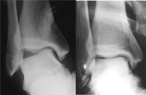
| We observed a decrease of the tibio-talar angle in the stress radiographs, with an average value of 6° (12-0°), being the average preoperative tibiotalar angle value of 18° (24-7°) (Figure 8). |
| In all cases the postoperative anterior drawer test (anterior sagittal translation of the talus on the tibia) was negative, being positive in 100% of the patients preoperatively. |
| Seven patients (77.7%) have been able to participate in sports at their preinjury level of activity, the 2 remaining patients (22.3), for personal reasons, did not practice sports after surgery. |
| 100% of the patients are satisfied and would recommend this procedure. |
| Discussion |
| The objective of chronic ankle instability treatment is to gain maximum stability with the lowest degree of limitation in functional outcome. |
| 87% of good and excellent results, without limitations to practice sport activities, have been reported with the modified Brostrom- Gould technique, but in our work group, we have had considerably fewer good results with a high percentage of recurrence rates. |
| Although the recurrence rate with the Chrisman-Snook technique has been lower, morbidity, such as saphenous nerve injury due to a wider approach, was significant. |
| Non-anatomic reconstruction techniques such as the Brostrom- Evans and the Chrisman-Snook are associated to some degree of tibiotalar and subtalar stiffness; although in our group of patients, this restriction in ankle inversion did not limit their day-to-day or athletic activities [9,10]. |
| According to the literature, the Brostrom-Evans procedure has been associated with complications such as persistent pain and swelling, eversion weakness and limitations in returning to their preinjury level of sport due to a wide surgical approach and less mechanical stability secondary to the tenodesis. |
| In our experience, with a two-year mean follow up, we have observed a hypertrophic scar in all our patients and a permanent saphenous nerve injury in one patient [11,12]. |
| Conclusion |
| The Brostrom-Evans procedure is moderately demanding and care should be taken with the saphenous nerve, the length of the hemitendon, the bone tunnel orientation, as well as with the tension given to the suture. We believe that it solves the chronic ankle instability problem at the expense of a decrease in normal hindfoot movement although this will not cause limitation for the patients. In those cases in which a calcaneus varus is associated to instability, we believe it is advisable to resolve the hindfoot varus and then perform the Brostrom-Evans plasty. |
References |
|
Figures at a glance
 |
 |
 |
 |
| Figure 1 | Figure 2 | Figure 3 | Figure 4 |
 |
 |
 |
 |
| Figure 5 | Figure 6 | Figure 7 | Figure 8 |
Relevant Topics
- Electrical stimulation
- High Intensity Exercise
- Muscle Movements
- Musculoskeletal Physical Therapy
- Musculoskeletal Physiotherapy
- Neurophysiotherapy
- Neuroplasticity
- Neuropsychiatric drugs
- Physical Activity
- Physical Fitness
- Physical Medicine
- Physical Therapy
- Precision Rehabilitation
- Scapular Mobilization
- Sleep Disorders
- Sports and Physical Activity
- Sports Physical Therapy
Recommended Journals
Article Tools
Article Usage
- Total views: 15925
- [From(publication date):
specialissue-2013 - Aug 30, 2025] - Breakdown by view type
- HTML page views : 11356
- PDF downloads : 4569
