ISSN: 2165-7025
Journal of Novel Physiotherapies
Make the best use of Scientific Research and information from our 700+ peer reviewed, Open Access Journals that operates with the help of 50,000+ Editorial Board Members and esteemed reviewers and 1000+ Scientific associations in Medical, Clinical, Pharmaceutical, Engineering, Technology and Management Fields.
Meet Inspiring Speakers and Experts at our 3000+ Global Conferenceseries Events with over 600+ Conferences, 1200+ Symposiums and 1200+ Workshops on Medical, Pharma, Engineering, Science, Technology and Business

Co-contraction Role on Human Motor Control. A Neural Basis

Marlene Rosa*
Autonomous Unit of Health Sciences, University of Aveiro, Portugal
Corresponding Author : Marlene Rosa
Autonomous Unit of Health Sciences
University of Aveiro, Portugal
Tel: 35-1968918915
E-mail: marlenerosa@ua.pt
Received: November 13, 2014; Accepted: February, 2015; Published: February 9, 2015
Citation: Rosa M (2015) Co-contraction Role on Human Motor Control. A Neural Basis. J Nov Physiother 5:248. doi: 10.4172/2165-7025.1000248
Copyright: © 2015 Marlene Rosa. This is an open-access article distributed under the terms of the Creative Commons Attribution License, which permits unrestricted use, distribution, and reproduction in any medium, provided the original author and source are credited.

Visit for more related articles at Journal of Novel Physiotherapies

Abstract

The aim of this overview is to understand the role of the co-contraction on movement accuracy and the respective neural processes involved in healthy people and in subjects after a Central Nervous System Lesion (CNS). First, it will be discussed the relevance of co-contraction to the motor control, based on Internal Model Theory and Equilibrium Point Theory. Secondly, it will be discussed the muscle co-contraction neural regulation at a spinal level control and how the agonist-antagonist muscles work on healthy and neurologic impairment subjects.

Keywords

Co-contraction; Motor control; Neural regulation; Healthy people; Stroke

Co-contraction and the motor control phenomena

Co-contraction is a strategy used by the Central Nervous System to achieve movement accuracy [1] especially during the learning process of a novel task [2-6]. The co-contraction is defined as a simultaneously contraction of two or more muscles around a joint [7] and it is a determinant factor to the evaluation of motor control (measuring muscle synergism [8] motor learning and dynamic joint stability (minimising the perturbing effects of different environments) [6,9].

The co-contraction is central programmed [6,10,11] but it is reinforced at a spinal level by direct commands to agonist and antagonist muscles. Therefore, the neurophysiology of muscle co-contraction could be first understood based on the “Motor Control Theories” [2,10,12] (Theory of Internal Models [3,13] and (ii) the Equilibrium Point Theory [10,13] but then it needs to be completed using the explanatory models for spinal regulation ( Common Drive Model) [2] and The Disynaptic Reciprocal Ia inhibition [14].

Motor Control Theories

Theory of internal models

Considering the Theory of Internal Models [2,5,10], the subjects are repeatedly exposed to sensory signals, while they are moving their limbs to interact with the environment during the learning of a task. To generate the muscle activation, all sensory signals are conducted to the motor areas of the CNS. This process generates an internal model at the cerebellum level, considering two important phenomena: (i) changes on synaptic neurons strength (ii) new connectivities of a group of neurons and both are responsible for generating neural impulses, contributing to motor learning and/or dynamics control [3].

Through the years, the Internal Model theory [12] become a combination of other new neurophysiologic principles [12,15]. In this context, the “force” was then presented as being the main concept in neuromuscular programming and a new component of the Internal Model Theory was developed - the Force Control Model [15]. This model defended that the movement trajectory is planned in terms of spatial coordinates and that the internal dynamical equations of motion were created based on forces and torques, while the body is interacting with the environment [13].

Equilibrium point theory

The Equilibrium Point Theory, also known as Lambda Model, supports the idea of intentional motor actions, without any programming. Instead, this theory defends that the CNS controls (by proprioceptive feedback to motoneurons), the thresholds muscle lengths or joint angles are determinant components of motor control [16]. Therefore, based on this theory, the CNS induces the muscle contraction, programming the spatial coordinates through the difference/ratio between the actual position of the body and the muscles/joints thresholds (which are central programmed) [17].

Theory of internal models and the equilibrium point theory - an integral model based on common concepts

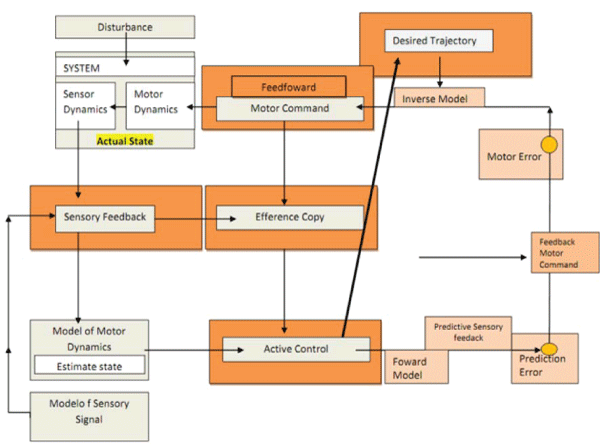
Feedback and Feedfoward control modes: On the basis of all motor control theories are the Feedback and Feedfoward control modes, used by the CNS on the control of musculoskeletal system.

The feedforward model is capable of achieving a causal relationship between inputs to the system (the distance of an object; the coordinates of the arm position) and it is important in motor learning situations (e.g., during an arm movement, it could predicts the next state – position and velocity – through the current state and motor command). Despite the feedfoward components not being directly related to sensorial information, it can be influenced by feedback signals [18].

The feedback components depend on sensorial information (the position of the arm), whereas the feedfoward components are based on system dynamics knowledge (which should be the adequate position to reach an object) [18]. The feedback control, somehow acts as an inverse model. The inverse model inverts the system. First, it is provided the motor command, causing a desired change in the state (elbow extension). Then, the inverse model act as a controller mechanism, providing the necessary motor commands and achieving the desired state transition [19].

Mechanical impedance and viscoelasticity - key properties of the CNS: Both theories of motor control are based on the ability of the CNS easily adapt to sudden changes of the environment. This ability depends on some key properties of the CNS, such as the mechanical impedance (a basic concept on the equilibrium point theory) and the viscoelasticity (basic concept on both theories).

The mechanical impedance is an important dynamic relation between small forces and position variations and it is a basic concept on the Equilibrium Point Theory. The mechanical impedance of the neuromuscular system determines the reaction forces on the hand in response to perturbations from the manipulated object and choosing an adequate mechanical impedance may be one of the ways the CNS controls the behavior of the complete system (hand+object) [20]. The mechanical impedance could be improved trough viscoelastic changes by feedfoward motor commands way, specifically in cases on rapid changes in the magnitude or nature of external forces [21]. The viscoelasticity is a property of biological materials that are both solid and fluid-like, such as tendons and ligaments. These materials possess time-dependent stress-strain relations, that change as the loading speed changes [22].

Based on these key properties of the CNS, some authors [5] suggested the integration of the Equilibrium Point Theory and Internal Model Theory in movement control. They defended that the CNS relies on the viscoelasticity property when the Internal Models are imperfect or the environment is unstable [23,24]. In this new model, the viscoelasticity depends on feedback controller, as while the internal models are the result of forward controller. On the following graph (Figure 1) it is presented a model created to explain how the motor control could be programmed, based on new integral model.

At least two different models are referred on the literature to explain the neural regulation of agonist-antagonist muscle activation at a spinal level: the Common Drive Model and the Disynaptic Reciprocal Ia Inhibition.

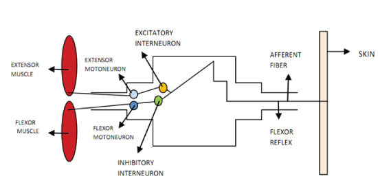
Common drive model : The nervous system does not control the firing rates of motor units individually, to generate the muscle synergy [5], instead the CNS programmed the excitation of the motoneuron pool [25]. A motoneuron pool is a group of motor neurons with common targets and afferent inputs [26]. In this muscle activation mode, a flexor muscle and a extensor muscle around one specific joint are controlled as if they were one muscle. The way a group of agonist-antagonist muscles are activated depends on the function to be performed but also depends on the spine origin proximity of their nerve roots. This model defends that the “flex” and “extend” commands channels in the CNS are reciprocally organised and its main characteristic is that the inhibition of the antagonist muscle happens prior to excitation of the agonist muscle – co-contraction phenomena [26]. The co-contraction is important in two specific situations both depending on the environment conditions: (i) during states of uncertainty or (ii) when it was required a compensatory force correction [25] (e.g., if they occurs destabilizing forces during a upper limb dynamic task etc. [11] (Figure 2).

The disynaptic reciprocal Ia inhibition: This model was proposed [14] to explain the reciprocal pattern of muscles activation on voluntary movements [27]. The brain controls agonist α-motoneurones and Ia inhibitory interneurones, which have monosynaptic projections to motoneurones of the antagonists in parallel [14,27]. Despite this mechanism involving only a single interneuron it has a special characteristic - their reciprocal organisation [27]. This organisation allows that during a simultaneous activation of two antagonist muscles, the muscular contractions generate less force. During this process occurs a “double action of the reciprocal inhibitory action” and the a-motoneurones supplying the antagonistic muscles is simultaneously depressed [14] (Figure 3).

The main difference between these two models is related to the processing mechanism: the Disynaptic Reciprocal Ia inhibition depends on a single interneuron with monosynaptic projections to motoneurones of the antagonists and the Common Drive Model defends that exist different motoneurons (one for the agonist and other for the antagonist muscles) linked by common targets and afferent inputs. Therefore, it remains unclear how is the co-contraction programmed by the CNS and further research should clarify these mechanisms related to specific tasks (e.g. postural or dynamics) and specific environment conditions (e.g. presence of perturbing forces) [2,14].

Co-contraction and the Motor Control in Healthy People

The muscle co-contraction role on movement control, considering healthy people, seems to be important to promote joint stability [11,5,6] especially on disturbing environment conditions speed changes [10,4,11] positioning instability [11] or vibration [11] improving joint stiffness and stability on disturbing environments and generating feedfoward control [2,6,28,29]. The muscle co-contraction is therefore an important an important reactive response during the learning process of a motor task.

The Co-contraction and the Posture

In order to understand the role of co-contraction on posture control, some studies have been conducted. Milner [28] and Perreault and Kirsch [29] both tested the joint stiffness regulation on healthy people, using robotic manipulators. These robotics aimed to measure the static stiffness, by the control of the relationship of the upper limb external displacement and elastic forces generated in response. These experiments demonstrated that the stiffness, at single and multi joint levels, increases if the co-contraction also increases, which led these authors to test this causal relation during postural tasks. During these studies, the participants (healthy people) were submitted to mechanical instabilities and they have to maintain a defined posture. Perreault and Kirsch [29] claimed that co-contraction is posture-independent, Milner [28] concluded that in subjects with high co-contraction levels demonstrated lower stiffness than predicted. That cases were explained by the reciprocal inhibition that was produced by the simultaneously antagonists activation, which may decrease the potential of maximum activation. Nielsen et al. [14] investigated two antagonist muscles (soleus and tibialis anterior) and its behavior during tonic contractions of the foot, whilst this joint was supported by a torquemeter. Twelve healthy participants were asked to perform different degrees of force. The authors measured the disynaptic reciprocal inhibition (spinal level motor channels) during the activation of ankle dorsiflexors, ankle plantarflexors and a combined activation of both muscle groups. They concluded that the dissynaptic reciprocal inhibition decreases during co-contraction (minimum potential of activation), but increases during agonist contraction (maximum potential of activation). At a final stage they defended that muscle co-contraction is centrally programmed, following an internal model to flexion and extension motor programmes [14] and that the muscle co-contraction may not be essential on the regulation of postural stability as it can decrease muscle efficiency. However, considering the high complexity of the neural processes involved on muscle co-contraction, more research is needed in this topic [11,14].

The Co-contraction in Dynamic Tasks

During dynamic tasks, no consensus exists on the neural mechanisms that regulate the co-contraction and the most adequate motor control theory within this topic is still uncertain [6].

The Equilibrium-point theory [10] was tested by creating dynamic conditions and simultaneously assessing gradual changes in a joint. Nine healthy right handed subjects used a maniple mounted on a platform in order to keep the forearm stable. They were instructed to maintain the wrist in a target window during three different disturbing conditions, in order to create a degree of uncertainty, avoiding the learning effect. The main conclusions of this study were: the co-contraction commands seem to be independent of peripheral feedback, which can suggest that they are centrally programmed, supporting the Internal Models Theory and not the Equilibrium-point theory.

In other experiment, Osu et al. [5] tested the integration of the Internal Models Theory and of the Equilibrium Point Hypothesis as if it were just one theory of motor control. Six healthy subjects were tested while they grip the handle of a force sensor. The investigators [5] requested the participants to keep the head of the force on a defined target window (observed on an EMG screen) while they were submitted to 8 randomised perturbing force directions. It was concluded that the detected changes in viscoelasticity were consistent with the learning effect and it proves that viscoelasticity may be dependent on the development of an internal model. These authors suggested therefore that the learning improves the internal models and that the CNS relies on viscoelasticity when the internal models are imperfect. The resultant hypothesis was that the CNS adapt an internal model to the new motor task (having a faster and more efficient reactive response)after the learning effect, and only then the muscle action starts being central programmed [5,6,10].

Co-contraction - a Focus on Gait Research

The majority of the studies about the role of co-contraction on motor control have been focused their attention on the upper limb: forearm [2,5], elbow and shoulder [4,6], elbow [30], wrist joint [10,11,28], upper arm and forearm [20,29]. There are few studies about the co-contraction regulation on motor control of the lower limb [14,31] and the ankle is the joint more frequently explored in the literature. Literature on motor control during gait is also scarce and its major impact is on development of robotic devices [32,33].

Robotics research is of special interest in this paper as it has been progressing through the development of algorithms based on the “Internal Models Theory”. Forward and inverse models are defended as absolutely necessary during the walking performance, considering these robots experiments. Also, the two classes of control systems (feedback and feedfoward controls) are both important to understand the sensory input and guide the movement during walking [33]. The relevance of the Internal Models Theory with feedback and feedfoward controls on gait neural programming was also defended by McFadyen et al. [34]. In particular, this author refers that the toe and the heel are the body endpoints which interact more intensively with the environment. Therefore, considering the many different ways that lower limb joints have to interact with the environment, researchers have been focused on the development of algorithms able to plan and execute a specific movement action. This will be a step forward to further understand gait movement control. One of the main conclusions of this author [34] is about the relevance of “anticipatory adjustments” to explain how the human body reacts to different environment conditions [35] (e.g., obstacles, inclined surfaces, vibration) and how it activates different strategies (e.g. muscle co-contraction), improving joint stability and the walking performance [14]. In summary, gait performance depends on the improvement – based on experience and training – of internal models whilst subjects are repeatedly exposed to different environment conditions. When a motor task is repeated it seems to enhance the feedfoward control and the anticipatory response. These are the neurophysiologic principles underlying the gait adaptation to different environmental conditions (obstacles course, changes on movement speed) [36].

Contraction after a Central Nervous System Lesion

After a CNS lesion (stroke, cerebral palsy, sensorimotor dysfunctions [37,38], a set of pathological events could be classified as positive or negative signs [39]. Loss of voluntary motor behavior, loss of force production and decrease of dexterity of movement are the most common negative signs after a CNS lesion [39]. Increase of stretch reflexes, increase spasms and increase of muscle co-contraction are the positive signs that characterized a CNS lesion [39]. The Stroke [40,41] and the Cerebral Palsy [42-44] are the two neurologic pathologies most studied within this topic, but in general, little is known about the co-contraction after a CNS lesion. Apparently, the abnormal muscle co-contraction after a CNS lesion could explain the movement abnormalities such as (i) prolonged movement times and (ii) lacking of muscle coordination [37]. Also, in the early rehabilitation period of neurologic pathologies, the majority of the motor tasks (e.g., grasping or walking) need to be learnt again. The co-contraction has an important role on movement accuracy during learning periods, until the CNS acquired an Internal Model.

The loss of the Common Drive mechanism by the interruption of information flow in corticospinal pathway as the best model to explain the modification of co-contraction post-stroke has a weak evidence [41]. Most of the literature explained the pathologic co-contraction after a neurologic lesion using the Disynaptic Reciprocal Ia inhibition. The Disynaptic Reciprocal Ia Reciprocal Inhibition model seems to explain not only the pathologic co-contraction, but also Hyperactive Strech Reflex [39]. The Strech Reflex normally is latent but in the case of neurologic pathology it become obvious and generates spasticity [45,46]. The spasticity has been commonly defined as “velocity dependent increase in tonic stretch reflexes” and could be influenced by the muscle co-contraction [44]. Therefore, on subjects with CNS lesion, the co-contraction and spasticity neural mechanisms are not easily to investigate separately because they are closely related. Priori et al. [47] studied this relationship between spasticity and co-contraction. They explained that the interneuronal dysfunction that origin the spasticity implies the abnormal inhibition of the antagonist muscles, causing abnormal co-contraction. The functional impairment is a result of both (i) co-contraction impairment and (ii) increase stretch reflex [47], Pierce et al. [44], studied the relation between spasticity and passive resistive torque, reflex activity, coactivation and reciprocal facilitation. Twenty children with Cerebral Palsy were studied whilst they were doing passive movements with the knee. The vastus lateralis and medial hamstrings activity were measured using the surface electromyography. Taken into account all the criteria evaluated, the reflex activity showed to play a less prominent role [44]. Levin et al. [40] clarified this relationship investigating the electromiographic co-contraction ratios during dorsiflexion in spastic hemiparetic people and healthy subjects. They concluded that the co-contraction ratios were inversely correlated with the force produced by the paretic dorsiflexors in people with stroke [40]. They also assumed that spasticity and co-contraction could be programmed by the same neural process once they are usually coexistent factors on subjects after a CNS [45,46,48].

Discussion and Conclusion

This overview explored the mechanisms underlying the muscle co-contraction phenomenon and its role in movement accuracy. The movement accuracy depends on the co-contraction especially during the learning of a motor task – trough the viscoelasticity changes (Equilibrium Point Hipothesis) - or in unpredictable environment conditions. After this learning period, the co-contraction is controlled by an Internal Model (Internal Model Theory) acquired by the CNS. The agonist-antagonist regulation also happens at a spinal level. The Disynaptic Reciprocal Ia inhibition is more studied in the literature and seems to control both co-contraction and the stretch reflex.

Few studies exist about the muscle co-contraction on subjects post CNS lesion and most of them have been focused mainly in the arm movement tasks. Despite the importance for develop robotic gait orthosis, little is known about the co-contraction profiles in locomotion. Further research in this topic should be developed to help the health professionals understanding the role that co-contraction plays in the CNS recovery process and define mechanical and manual strategies to its neural modulation.

References


  1. Gordon J, Ghez C (1984) EMG patterns in antagonist muscles during isometric contraction in man: relations to response dynamics.Exp Brain Res 55: 167-171.
  2. De Luca CJ, Mambrito B (1987) Voluntary control of motor units in human antagonist muscles - coactivation and reciprocal activation. Journal of Neurophysiology 58: 525-542.
  3. Bizzi E, Mussa-Ivaldi FA (1998) Neural basis of motor control and its cognitive implications.Trends CognSci 2: 97-102.
  4. Gribble PL, Ostry DJ (2000) Compensation for loads during arm movements using equilibrium-point control.Exp Brain Res 135: 474-482.
  5. Osu R, Franklin DW, Kato H, Gomi H, Domen K, et al. (2002) Short- and long-term changes in joint co-contraction associated with motor learning as revealed from surface EMG.J Neurophysiol 88: 991-1004.
  6. Gribble PL, Mullin LI, Cothros N, Mattar A (2003) Role of cocontraction in arm movement accuracy.J Neurophysiol 89: 2396-2405.
  7. Fonseca STD, Silva PLPD (2008) Analysis of a electromyographic method for quantification of muscle co -contraction .
  8. Faria CD, Teixeira-Salmela LF, Gomes PF (2009) Applicability of the coactivation method in assessing synergies of the scapular stabilizing muscles.J Shoulder Elbow Surg 18: 764-772.
  9. Kelso JAS (1982) Human Motor Bahvior - an introduction, Lawrence Erlbaum Associates, Inc.
  10. Levin MF, Feldman AG, Milner TE, Lamarre Y (1992) Reciprocal and coactivation commands for fast wrist movements.Exp Brain Res 89: 669-677.
  11. Milner TE1 (2002) Adaptation to destabilizing dynamics by means of muscle cocontraction.Exp Brain Res 143: 406-416.
  12. Ito M (1970) Neurophysiological aspects of the cerebellar motor control system.Int J Neurol 7: 162-176.
  13. Ostry DJ, Feldman AG (2003) A critical evaluation of the force control hypothesis in motor control.Exp Brain Res 153: 275-288.
  14. Nielsen J, Kagamihara Y (1992) The regulation of disynaptic reciprocal Ia inhibition during co-contraction of antagonistic muscles in man.J Physiol 456: 373-391.
  15. Hollerbach J M (1982) Computers, brains and the control of movement. Trends in Neurosciences 5: 189-192.
  16. Fel'dman AG (1966) On the functional tuning of the nervous system in movement control or preservation of stationary pose. II. Adjustable parameters in muscles.Biofizika 11: 498-508.
  17. Latash ML, Levin MF, Scholz JP, Schöner G (2010) Motor control theories and their applications.Medicina (Kaunas) 46: 382-392.
  18. Stroeve S (1997) A learning feedback and feedforward neuromuscular control model for two degrees of freedom human arm movements. Human Movement Science 16: 621-651.
  19. Wolpert DM, Kawato M (1998) Multiple paired forward and inverse models for motor control.Neural Netw 11: 1317-1329.
  20. Hogan N (1984) Adaptative control of mechanical impedance by coactivation of antagonist muscles.IEEE Trnasactions on automatic control 29: 681-689.
  21. Milner TE, Franklin DW (2005) Impedance control and internal model use during the initial stage of adaptation to novel dynamics in humans.J Physiol 567: 651-664.
  22. Zhang G (2005) Evaluating the viscoelastic properties of biological tissues in a new way.J Musculoskelet Neuronal Interact 5: 85-90.
  23. Gibbs J, Harrison LM, Stephens JA (1995) Organization of inputs to motoneurone pools in man.J Physiol 485 : 245-256.
  24. Manto M1 (2008) Tremorgenesis: a new conceptual scheme using reciprocally innervated circuit of neurons.J Transl Med 6: 71.
  25. De Luca CJ, Erim Z (2002) Common drive in motor units of a synergistic muscle pair.J Neurophysiol 87: 2200-2204.
  26. Price SR, De Marco Garcia NV, Ranscht B, Jessell TM (2002) Regulation of motor neuron pool sorting by differential expression of type II cadherins.Cell 109: 205-216.
  27. Nielsen JB (2004) Sensorimotor integration at spinal level as a basis for muscle coordination during voluntary movement in humans.J ApplPhysiol (1985) 96: 1961-1967.
  28. Milner TE, Cloutier C, Leger AB, Franklin DW (1995) Inability to activate muscles maximally during cocontraction and the effect on joint stiffness.Exp Brain Res 107: 293-305.
  29. Perreault EJ, Kirsch RF, Crago PE (2002) Voluntary control of static endpoint stiffness during force regulation tasks.J Neurophysiol 87: 2808-2816.
  30. Bennett DJ (1993) Torques generated at the human elbow joint in response to constant position errors imposed during voluntary movements. Experimental Brain Research 95: 488-498.
  31. Nielsen J, Kagamihara Y (1994) Synchronization of human leg motor units during co-contraction in man.Exp Brain Res 102: 84-94.
  32. Koopman B, Grootenboer HJ, de Jongh HJ (1995) An inverse dynamics model for the analysis, reconstruction and prediction of bipedal walking.J Biomech 28: 1369-1376.
  33. Azevedo C, Espiau B, Amblard B, Assaiante C (2007) Bipedal locomotion: toward unified concepts in robotics and neuroscience.BiolCybern 96: 209-228.
  34. McFadyen BJ, Winter DA, Allard F (1994) Simulated control of unilateral, anticipatory locomotor adjustments during obstructed gait.BiolCybern 72: 151-160.
  35. Cochrane DJ1 (2011) The potential neural mechanisms of acute indirect vibration.J Sports Sci Med 10: 19-30.
  36. Bhatt T, Pai YC (2005) Long-term retention of gait stability improvements.J Neurophysiol 94: 1971-1979.
  37. Johnson M P B G R (2001) Upper motor neuron syndrome and spasticity: clinical management and neurophysiology, Cambridge USA.
  38. Gracies JM1 (2005) Pathophysiology of spastic paresis. II: Emergence of muscle overactivity.Muscle Nerve 31: 552-571.
  39. Zafonte N D Z D I K R D (2007) Brain Injury Medicine.
  40. Levin M F,Hui-Chan C (1994) Ankle spasticity is inversely correlated with antagonist voluntary contraction in hemiparetic subjects. Electromyography and clinical neurophysiology 34: 415-425.
  41. Kisiel-Sajewicz K, Fang Y(2011) Weakening of Synergist Muscle Coupling During Reaching Movement in Stroke Patients. Neurorehabilitation and Neural Repair 25: 359-368.
  42. Harrison A (1988) Spastic cerebral palsy: possible spinal interneuronal contributions. Developmental Medicine and Child Neurology 30: 769-780.
  43. Gordon LM, Keller JL, Stashinko EE, Hoon AH, Bastian AJ (2006) Can spasticity and dystonia be independently measured in cerebral palsy?PediatrNeurol 35: 375-381.
  44. Pierce SR, Barbe M F (2008) Roles of Reflex Activity and Co-contraction During Assessments of Spasticity of the Knee Flexor and Knee Extensor Muscles in Children With Cerebral Palsy and Different Functional Levels. Physical Therapy 88: 1124-1134.
  45. Corcos DM, Gottlieb GL, Penn RD, Myklebust B, Agarwal GC (1986) Movement deficits caused by hyperexcitable stretch reflexes in spastic humans.Brain 109 : 1043-1058.
  46. Leonard CT (1994) Motor Behavior and Neural Changes Following Perinatal and Adult-Onset Brain Damage: Implications for Therapeutic Interventions.Physical Therapy 74: 753-767.
  47. Priori A, Cogiamanian F (2006) Pathophysiology of spasticity. Neurological Sciences 27: s307-s309.
  48. Pereira MP, Gonçalves M (2011) Muscular coactivation (CA) around the knee reduces power production in elderly women.Arch GerontolGeriatr 52: 317-321.
--

Figures at a glance

Figure Figure Figure
Figure 1 Figure 2 Figure 3
Post your comment

Share This Article

Article Usage

  • Total views: 18652
  • [From(publication date):
    February-2015 - Sep 23, 2024]
  • Breakdown by view type
  • HTML page views : 14015
  • PDF downloads : 4637
Top