Short Communication Open Access
Diagnosis of Spondylodiscitis with 18F-Fluorodeoxyglucose Positron Emission Tomography/Computed Tomography Scan in a Patient with Bacteriemia
Luisa Fernanda León-Ramírez1, Ana Jiménez-Ballve*, María Jesús Pérez-Castejón1, Roberto Delgado-Bolton2, Cristina Sánchez-Enrique3, Isidre Vilacosta3 and José L. Carreras Delgado1
1Nuclear Medicine Department, Hospital Clínico San Carlos, Instituto de Investigación Sanitaria Hospital Clínico San Carlos, Madrid, Spain
2Department of Diagnostic Imaging (Radiology) and Nuclear Medicine, San Pedro Hospital and Centre for Biomedical Research of La Rioja (CIBIR), University of La Rioja, Logroño (La Rioja), Spain
3Cardiology Department, Hospital Clínico San Carlos, Instituto de Investigación Sanitaria Hospital Clínico San Carlos, Madrid, Spain
- Corresponding Author:
- Ana Jiménez-Ballvé
C/ del Professor Martín Lagos S/N
28040, Madrid, Spain
Tel: (+34) 637304314
E-mail: anajimenezb@hotmail.com
Received date: February 29, 2016; Accepted date: April 18, 2016; Published date: April 22, 2016
Citation: León-Ramírez LF, Jiménez-Ballvé J, Pérez-Castejón MJ, Delgado-Bolton R, Sánchez-Enrique C, et al. (2016) Diagnosis of Spondylodiscitis with 18F-Fluorodeoxyglucose Positron Emission Tomography/Computed Tomography Scan in a Patient with Bacteriemia. J Clin Infect Dis Pract 1:104. doi: 10.4172/2476-213X.1000104
Copyright: © 2016 Leon-Ramirez LF, et al. This is an open-access article distributed under the terms of the Creative Commons Attribution License, which permits unrestricted use, distribution, and reproduction in any medium, provided the original author and source are credited.
Visit for more related articles at Journal of Clinical Infectious Diseases & Practice
Introduction
A 45 year-old man presented fever, headache, hepatosplenomegaly and positive meningeal signs. Antibiotic treatment with Cloxacillin was initiated (1g intravenously every 6 hours) and then stopped when bacterial meningitis was excluded. Fever of unknown origin was considered and Cloxacillin was re-started with a higher dose (2g intravenously every 6 hours) after positive haemocultures for multisensitive Staphylococcus aureus and echocardiography showed possible vegetation on the tricuspid valve.
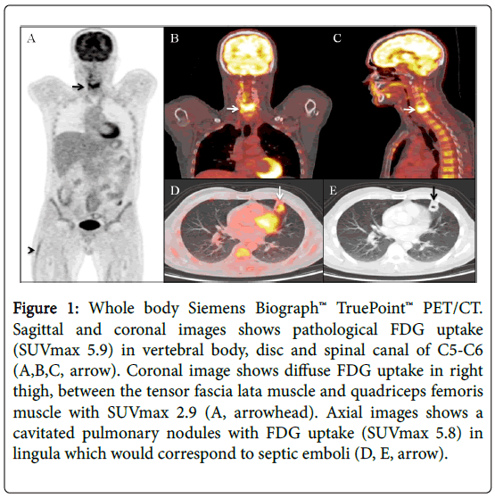
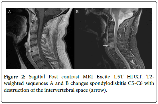
A whole body positron emission tomography/computed tomography (PET/CT) scan with 18F-Fluorodeoxyglucose (FDG) was performed looking for infective endocarditis (Figure 1). No pathologic FDG uptake was evident in the valve, probably because of the time-lapse since the initiation of antibiotic treatment. However, PET/CT showed intense FDG pathological uptake in the vertebral bodies, disc and spinal canal of C5-C6 (Figures 1A-1C), diffuse FDG uptake in the right thigh between the tensor fascia lata and quadriceps femoris muscles (Figure 1A), and lung parenchyma (Figures 1D and 1E), all suggesting embolic infectious strokes. Magnetic resonance imaging (MRI) was indicated (T1 sequence Figure 2A, T2 sequence Figure 2B), confirming cervical spondylodiscitis changes in C5-C6 vertebral bodies with destruction of the intervertebral space and increased soft tissue in the anterior epidural space and paravertebral space with laminar morphology. Because of the imaging findings, two new antibiotics were added, Meropenem (2g IV every 8 hours for 7 days) and Gentamicin (2 mg/kg loading dose, followed by 5 mg/kg IV every 24 hours for 7 days). After three months of antibiotic treatment, a PET/CT with complete resolution of the pathological uptakes confirmed the favourable evolution of the embolic infection. Additionally, the CT scan evidenced the fusion of C5-C6 vertebral bodies, being the most frequent complication of spondylodiscitis.
Figure 1: Whole body Siemens Biograph™ TruePoint™ PET/CT. Sagittal and coronal images shows pathological FDG uptake (SUVmax 5.9) in vertebral body, disc and spinal canal of C5-C6 (A,B,C, arrow). Coronal image shows diffuse FDG uptake in right thigh, between the tensor fascia lata muscle and quadriceps femoris muscle with SUVmax 2.9 (A, arrowhead). Axial images shows a cavitated pulmonary nodules with FDG uptake (SUVmax 5.8) in lingula which would correspond to septic emboli (D, E, arrow).
Spondylodiscitis, vertebral osteomyelitis or infectious spondylodiscitis is an uncommon disease (<4%) that usually affects male adults. The most frequent way of developing the disease is via hematogenous transmision caused by extraspinal infections. Postsurgical and postraumatic are other ways of transmission caused by direct bacterial inoculation. Finally, nearness is another way of transmission, when an infection extends to the nearby tissues [1]. Staphylococcus aureus is the most frequent cause of the disease, being involved in more than 50% of cases. Back pain is the most frequent symptom, affecting the lumbar region in 75% of cases. MRI is a powerful diagnostic tool that can be used to help evaluate spinal infection and help distinguish between an infection, the extension and local effects and other clinical conditions [2].
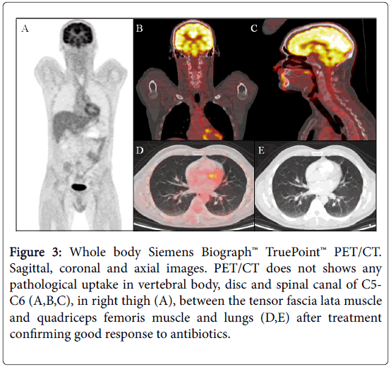
FDG PET/CT is an emerging tool for the diagnosis of infective endocarditis, as FDG accumulates in areas of increased glucose metabolism, such as inflammatory tissues. FDG PET/CT has demonstrated a high efficacy for the detections of complications such as spondylodiscitis and other embolic strokes located in lungs or spleen. Possible indications would be to confirm or discard the infection in a patient with bacteremia caused by gram positive microorganisms, to estimate its extension, and to detect associated complications. Therefore, PET/CT may have an impact on the therapeutic treatment, indicating the initiation of antibiotic therapy and allowing a precise monitoring of the disease [3] (Figure 3).
Figure 3: Whole body Siemens Biograph™ TruePoint™ PET/CT. Sagittal, coronal and axial images. PET/CT does not shows any pathological uptake in vertebral body, disc and spinal canal of C5-C6 (A,B,C), in right thigh (A), between the tensor fascia lata muscle and quadriceps femoris muscle and lungs (D,E) after treatment confirming good response to antibiotics.
References
- Pintado-García V (2008) Infectious spondylitis. Enferm Infec c Microbiol Clin 26: 510-517.
- Hong SH, Choi JY, Lee JW, Kim NR, Choi JA, et al. (2009) MR imaging assessment of the spine: infection or an imitation?. Radiographics 29: 599-612.
- Vos FJ, Bleeker-Rovers CP, Sturm PD, Krabbe PF, van Dijk AP, et al. (2010) 18F-FDG PET/CT for detection of metastatic infection in gram-positive bacteremia. J Nucl Med 51: 1234-1240.
Relevant Topics
- Antibiotics and Resistance
- Antifungal
- Antiviral therapy
- Bacteremia
- Bacterial diseases
- Broad Spectrum of Antibiotics
- Clinical Infectious Diseases
- Diagnosis of Pathogenic microorganisms
- Emerging infections
- Natural Antibiotics
- Opportunistic Pathogens
- Parasitic Diseases
- Pertussis Vaccines
- Prevention of infection
- Septicemia
- Viral Infections
- Viremia
Recommended Journals
Article Tools
Article Usage
- Total views: 10666
- [From(publication date):
August-2016 - Aug 29, 2025] - Breakdown by view type
- HTML page views : 9751
- PDF downloads : 915


