Research Article Open Access
Effect of Dynamic Knee Motion on Paralyzed Lower Limb Muscle Activityduring Orthotic Gait: A Test for the Effectiveness of the Motor-Assisted Knee Motion Device
| Hiromi Akahira1,2, Yuko Yamaguchi1, Kimitaka Nakazawa3, Yuji Ohta1and Noritaka Kawashima4* | |
| 1Faculty human life and environmental science, Ochanomizu University, Japan | |
| 2Products Research and Development, Research Institute, TOTO Ltd, Japan | |
| 3Graduate School of Arts and Sciences, University of Tokyo, Japan | |
| 4Department of Rehabilitation for the Movement functions, Research Institute, National Rehabilitation Center for Persons with Disabilities, Japan | |
| Corresponding Author : | Noritaka Kawashima Department of Rehabilitation for the Movement Functions Research Institute National Rehabilitation Center for Persons with Disabilities 4-1, Namiki, Tokorozawa, Saitama, Japan Tel: +81-4-2925-3100 Fax: +81-4-2995-3132 E-mail: kawashima-noritaka@rehab.go.jp |
| Received August 29, 2012; Accepted September 22, 2012; Published September 25, 2012 | |
| Citation: Akahira H, Yamaguchi Y, Nakazawa K, Ohta Y, Kawashima N (2012) Effect of Dynamic Knee Motion on Paralyzed Lower Limb Muscle Activity during Orthotic Gait: A Test for the Effectiveness of the Motor-Assisted Knee Motion Device. J Nov Physiother S1:004. doi: 10.4172/2165-7025.S1-004 | |
| Copyright: © 2012 Akahira H, et al. This is an open-access article distributed under the terms of the Creative Commons Attribution License, which permits unrestricted use, distribution, and reproduction in any medium, provided the original author and source are credited. | |
Visit for more related articles at Journal of Novel Physiotherapies
Abstract
Orthotic gait in paraplegic persons is a “stiff-leg” gait, which is a gait with the knee locked in full extension position. We developed a motor-assisted knee motion device with the use of a pair of linear electric actuator attached to the knee joint of a conventional reciprocal gait orthosis (Advanced Reciprocating Gait Orthosis: ARGO). The purpose of this study was to examine the effect of dynamic knee motion on lower limb muscle electromyographic (EMG) activity during orthotic gait. Six motor complete spinal cord injured persons participated, and the subjects were asked to walk on a treadmill with two types of orthoses; Knee-ARGO and Normal-ARGO. The results demonstrated that magnitude of EMG activity in the gastrocnemius and the rectus femoris muscles was significantly increased by an accomplished dynamic knee motion. These changes might be attributed to the occurrence of an additional afferent neural input with the knee motion. The present results suggest that the assisted knee motion by generating powered device have a potential to activate the neuromuscular function in the paralyzed lower limb.
| Keywords |
| Spinal cord injury; Orthotic gait; Knee dynamic motion; Powered orthosis; Paralyzed muscle activities |
| Abbreviations |
| SCI: Spinal Cord Injury; EMG: Electromyography; GRF: Ground Reaction Force; ARGO: Advanced Reciprocating Gait Orthosis |
| Introduction |
| Motor paralyses following spinal cord injury (SCI) consequently lead patients to use wheelchairs instead of bipedal walking for transportation. It is well recognized that a long-term isolation from the upright standing and walking cause physical deterioration, such as, the muscle atrophy and the reduction of the bone mineral density [1,2]. Moreover, it is probable that a reduced physical activity level may increase a risk of lifestyle disease [3]. To prevent such disuse syndrome, walking and standing training with gait orthosis are usually prescribed for SCI patients in their therapeutic phase. On the other hand, previous researches have pointed out that the gait trainings are frequently abandoned presumably due to a physiological burden of orthotic gait. It is no doubt that the energy cost for orthotic gait is far worse than normal walking because they need to produce complementary upper limb and trunk motion in order to swing their paralyzed lower limb [4-7]. A remarkable difference between orthotic gait and normal walking is whether there are dynamic knee motions or not. Because of motor paralysis of the lower leg, the knee joint should be locked in full extension position to maintain upright standing posture and to accomplish orthotic gait. Previous investigations have attempted to accomplish an effective orthotic gait performance, for example, use of functional electrical stimulation (FES) [8,9] and development of the knee-moved orthosis [10,11], these are not sufficient as a solution to the above problem. |
| We previously developed a novel motor-assisted knee motion devise for orthtic gait which accomplish dynamic knee motions during swing phase. In the previous study, we evaluated it from the viewpoint of the gait kinematics [12], and suggested that the knee dynamic motions were suitably accomplished with maintaining the ordinary gait speed. In the present study, we aimed to examine the effect of an additional dynamic knee motion on the muscle activities in the paralyzed lower limb during orthotic gait. It is well known that locomotor-like coordinated muscle activity can be induced in paralyzed lower limb muscles during passive stepping movement on the treadmill [13-16] and during orthotic gait [17-19]. We hypothesized that a dynamic knee motion accomplished by our newly-developed device would alter the muscular activity in the paralyzed lower limb muscles because the knee motion may result in an induction of afferent inputs to spinal motoneurons. The purpose of this study was therefore to compare the lower limb muscle activity and examine the effect of a knee motion on the paralyzed lower limb muscle activities. |
| Methods |
| Subjects |
| Six paraplegic persons (19-34 years) with traumatic SCI voluntarily participated in this study. All subjects had injured at thoracic level (Th5−Th12), and had complete motor paralysis in the lower limb muscles (ASIA classification; grade A or B). No subjects were currently taking medication for spasticity. The physical characteristics of the subjects are summarized in table 1. All subjects had participated in the basic rehabilitation process, and had undergone at least 10 weeks of orthotic gait training using the original (knee locked in full extension position) advanced reciprocating gait orthosis (ARGO, Steeper Inc., UK). At the time of the experiment, each subject could make independent walking at least for 20 min (Subject 1 and 2 still needed a slight support to avoid falling). All subjects gave their informed consent to the experimental procedures, which were conducted in accord with the Helsinki Declaration of 1975 and approved by the local biological ethics committee of the National Rehabilitation Center for Persons with Disabilities (Tokorozawa, Japan). |
| The powered orthosis |
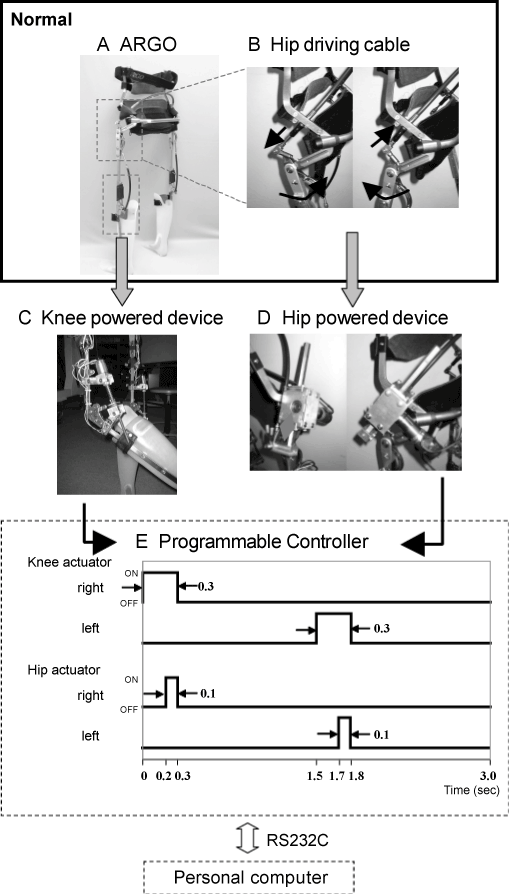
| The ARGO was originally designed for SCI persons to make gait more comfortably. It features the reciprocal cable which connects both sides of the leg frame. With this cable, a torque exerted by the right (or left) hip joint can be mechanically transmitted to the other and each leg is propelled forward reciprocally. Figure 1 shows schematic representations of the newly developed power-assisted device. To accomplish dynamic knee motion during swing phase, a pair of linear actuators consisting of a low-inertia DC motor (3557; Faulhaber, Switzerland) and a ball screw (lead of 2 mm; Kuroda Precision Industries Ltd., Japan) were mounted at the back of the knee joint of the ARGO. The stroke length of the linear actuator was set at 15 cm, which created a range of knee rotational angles from 0 degree (extension) to 70 degree (flexion). The original knee locking mechanism of the ARGO was retained to maintain the extended position during the stance phase. At the beginning of flexion (i.e. when shifting from the stance phase to the swing phase), the actuator unlocked the knee by lifting up the locking handle. At heel strike, the knee joint was fixed again by the lock at the fully extended position. To link the hip motion to the knee motion, another DC motor (3557; Faulhaber, Switzerland) and a rack-and-pinion gear (EP150; Asahi Seiko Co. Ltd., Japan) were mounted at the end of the hip driving cable (Figure 1B). Photographs of the powered orthotic gait device mounted with both actuators are shown in figure 1C and 1D. In total, three actuators were mounted on the orthosis, and they were sequentially controlled in accordance with the timing chart (Figure 1E), by using a sequencer (EPS; Tristate Ltd., Japan) (CTC Tool; Dyadic Systems Co. Ltd., Japan) that controlled the operation phase and period of the motors. In the following description, we use the terms “Knee-ARGO” for the newly developed orthosis (linkage of hip and knee motor) and “Normal-ARGO” for the conventional orthosis (only hip motor). |
| Experimental protocol |
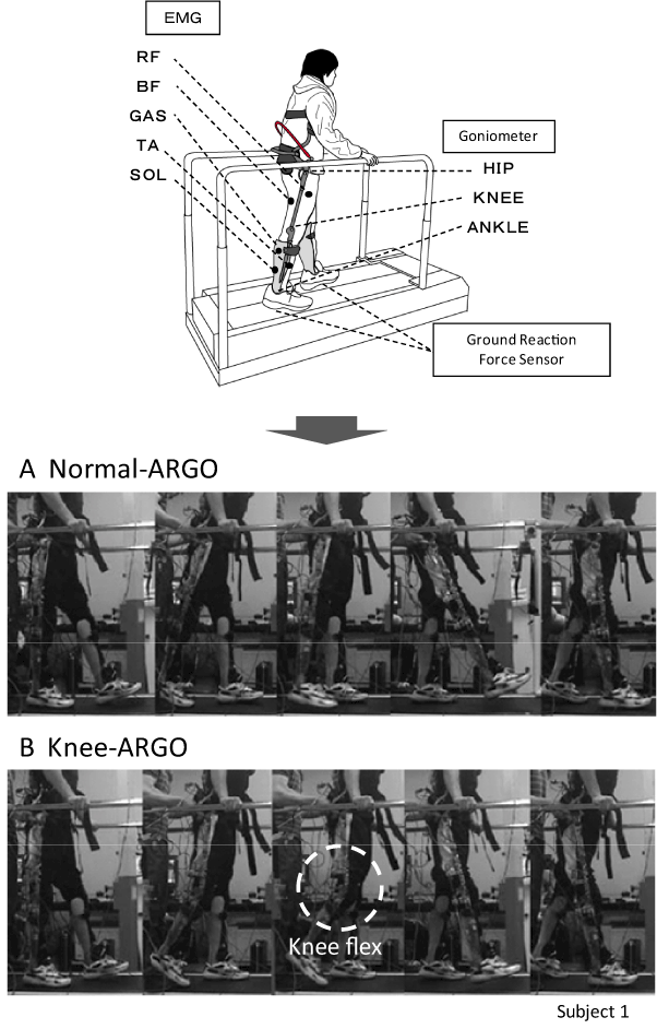
| In order to regulate the gait speed for both of Normal and Knee-ARGO, all experiments were conducted on the treadmill. The treadmill system (ADAL-3DC, Tecmachine) consists of two laterallymounted belts and force plates, which could yield the dataset of the ground reaction forces (GRF) produced by each leg. All the subjects walked on the treadmill without weight support, using a right and left bar of the treadmill only for the balance support. The belt speed of the treadmill was set at a comfortable gait speed for each subject. The walking velocity was the same for both orthosis. Based on the speed, the phase and duration of the knee flexion were set as shown in figure 1E. The experimental settings for this experiment are shown in figure 2. |
| Data recording and statistics |
| The electromyograms (EMG) were recorded from the soleus (SOL), medial head of the gastrocnemius (GAS), tibialis anterior (TA), rectus femoris (RF), and long head of the biceps femoris (BF) muscles of both legs by using bipolar electrodes. The skin was washed with a scrub gel and rubbed with sandpaper to reduce the resistance of the skin in order to exclude any artifact in the EMG signal. All data were digitized with a sampling frequency of 1 kHz. The amplifier (Bagnoli-8 EMG SYSTEM, DELSYS LTD, USA), modified the EMG signal with band pass filtering between 20 and 450 Hz. The kinematical motions of the knee, hip, and ankle joint were recorded by an electrogonioeter (Goniometer System, Biometrics Ltd., UK). The ground reaction force of each leg was measured by the force plate built in the treadmill. |
| These signals were obtained at least for one minute during stable walking. The data of the first one minute were discarded because the gait performance was not stable. The digitized EMG signal was full-wave rectified after the DC component was subtracted. It was then averaged ten locomotion cycles. The size of muscle activity was quantified using the integrated value and the mean amplitude of the EMG signals. For a comparison of the kinematic and kinetic variables and the EMG data between two orthosis, paired t-test was used. Significance was accepted at p<0.05. All values are shown as mean ± SD. |
| Results |
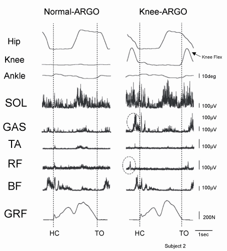
| Figure 2 shows a sequential photograph of walking with Normal and Knee-ARGO. As shown in this figure, Knee-ARGO could make dynamic knee motions during swing phase of walking. A similar result was obtained as subjects at all the lesion levels though this photograph was subject 1. Figure 3 shows a typical data of the kinematics (hip, knee, and ankle joint motion), the EMG activities (SOL, GAS, TA, RF, BF), and the GRF during both two types of orthotic gait obtained from a subject. Except for subject 6, all subjects showed the EMG activities, which was well synchronized with the gait cycle in the lower limb muscles during orthotic gait. |
| Kinetics and kinematics measurements |
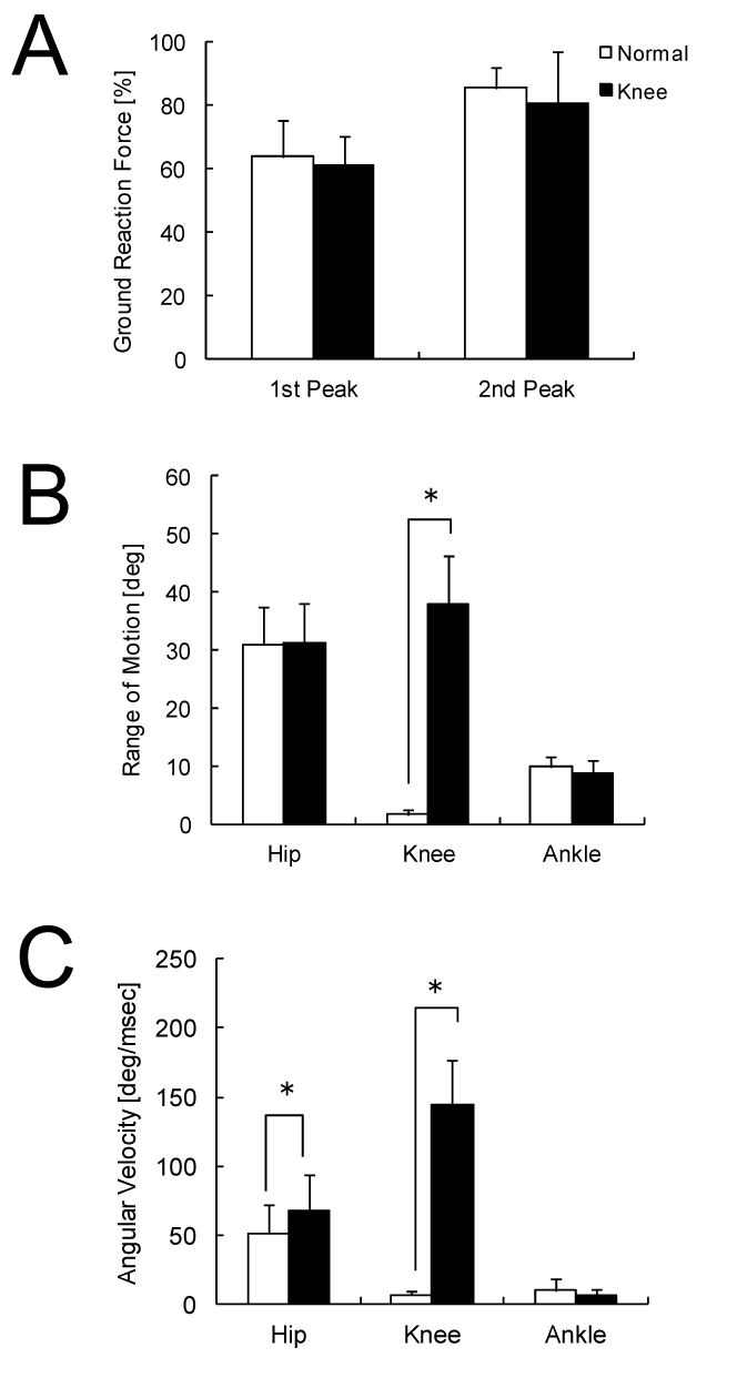
| As shown in the figure 3, the GRF shows two peaks during one gait cycle. Consequently, the GRF data were analyzed by averaging the torque value of the 1st and 2nd peak among six subjects. The 1st peak of Normal and Knee-ARGO was 40.2 ± 17.5 N and 38.9 ± 12.6 N, and the 2nd peak of them was 53.6 ± 9.5 N and 50.2 ± 9.6 N, respectively. As shown in the figure 4A, either 1st or 2nd peak showed no statistical difference between two orthoses. |
| Figure 4B and 4C show the comparison of the range of motion and angular velocity in each hip, knee and ankle joint between two types of orthoses. During gait with Knee-ARGO, knee range of motion and the angular velocity of the flexion phase were found to be 37.8 ± 8.5 deg and 144.4 ± 37.7 deg/sec, respectively. As shown in these figures, there were no significant differences between two orthoses in hip and ankle range of motions. On the other hand, angular velocity of hip joint during gait with Knee-ARGO was greater than that during gait with Normal ARGO (p<0.05), which could be attributed to the decrease in the inertia moment due to the knee flexion. |
| EMG activity of the paralyzed lower limb muscles |
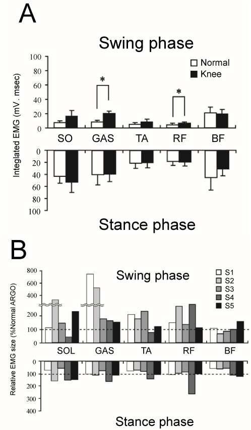
| Figure 5 shows the results of EMG activities recorded through the gait experiments for five subjects with the two orthosis. Figure 5A shows the EMG from each lower muscle during swing and stance phase. EMG data were analyzed by integrating. Figure 5B shows the breakdown of the above figure. Each data was normalized by the mean value of the normal ARGO and shown in percentage. |
| Regarding the EMG activity due to the knee rotation, there was a trend that muscles, such as RF and GAS, whose lengths change due to knee swing showed significant increase in EMG activities. The integrated EMG of the GAS and RF muscle in the swing phase during gait with Knee ARGO was significantly larger than that during gait with Normal ARGO (Normal ARGO vs. Knee ARGO: 8.4 ± 3.2 vs. 19.6 ± 5.6 in GAS and 3.8 ± 3.1 vs. 6.5 ± 1.3 in RF). That is, subject 1, 2, 3, and 4 showed the EMG activities of GAS during swing phase, and subject 2 and 4 showed those of RF, as shown in the figure 5B. |
| Discussion |
| In the present study, we examined the effect of knee motions on muscular activity in the paralyzed lower limb of the SCI persons during orthotic gait. The developed orthosis, Knee-ARGO, was able to accomplish dynamic knee motions during swing phase of gait cycle, without any obvious changes of the characteristics of GRF and hip and ankle joint motions. We found that the amplitude and phase of EMG activities of GAS and RF muscles were significantly changed (enhanced and prolonged) by induction of the knee motions. In the following section, possible neural mechanism and the implication for gait rehabilitation of SCI patients will be discussed. |
| Alteration of the EMG activity by dynamic knee motions |
| In the previous study, many researchers have been reported the induction of the muscle activity in the paralyzed muscle during stepping movement. Although the types of therapeutic approaches and devises are different among those studies, there is a consensus that the locomotor-like EMG activity obtained from SCI persons can be regarded as an resultant output of an interaction of the central pattern generator (CPG) in the spinal cord with the sensory input rather than a mere reflex response [13,14,20-22]. We previously examined the characteristics of the locomotor-like muscle activity during orthotic gait, and suggested that the magnitude of the muscle activities in the paralyzed lower limb muscles are largely affected by ground reaction force and joint kinematics pattern [17-19]. In case of orthotic gait, while patients should employee a strategy of “stiff-leg” gait, full body load can be supplied during stance phase. Such characteristics of the gait pattern would largely affect the magnitude of locomotor-like muscle activity. |
| During gait with Knee-ARGO, all subjects showed remarkable EMG activity of the GAS muscle in end of swing phase which is identical to knee extension phase. Similarly, the RF muscle showed remarkable EMG activity during middle to end of swing phase which is identical to the timing just after knee flexion. Because the acting phase of both muscles were corresponding to the phase in which muscles were stretched by knee flexion, it is possible that the induced muscle activities were mediated by stretch reflex pathway. On the other hand, the duration of the EMG activity in the GAS muscle was too long to regard as the simple stretch reflex. Moreover, the SOL muscle which does not have direct contribution to knee joint motion also showed alteration of the magnitude of EMG activity in some subjects. |
| Since acting phase of the EMG activity was almost identical to those reported in the previous study [15,18-20], similar neurological behinds can be presumed. As shown in the figure 3, GAS and RF muscle activities were remarkably enhanced by inducing dynamic knee motions. Since we confirmed that the kinetic and kinematics variables except for knee motions showed almost identical between Normal and Knee-ARGO, the EMG changes observed in this study might attribute to the occurrence of an additional afferent sensory input with knee motions. Although Dietz et al. [23] reported knee motion is not critical factor for the induction of the locomotor-like EMG activity in SCI persons, our results suggested that dynamic knee motion induce alteration of the muscle activity in the knee muscles. |
| Feasibility of the motor-assisted device for orthotic gait |
| In this study, we paid attention to whether the dynamic knee motion would alter muscle activity of the paralyzed lower limb muscles. This is because knee motion would result in sending the afferent inputs from the muscles and the joint to spinal cord. The present results suggested that the knee dynamic motion have a potential to facilitate the neuromuscular function in the paralyzed lower limb. This viewpoint will provide a new aspect for the development of a rehabilitation alternatives and good agreement with the concept of neurorehabilitation. |
| While we here paid attention to neuromuscular changes rather than the improvement of the gait motion, it is possible that the working cost and an extent of the foot clearance are also improved. Needless to say, the energy cost of orthotic gait is far worse than normal walking because the knees are fixed at extension position in the conventional gait orthoses. It is, therefore, quite important to find a way for reducing the excess physiological load during orthotic gait. In the previous study, many biomechanical trials have been conducted to establish dynamic knee motion during orthotic gait while decreasing the foot-floor clearance or walking cost [10,11]. The idea of these studies seemed to be from the biomechanics standpoint, for example, taking an adequate foot-floor clearance or reduction in the working cost for orthotic gait. To confirm these points, we need to do more precise motion analysis and an evaluation of the energy cost in future studies. Furthermore, there is still limitation for the setting value of the sequence controller we used in this study. For future study, we have to examine about optimal timing and duration for working each of hip and knee actuator. |
| Conclusion |
| In the present study, we evaluated an effectiveness of dynamic knee motions from the viewpoint of the alteration of neuromuscular activity in the paralyzed muscles. The results showed that the developed device could make knee dynamic motions with keeping ordinary gait character, and induce an alteration of the muscular activity of the lower limb muscle. The present results indicate that the accomplished dynamic knee motion could alter motor output in the below level of the injured cords which are completely isolated from brain. It might be possible that physiologically and biomechanically suitable knee dynamic motion realized by powered devise have a potential to facilitate the neuromuscular function in the paralyzed lower limb. This viewpoint will provide a new aspect for the development of a rehabilitation alternative. |
| Acknowledgement |
| A part of this study was supported by the health and labour sciences research grants of the Ministry of Health, Labour and Welfare (Japan), and Heiwa Nakajima Foundation. |
References
- Frey-Rindova P, de Bruin ED, St├?┬╝ssi E, Dambacher MA, Dietz V (2000) Bone mineral density in upper and lower extremities during 12 months after spinal cord injury measured by peripheral quantitative computed tomography. Spinal Cord 38: 26-32.
- Lotta S, Scelsi R, Alfonsi E, Saitta A, Nicolotti D, et al. (1991) Morphometric and neurophysiological analysis of skeletal muscle in paraplegic patients with traumatic cord lesion. Paraplegia 29: 247-252.
- Noreau L, Proulx P, Gagnon L, Drolet M, Laram├?┬ęe MT (2000) Secondary impairments after spinal cord injury: a population-based study. Am J Phys Med Rehabil 79: 526-535.
- Gordon EE, Vanderwalde H (1956) Energy requirements in paraplegic ambulation. Arch Phys Med Rehabil 37: 276-285.
- Chantraine A, Crielaard JM, Onkelinx A, Pirnay F (1984) Energy expenditure of ambulation in paraplegics: effects of long term use of bracing. Paraplegia 22: 173-181.
- Bernardi M, Canale I, Castellano V, Di Filippo L, Felici F, et al. (1995) The efficiency of walking of paraplegic patients using a reciprocating gait orthosis. Paraplegia 33: 409-415.
- Kawashima N, Taguchi D, Nakazawa K, Akai M (2006) Effect of lesion level on the orthotic gait performance in individuals with complete paraplegia. Spinal Cord 44: 487-494.
- Hirokawa S, Grimm M, Le T, Solomonow M, Baratta RV, et al. (1990) Energy consumption in paraplegic ambulation using the reciprocating gait orthosis and electric stimulation of the thigh muscles. Arch Phys Med Rehabil 71: 687-694.
- Solomonow M, Baratta RV, D'Ambrosia R (2000) Standing and walking after spinal cord injury: Experience with the reciprocating gait orthosis powered by electrical muscle stimulation. Topics in spinal cord injury rehabilitation 5: 29-53.
- Greene PJ, Granat MH (2003) A knee and ankle flexing hybrid orthosis for paraplegic ambulation. Med Eng Phys 25: 539-545.
- Greene PJ, Granat MH (2000) The effects of knee and ankle flexion on ground clearance in paraplegic gait. Clin Biomech (Bristol, Avon) 15: 536-540.
- Ohta Y, Yano H, Suzuki R, Yoshida M, Kawashima N, et al. (2007) A two-degree-of-freedom motor-powered gait orthosis for spinal cord injury patients. Proc Inst Mech Eng H 221: 629-639.
- Dietz V, Colombo G, Jensen L (1994) Locomotor activity in spinal man. Lancet 344: 1260-1263.
- Dietz V, Colombo G, Jensen L, Baumgartner L (1995) Locomotor capacity of spinal cord in paraplegic patients. Ann Neurol 37: 574-582.
- Dobkin BH, Harkema S, Requejo P, Edgerton VR (1995) Modulation of locomotor-like EMG activity in subjects with complete and incomplete spinal cord injury. J Neurol Rehabil 9: 183-190.
- Wernig A, M├?┬╝ller S, Nanassy A, Cagol E (1995) Laufband therapy based on 'rules of spinal locomotion' is effective in spinal cord injured persons. Eur J Neurosci 7: 823-829.
- Kojima N, Nakazawa K, Yano H (1999) Effects of limb loading on the lower-limb electromyographic activity during orthotic locomotion in a paraplegic patient. Neurosci Lett 274: 211-213.
- Nakazawa K, Kakihana W, Kawashima N, Akai M, Yano H (2004) Induction of locomotor-like EMG activity in paraplegic persons by orthotic gait training. Exp Brain Res 157: 117-123.
- Kawashima N, Nakazawa K, Akai M (2008) Characteristics of the locomotor-like muscle activity during orthotic gait in paraplegic persons. Neurol Res 30: 36-45.
- Kawashima N, Nozaki D, Abe MO, Akai M, Nakazawa K (2005) Alternate leg movement amplifies locomotor-like muscle activity in spinal cord injured persons. J Neurophysiol 93: 777-785.
- L├?┬╝nenburger L, Bolliger M, Czell D, M├?┬╝ller R, Dietz V (2006) Modulation of locomotor activity in complete spinal cord injury. Exp Brain Res 174: 638-646.
- Gorassini MA, Norton JA, Nevett-Duchcherer J, Roy FD, Yang JF (2009) Changes in locomotor muscle activity after treadmill training in subjects with incomplete spinal cord injury. J Neurophysiol 101: 969-979.
- Dietz V, M├?┬╝ller R, Colombo G (2002) Locomotor activity in spinal man: significance of afferent input from joint and load receptors. Brain 125: 2626-2634.
Tables and Figures at a glance
| Table 1 |
Figures at a glance
 |
 |
 |
 |
 |
| Figure 1 | Figure 2 | Figure 3 | Figure 4 | Figure 5 |
Relevant Topics
- Electrical stimulation
- High Intensity Exercise
- Muscle Movements
- Musculoskeletal Physical Therapy
- Musculoskeletal Physiotherapy
- Neurophysiotherapy
- Neuroplasticity
- Neuropsychiatric drugs
- Physical Activity
- Physical Fitness
- Physical Medicine
- Physical Therapy
- Precision Rehabilitation
- Scapular Mobilization
- Sleep Disorders
- Sports and Physical Activity
- Sports Physical Therapy
Recommended Journals
Article Tools
Article Usage
- Total views: 6133
- [From(publication date):
specialissue-2012 - Aug 30, 2025] - Breakdown by view type
- HTML page views : 1532
- PDF downloads : 4601
