Research Article Open Access
EMG Asymetricity of Selected Knee Extensor Muscles in Sustained Squat Posture (A Yogic Posture) of Athletes in Relation to their Gender and Performance
Manvinder Kaur1, Sanjeev Nara1, Dhananjoy Shaw2 and Dinesh Bhatia3*1Department of Biomedical Engineering, Deenbandhu Chhotu Ram University of Science & Technology, Murthal131039, Haryana, India
2Biomechanics Laboratory, Indira Gandhi Institute of Physical Education and Sports Sciences, University of Delhi, Delhi-110018, India
3Biomedical Engineering Department, North Eastern Hill University (NEHU), Shillong-793022, Meghalaya, India
- *Corresponding Author:
- Dinesh Bhatia
Associate Professor and Head
Biomedical Engineering Department
North Eastern Hill University (NEHU)
Shillong-793022, Meghalaya, India
Tel: +91 364 2723853
E-mail: bhatiadinesh@rediffmail.com
Received date: November 11, 2016; Accepted date: December 15, 2016; Published date: December 24, 2016
Citation: Kaur M, Nara S, Shaw D, Bhatia D (2016) EMG Asymetricity of Selected Knee Extensor Muscles in Sustained Squat Posture (A Yogic Posture) of Athletes in Relation to their Gender and Performance. J Nov Physiother 6:322. doi: 10.4172/2165-7025.1000322
Copyright: © 2016 Kaur M, et al. This is an open-access article distributed under the terms of the Creative Commons Attribution License, which permits unrestricted use, distribution, and reproduction in any medium, provided the original author and source are credited.
Visit for more related articles at Journal of Novel Physiotherapies
Abstract
Squat is a popular yogic exercise or posture, most frequently used for quadriceps strengthening and rehabilitation. It is used for evaluation process or test by physiotherapists/clinicians/others associated with rehabilitation of athletes to strengthen their lower-body muscles and connective tissues after joint-related injury. It has been used extensively for therapeutic treatment of ligament lesions, patellofemoral dysfunctions, patellar subluxation and patellar dislocation etc. The purpose of this study was to compare the effect of sustained squat posture (a yogic posture) of athletes among male (n1=32) and female (n2=18) with high and low performance (based upon median split method) on EMG activity of Vastus Medialis (VM) and Vastus Lateralis (VL) muscles for 120 seconds. Squatting was performed at an angle of 135º-150º because strength of knee extensor (muscles) varies between these angles and is an important consideration for the performance of numerous physical daily routine activities such as walking, running, cycling, stepping and driving including development of fundamental skills for different sports across all ages and sex. VM and VL from the Quadriceps muscle group were chosen for recording the physiological signal simultaneously from right and left leg of the subjects, as these muscles determines stress on the patellofemoral joint and stabilizes the patellar bone during squatting. Frequency and time domain variables were selected to extract meaningful information from the EMG signal using the algorithm developed in MATLAB. The selected variables were Median Frequency (MDF), Mean Frequency (MNF), Root Mean Square (RMS) and Integrated EMG (IEMG). Statistical analysis showed significant differences among gender and performance level along with significant bilateral asymmetry in these athletes. Understanding from the findings is pivotal for achieving optimal muscular strength, for the development of strength endurance and reducing the prospect of training-related injuries
Keywords
Electromyography; Fatigue; Squat; Gender; Performance
Introduction
Squat Posture (a yogic posture) is considered as one of the closed kinetic exercises that is used for evaluation process or test by physiotherapists/clinicians/others associated with rehabilitation of athletes to strengthen the lower-body. It is used most frequently in strength and conditioning exercises. A wide range of athletic movements have biomechanical and neuromuscular similarity to squat therefore it is included as a core exercise in many sports routines that are designed to enhance athletic performance [1,2]. Benefits related to squat activity is not limited to athletes only and most of the squatting movements have close specificity to many routine activities of daily living among individuals such as vehicle driving, lifting packages and picking up children etc. Moreover, in developing countries such as India squatting tasks are more prominent since people do most of the work for comfort and ease by sitting on the floor. Hence, it has become increasingly popular in clinical procedures, with physiotherapist trainings and rehabilitation procedures as a means to strengthen lower-body muscles and connective tissues after joint-related injury [3,4].It is extensively used for therapeutic treatment of ligament lesions, patellofemoral dysfunctions, total joint replacement, and ankle instability [4]. Therefore, it is crucial to understand and compare the muscular activities, which occur in the knee joint during performance of squat exercises or posture in order to determine positions of better muscular balance, ligament tension and articular compression.
The main purpose of this study is to compare the effect of sustained squat posture (a yogic posture) in quadriceps muscles in relation to gender and performance level among athletes. The EMG activity of Vastus Medialis (VM) and Vastus Lateralis (VL) muscles (quadriceps group) are considered for this study primarily due to the fact that they determine the amount of stress on the patellofemoral joint and support stabilization of the patellar bone during the squat posturing [5]. Moreover, these muscles carry out concentric knee extension, as well as eccentrically resisting knee flexion during squatting [5,6]. In this study, frequency and time domain variables were selected to extract desired information from the physiological (EMG) signal. We employed EMG because it helps to reflect the degree of muscular activation at a given moment of isometric contraction [7]. In addition to this, EMG is widely used in analysis studies of the human movement with the purpose to investigate muscular function through acquisition and analysis of electric signals produced by the muscles [7,8]. Understanding from the findings is pivotal for achieving optimal muscular strength and strength endurance development as well as reducing the prospect of training-related injuries to sportspersons.
Materials and Method
Fifty (50) healthy sportsperson engaged in different sports were recruited as volunteers. Inclusion criteria for the study was subjects above 18 years of age, non-alcoholic, willing to participate in the study and not suffering from any other medical illness which might hamper functioning of their nerves and muscles, so as to make them unfit to participate in the study. The study population consisted of 32 Males and 18 Females. Mean (± SD) age, height and weight calculated is shown in Table 1.
| Category | Age(Years) | Height(cm) | Weight (Kg) |
| Female | 19.3±1.8 | 156.6±6.9 | 52.7±11.7 |
| Male | 19.8±2.2 | 170.7± 8.6 | 70.4±13.8 |
Table 1: Characteristics of female and male subjects.
The exclusion criterion for the study was subjects having any medical conditions/ contractures/ deformities in the joint or suffering from skin condition which might impede the fixation of the electrodes on the body surface. Before participating in the study each participant was explained about the purpose and protocol to be followed for the study and they were asked to undergo few pre-data collection trails so as to make them familiar with study. As per ethical requirements, all subjects provided voluntary written informed consent before participating in the study. The methodology followed in the study along with the positioning of electrodes on quadriceps muscles is shown schematically in Figure 1.

Figure 1: Schematic illustration of methodology.
Data acquisition
EMG data were acquired using a 4-Channel Wireless EMG BIOPAC Inc. MP 150 system (CMRR: 110dB at 50/60 Hz and Gain: 5-50,000, Input Impedance: 2 MΩ). This data acquisition was carried out in a quiet laboratory room for all the subjects. The research team could closely monitor the activity of the subjects from adjacent room through a glass window and acquire data wirelessly. Before data acquisition, the skin of subjects was cleaned with cotton containing alcohol to minimize the skin impedance, thereby improving signal acquisition. Disposable electrodes (44 × 32 × 1 mm) were placed on the VM and VL muscles of the participants based on Seniam: European Recommendations for Surface Electromyography [9]. The EMG data was acquired simultaneously from both the legs. A reference electrode was placed on knee of the subject to act as ground and as per safety requirements of the equipment [10]. The inter-electrode distance was 20 mm, center to center.
The schematic of electrode placement is shown in Figure 1. After the subject preparation was complete, they were asked to perform squatting posture in order to become familiar with the protocol. Squatting posture was performed at an angle of 135º-150º because strength of knee extensor (muscles) varies between these angles and is an important consideration for the performance of numerous physical daily routine activities such as walking, running, cycling, stepping and driving including development of fundamental skills for different sports across all ages and sex [6]. The data acquisition software (Acqknowledge 4.1, BIOPAC systems Inc.) was set to sampling frequency of 2000 Hz to avoid the aliasing effects as per Nyquist criteria.
Data segmentation
The study group of male and female subjects was further sub-divided into low and high performance groups on the basis of the duration of time they were able to sustain the squat posture using median split method. These groups were named as Female low performance (FLP), female high performance (FHP), Male low performance (MLP) and Male high performance (MHP). All these groups were able to perform minimum of 120 seconds squat posture, therefore to better understand the influence of gender and performance on EMG, the data is further segmented into 30, 60, 90 and 120 seconds.
Data processing
The raw EMG signals acquired from the subjects were quantified with the help of MATLAB. The signal processing applied onto the raw EMG signals involves two parts-pre-processing and feature extraction as shown in Figure 2. For pre- processing of EMG signal, digital filters were employed using MATLAB. Notch filter was applied in order to remove 50 Hz noise interference from the signal. Cascaded low-pass (20 Hz) and a high-pass filter (450 Hz) were applied in order to remove other noise sources from the signal. Time and frequency domain variables were extracted from the filtered signal namely-Root Mean Square (RMS), Integrated EMG (IEMG), Median Frequency (MDF) and Mean Frequency (MNF) respectively for each subject. These variables were chosen because these are the most frequently opted variables for determining the changes due to fatigue in muscles.

Figure 2: Signal processing steps.
Root mean square(RMS)
RMS is the square root of the average power of EMG signal for a given period of time. It is most frequently used parameter for the EMG analysis in study of muscular fatigue. It reflects the mean power of the signal. For calculating RMS value, usually the square root of the mean of the squares of the values of EMG signal is computed, which gives a better picture than doing an integration EMG analysis [11]. It can be represented as shown in equation 1. It is considered as one of the parameter to be analyzed here because it reflects the level of physiological activity in the motor unit during sustained muscle contraction [10].
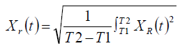 (1)
(1)
where, xr(t)= RMS signal and xR(t)= Rectified signal
Integrated EMG (IEMG)
IEMG is the area under curve of the rectified EMG signal. It is basically the mathematical integral of the absolute value of the raw EMG signal [11] and is represented by equation 2. Increase in IEMG value during muscle contraction is related to higher muscle fiber recruitment for a fixed external force [12]. Hence, its analysis needs to be understood to determine the development of fatigue in muscle fiber(s) during contraction.
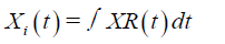 (2)
(2)
where, xi(t)= Integrated signal and xR(t)= Rectified signal
Median frequency (MDF)
MDF is a frequency at which the EMG power spectrum is divided into two regions with equal amplitude [10]. It is divided into two regions because it is half of the total power. It is calculated in two stages as explained below: First, the intensity of the signal in the whole spectrum is summed, and divided by two [2]. In the second stage, a frequency is selected at which cumulative intensity (i.e. all intensity values for frequencies lower than the selected frequency including the focal intensity) exceeds the value calculated in step 1. Muscle fatigue results in a downward shift of frequency spectrum of the EMG signal [13,14]. It is the most useful frequency-domain feature frequently used to describe fatigue behavior of the muscle and is represented by equation 3.
MDF M M
 (3)
(3)
j=1 j=MDF j=1
where, Pj= EMG power spectrum at the frequency bin j and M= length of frequency bin
Mean frequency (MNF)
MNF value is the product of frequency and amplitude of spectrum which is equal to the average of all such products throughout the spectrum as shown in equation 4. MNF of EMG activity is twice when the muscle is at rest as compared to EMG activity when the muscle is under fatigue [13]. It is analyzed here because it is one of the important frequency domain parameter for fatigue detection in muscles.
where, fj=frequency value of EMG power spectrum at the frequency bin j, Pj=EMG power spectrum at the frequency bin j and M= length of frequency bin
Statistical analysis
A One-Way ANOVA was carried out to determine if the influence of gender and performance is statistically significant during squat posture in VM and VL muscles in both the legs of participants. Least significant differences (LSD) among the different groups for the selected muscles of both the legs were conducted as post hoc tests. The level of significance was set to p<0.05. All the statistical analysis was carried out using SPSS Software (IBM).
The summary of findings related to the different frequency domain parameters- MDF and MNF and time domain parameters- RMS and IEMG are shown in Table 2. These findings suggest the possible gender and performance related differences in VMR, VLR, VML and VLL muscles during isometric contractions and are described as follows.
| Variable | Muscle | Comparison | 0-30 sec | 30-60 sec | 60-90 sec | 90-120 sec |
| MDF | VMR | 1,2 | ||||
| 2,4 | ||||||
| 3,4 | * | * | ||||
| VLR | 1,2 | |||||
| 2,4 | ||||||
| 3,4 | * | |||||
| VML | 1,2 | |||||
| 2,4 | ||||||
| 3,4 | ||||||
| VLL | 1,2 | |||||
| 2,4 | ||||||
| 3,4 | ||||||
| MNF | VMR | 1,2 | ||||
| 2,4 | ||||||
| 3,4 | * | * | * | |||
| VLR | 1,2 | |||||
| 2,4 | ||||||
| 3,4 | * | |||||
| VML | 1,2 | |||||
| 2,4 | ||||||
| 3,4 | * | |||||
| VLL | 1,2 | |||||
| 2,4 | ||||||
| 3,4 | * | |||||
| RMS | VMR | 1,2 | * | |||
| 2,4 | ||||||
| 3,4 | * | |||||
| VLR | 1,2 | |||||
| 2,4 | ||||||
| 3,4 | * | * | ||||
| VML | 1,2 | * | * | * | * | |
| 2,4 | ||||||
| 3,4 | ||||||
| VLL | 1,2 | |||||
| 2,4 | ||||||
| 3,4 | * | |||||
| IEMG | VMR | 1,2 | * | |||
| 2,4 | ||||||
| 3,4 | * | * | * | * | ||
| VLR | 1,2 | |||||
| 2,4 | ||||||
| 3,4 | * | * | * | |||
| VML | 1,2 | * | * | * | * | |
| 2,4 | ||||||
| 3,4 | ||||||
| VLL | 1,2 | |||||
| 2,4 | ||||||
| 3,4 | * | * | * |
Table 2: Summary of the findings for frequency and time domain variables.
1) MDF of VMR was significantly different between high and low performance male athletes at 60 seconds and 90 seconds.
2) MDF of VLR was significantly different between high and low performance male athletes at 60 seconds.
3) MNF of VMR was significantly different between high and low performance male athletes at 30, 60 and 90 seconds.
4) MNF of VLR was significantly different between male low performance and male high performance athletes at 60 seconds.
5) MNF of VML was significantly different between male low performance and male high performance athletes at 60 seconds.
6) MNF of VLL was significantly different between male low performance and male high performance athletes at 60 seconds.
7) RMS of VMR was significantly different among high and low performance female athletes at 60 seconds. It was also significantly different between high and low performance male athletes at 30 seconds.
8) RMS of VLR was significantly different among high and low performance male athletes at 30 and 120 seconds.
9) RMS of VML was significantly different between high and low performance female athletes at 30, 60, 90 and 120 seconds.
10) RMS of VLL was significantly different between male low performance and male high performance athletes at 60 seconds.
11) IEMG of VMR was significantly different between high and low performance female athletes at 60 seconds. It was also significantly different between high and low performance male athletes at 30, 60, 90 and 120 seconds.
12) IEMG of VLR was significantly different between high and low performance male athletes at 30, 90 and 120 seconds.
13) IEMG of VML was significantly different between high and low performance female athletes at 30, 60, 90 and 120 seconds.
14) IEMG of VLL was significantly different between high and low performance male athletes at 30, 90 and 120 seconds.
Results
Median and Mean Frequency values of VM and VL for both the legs are shown in Figure 3. It shows performance difference (high performance having higher value) for male and female athletes across the experimental protocol with regards to the activity of the VM and VL muscles for both the legs. For MDF and MNF, mean values of lower performance groups were found to be higher in terms of performance difference and male dominated female in terms of sex difference along the progression of time in both the muscles of subjects during squat posture, that may be attributed to males higher height and body weight thus higher body mass index and muscle mass.

Figure 3: Comparison of frequency domain parameters in relation to performance and sex difference.
RMS and IEMG for the VM and VL muscles for both the legs are shown in Figure 4. This figure demonstrates performance difference (high performance having higher value) for male and female athletes with regard to time domain parameters of both the muscles. The mean values of high performance groups were found to show higher time domain EMG activity. Female (having higher time domain EMG activity) dominated male in terms of sex difference along the progression of time in all the muscles of subjects during squatting except for the vastus lateralis left leg for which male dominated female along the progression of time. This result reflects that females show less fatigue development than that of male counterparts with regards to VL muscle of the left leg.

Figure 4: Comparison of time domain parameters in relation to performance and sex difference.
Discussion of Findings
The findings of this study collectively demonstrated gender and performance differences by showing significant changes in time and frequency domain parameters over period of time. Significant changes in both performance and gender differences have been found across the experimental protocol. EMG activity for frequency domain parameters (MDF and MNF) at 60 seconds dominated with significant gender differences during sustained squat posture. For MDF and MNF, with the progression of time, the number of significant differences with regards to gender differences decreases. Gender differences are predominating on the right side (leg) throughout the experimental protocol. EMG activity for time domain parameters (RMS and IEMG) at first 30 seconds dominated with significant gender differences during sustained squat posture. For RMS and IEMG, along with the progression of time the number of significant differences related to gender differences increases. Gender differences are predominating on right side (leg) which is found to be same as demonstrated by the frequency domain parameters. This dominance of right side in subjects suggests bilateral distribution of load related to fatigue and sex difference. It is clearly demonstrated that with progression of time, MDF and MNF values first increases and then decreases which can be attributed to the changes in the duration of the motor unit action potential, recruitment and deactivation of motor units, synchronous discharge of motor units and alteration in activity between synergistic muscles as per the established literature [14-17]. The time domain parameters (RMS and IEMG) demonstrated sharp decrease with time which could be related to the number of active motor units and shape of motor unit action potential. The possible gender related difference found in the study may be attributed to the differences in muscle fiber content. Fiber composition and size have been shown by researchers to influence both the frequency and amplitude content of EMG [14].
Understanding from these findings is pivotal for achieving optimal muscular strength and strength endurance development as well as reducing the prospect of a training-related injury.
Conclusions
The following are the conclusive remarks from the study:
1) The study found gender differences with regard to EMG activity of selected knee extensors (VM and VL) during squat posture (a yogic posture) in athletes.
2) Predominance of right side (leg) was found during the entire protocol.
3) Asymmetry in EMG activity of right and left knee, muscle to muscle as well as variables grouped/extracted by time domain and frequency domain analysis was observed.
4) Performance level of athletes demonstrated insignificant differences of EMG activity of the selected muscles.
Finally we are concluding that the present study is important for the understanding the significance of gender on selected variables during sustained squat posture. Such understanding is pivotal both for achieving optimal muscular strength and or strength endurance development as well as reducing the prospect of a training-related injury.
Acknowledgement
The authors acknowledge all volunteers and sportspersons from the Indira Gandhi Institute of Physical Education and Sports Sciences, University of Delhi for their voluntary participation in the study.
References
- Schoenfeld BJA (2010) Squatting kinematics and kinetics and their application to exercise performance. J Strength Cond Res 24: 3497-3506.
- Escamilla RF (2001) Knee biomechanics of the dynamic squat exercise. Med Sci Sports Exerc 33: 127-141.
- Escamilla RF, Fleisig GS, Zheng N, Lander JE, Barrentine SW, et. al. (2001) Effects of technique variations on knee biomechanics during the squat and leg. Med Sci Sports Exerc 33: 1552-1566.
- Senter C, Hame SL (2006) Biomechanical analysis of tibial torque and knee flexion angle: Implications for understanding knee injury. Sports Med 36: 635-641.
- Basmajian JVA, De Luca CJB (1985) Muscle Alive: Their Function Revealed by Electromyography. Willians & Wilkins, Baltimore, USA.
- Shaw DA (2000) Encyclopedia of Sports Injuries and Indian Sports Persons. KSK Publishers & Distributors, India.
- Hopkins JT, Ingersoll CD, Sandrey MA, BleggiSD (1999) An Electromyographic comparison of 4 closed chain exercises. J Athl Train 34: 353-357.
- Mathur S, Eng JJ, MacIntyre DL (2005) Reliability of surface EMG during sustained contractions of the quadriceps. J Electromyograp Kinesiol 15: 102-110.
- Seniam (1999) European recommendations for surface electromyography. Roessingh Research and Development, Enschede, Holland.
- Konard PA (2005) The ABC of EMG: A Practical Introduction to Kinesiological Electromyography. Noraxon Inc,USA.
- Kaur MA, Mathur SB, Bhatia DC, Verma SD (2015) siGnum: Graphical User Interface for EMG Signal Analysis. J Med Eng Technol 39: 19-25.
- Sharma CA, Duhan MB, Bhatia DC (2011) Study of Signal Processing Techniques for EMG Analysis. IJBBR 1: 141-148.
- Thongpanja S, Phinyomark A, Phukpattaranont P, LimsakuC (2013) Mean and Median Frequency of EMG Signal to Determine Muscle Force based on Time dependent Power Spectrum. Elektronikair Elektrotechnika 19: 51-56.
- Pincivero DM, Campy RM, Salfetnikov Y, Bright A, Coelho AJ (2001) Influence of contraction intensity, muscle, and gender on median frequency of the quadriceps femoris. J Appl Physiol 90: 804-810.
- Uzun S, Sayli O, Tatar Y, Cotuk B (2013) EMG evaluation of fatigue during isometric contractions in female rowers. J Med Biomed Sci 4: 7-12.
- Ipate MC (2011) Analysis of Electromyography Records During Voluntary Contraction and the Identification of Specific Characteristics of Muscular Activity. ATEE pp: 1-4.
- Bansal G, Bhatia D, Joshi D, Anand S, Tewari RP (2011) Coordination between Lower Limb Muscles in Different Locomotion Activities. IJBET 6: 129-141.
Relevant Topics
- Electrical stimulation
- High Intensity Exercise
- Muscle Movements
- Musculoskeletal Physical Therapy
- Musculoskeletal Physiotherapy
- Neurophysiotherapy
- Neuroplasticity
- Neuropsychiatric drugs
- Physical Activity
- Physical Fitness
- Physical Medicine
- Physical Therapy
- Precision Rehabilitation
- Scapular Mobilization
- Sleep Disorders
- Sports and Physical Activity
- Sports Physical Therapy
Recommended Journals
Article Tools
Article Usage
- Total views: 5474
- [From(publication date):
February-2017 - Aug 18, 2025] - Breakdown by view type
- HTML page views : 4570
- PDF downloads : 904