Case Report Open Access
Epidural Abscess Secondary to Paravertebral Pyomyositis in a Patient with a Sacrococcygeal Suppurated Cyst
| Dario Piazzalunga1, Federico Coccolini1, Nicola Colaianni1, Roberto Manfredi1, Alessandro Lunghi2, Alessandra Tebaldi3, Giulia Montori1,4*, Giulia Merigo1,4, Luca Baiocchi4, Nazario Portolani4 and Luca Ansaloni1 | ||
| 1General and Emergency Surgery Department, Ospedale Papa Giovanni XIII, Bergamo, Italy | ||
| 2Neuroradiology Department, Ospedale Papa Giovanni XIII, Bergamo, Italy | ||
| 3Infectious Diseases Department, Ospedale Papa Giovanni XIII, Bergamo, Italy | ||
| 4General Surgery III department, Spedale Civile, Brescia, Italy | ||
| Corresponding Author : | Giulia Montori General Surgery Department Ospedale Papa Giovanni XIII Piazza OMS 1 24100, Bergamo, Italy Tel: 039-3292244306 Email: giulia.montori@gmail.com |
|
| Received July 15, 2014; Accepted October 21, 2014; Published October 28, 2014 | ||
| Citation: Piazzalunga D, Coccolini F, Colaianni N, Manfredi R, Lunghi A, et al. (2014) Epidural Abscess Secondary to Paravertebral Pyomyositis in a Patient with a Sacrococcygeal Suppurated Cyst. J Infect Dis Ther 2:173. doi: 10.4172/2332-0877.1000173 | ||
| Copyright: © 2014 Piazzalunga D, et al. This is an open-access article distributed under the terms of the Creative Commons Attribution License, which permits unrestricted use, distribution, and reproduction in any medium, provided the original author and source are credited. | ||
Related article at Pubmed Pubmed  Scholar Google Scholar Google |
||
Visit for more related articles at Journal of Infectious Diseases & Therapy
Abstract
Pyomyositis is a sub-acute bacterial infection involving the skeletal muscle that can occur in a primitive form (rare, more common in tropical climates and so called "tropical myositis") or secondary to inflammatory phenomena of the skin, subcutaneous, or adjacent bone. It has been described the possibility that an epidural abscess could complicate a myositis of the paraspinal muscles. Moreover exists the description of paravertebral myositis complicating a suppurated sacrococcygeal pilonidal cyst. In this case, the patient presents a sacrococcygeal abscess evolving to a paravertebral myositis and further to an epidural abscess that has never been described before.
| Introduction |
| Pyomyositis is a sub-acute bacterial infection involving the skeletal muscle that can occur in a primitive form (rare, more common in tropical climates and so called "tropical myositis") or secondary to inflammatory phenomena of the skin, subcutaneous, or adjacent bone. |
| It has been described the possibility that an epidural abscess could complicate a myositis of the paraspinal muscles. Moreover exists the description of paravertebral myositis complicating a suppurated sacrococcygeal pilonidal cyst. In this case, the patient presents a sacro-coccygeal abscess evolving to a paravertebral myositis and further to an epidural abscess that has never been described before. |
| Case Report |
| A 33-year-old male, without medical history worthy of note. He complained gradually worsening back pain radiated to the back face of the lower right limb. He was previously admitted twice to an orthopedic unit and undergone to lumbosacral MR. He was diagnosed to be affected by right lumbosciatica and discharged with oral therapy with methylprednisolone and semi-rigid brace. He was evaluated by a neurosurgeon in an outpatient setting; oral indomethacin was added to the therapy. |
| One week after the first admission the patient referred to the Emergency Department of our hospital, where low-grade fever and increased acute-phase reactants (WBC 19.010/mmc, CRP 7.2) were detected with a redness and painful swelling in the sacro-coccygeal region with purulent secretion. The abscess was drained of pus and hairs. A sacro-coccygeal spine X-ray was negative for bone lesions. The patient was then admitted to the general surgery unit, suffering intensely by right lumbosciatica with Lasegue sign at 20-30° and unable to walk. |
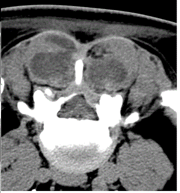
| Because of the important clinical neurological involvement, another lumbosacral MR has been performed. At the imaging we found an unrecognized inflammatory bilateral infiltration of the lumbosacral paravertebral muscles. A lumbosacral CT scan showed hypo-density and swelling of paravertebral muscles, with appearance of organized abscess. This lesion penetrated the vertebral canal at the lumbosacral passage and invaded the posterior epidural space causing compression of the dural sac (Figure 1). |
| A bacterial culture from the sacro-coccygeal secretion revealed Staphylococcus aureus and the needle aspiration of low lipoid material from paravertebral muscles exited in sterile culture. |
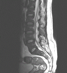
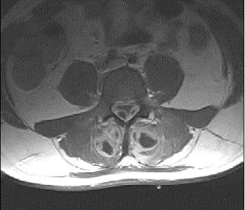
| A second MR pointed out a bilateral alteration of the paravertebral muscles from L2 to the sacrum. |
| This alteration was inhomogeneous on T1 and T2 scansions; contrast medium demarcated cystic necrotic areas. The MR also documented the presence of a posterolateral extradural collection with necrotic components compressing the dural sac and the roots of the cauda equina from L2 to L5-S1 disk (Figures 2 and 3). The absence of peripheral sensory and motor deficits (particularly the absence of bladder and rectal disturbance) induced to a conservative approach with antibiotic therapy for 15 days: first empirical broad-spectrum (Linezolid iv 1200 mg/day plus Meropenem iv 2 g/day), then targeted on microbiological findings (Oxacillin iv 12 g/day plus Levofloxacin iv 750 mg/day). The patient was discharge with others 10 days of antibiotic therapy (Augmentin 3 g/day and Levoxacin 500 mg/day). The intensity of the pain and the inflammation signs gradually decreased. The clinical and radiological follow-up demonstrated a progressive resolution of the disease. Lunbosacral MR was repeated at 15, 30 and 90 days. |
| The sacrococcygeal abscess was medicated until re-epithelialization and completes cleansing of the area. After 6 months we proceeded to radical surgery to clean the area with open treatment, with histological findings of fibrosis, hair glands, and rare macrophages. |
| Discussion |
| Pyomyositis is a rare sub-acute, deep bacterial infection involving the skeletal muscle usually accompanied by single or multiple abscesses. |
| Described for the first time by Scribe in 1885 [1], the first case in a temperate region has been described in 1971 by Levin [2]. Depending on their etiology could be identified a primitive forms (which are generally called "tropical myositis") and forms secondary to contiguous soft tissue, skin or bones infections. |
| Its rarity is considered to be due to resistance to infection of the intact skeletal muscle, even in the course of bacteremia. The reporting of the primitive forms in association with traumatic events or extreme muscle fatigue seems to support the hypothesis of the need for a muscle injury which overlay the hematogenous bacterial penetration under transient bacteremia. While extra-tropical forms are frequently observed in immunosuppressed patients, tropical myositis is typically a disease of young, immunocompetent patient [3]. The causative agent is Staphylococcus aureus in 90% of the tropical forms and in 75% of those extra-tropical, followed by group A streptococcus spp. [4]. |
| Cultures from pus are sterile in 15-30% of cases of tropical regions, with 90-95% of negative blood cultures [5,6]. A higher percentage of positive cultures (20-30%) is observed in the extra-tropical regions, perhaps due to better available microbiological assays. |
| In early stages of tropical pyomyositis muscles show oedematous separation of fibres, followed by patchly myocytolysis progressing to complete disintegration. The fibres are surrounded by lymphocytes and plasma cells. Muscle fibres may heal without abscess formation or degenerate, progressing to suppuration with bacteria and polymorphonuclear leucocytes. A single muscle group is usually involving, with multiple groups involvement in 12-40% of the cases. |
| Three developmental stages of the disease are described. The first one, invasive stage, characterized by sub-acute onset with swelling, pain, fever not always present, weak systemic signs, with or without erythema; the clinical picture is often interpreted as nonspecific pain or fibromyalgia. The second one, suppurative stage, between the second and third week, with progression to abscess formation in the context of the muscle. High fever with more severe systemic signs is present, and the diagnosis is usually made at this stage. The fluctuation may be missing, due to the tension of the surrounding muscle, also like the skin rash. The last one, late stage, with progression to the dissemination of the infection, septic shock, multi-organ failure and metastatic abscesses [3]. |
| Spinal epidural abscess is a rare but feared suppurative infection of the central nervous system, with an incidence of 0.2 to 2 cases/10000 hospitalized patients for year. Recently the association of paravertebral pyomyositis and epidural abscess has been described [7,8]. Epidural abscess secondary to pyomyositis is characterized by a dorsal involvement due to the proximity of the affected muscles. |
| The transmission would occur by contiguity through the intervertebral foramen from focal disruptions of the adjacent epimysium. |
| At the best of our knowledge only one report exists about paravertebral pyomyositis complicating a sacro-coccygeal cyst [9], and no cases of association between suppurated sacro-coccygeal cyst and epidural abscess have been described before. The case we report shows the possibility of the fearsome evolution of a suppurated sacrococcygeal pilonidal cysts, generally considered a minor disease and rarely worthy of hospitalization in acute. The reasons which led to an attitude of suspicion were the importance and worsening of symptoms on the lumbosciatica, the increased laboratory indices of inflammation despite the cyst was already burrowing at the time of access to the Emergency Department, the deep paravertebral swelling. Placing the suspicion of a nonrandom association between neurological symptoms and suppurative sacro-coccygeal inflammation, we could outline the diagnostic procedure with CT and MRI showing the spinal paravertebral pyomyositis and the epidural abscess. |
| Conclusion |
| A seemingly banal and routine situation, labeling as casual the atypical clinical elements, can hide dangerous pitfalls. An attitude of suspicion and an interdisciplinary approach allowed us to reach the correct definition of the framework. The uniqueness of the event and the initial monodisciplinary management of the symptoms parade resulted in a significant delay in diagnosis and therapy, which could have debilitating consequences. |
References
- Scriba J, Beitrangzur (1885) Aetiologie der myositis acuta, Deutsche ZeitChir 22:497-502
- Levin MJ, Gardner P, Waldvogel FA (1971) An unusual infection due to staphylococcus aureus.N Engl J Med 284: 196-198.
- Ansaloni L (1996) Tropical pyomyositis.World J Surg 20: 613-617.
- Chauhan S, Jain S, Varma S, Chauhan SS (2004) Tropical pyomyositis (myositis tropicans): current perspective.Postgrad Med J 80: 267-270.
- Sarubbi FA, Gafford GD, Bishop DR (1989) Gram-negative bacterial pyomyositis: unique case and review.Rev Infect Dis 11: 789-792.
- Gambhir IS, Singh DS, Gupta SS, Gupta PR, Kumar M (1992) Tropical pyomyositis in India: a clinico-histopathological study.J Trop Med Hyg 95: 42-46.
- Marshman LA, Bhatia CK, Krishna M, Friesem T (2008) Primary erector spinaepyomyositis causing an epidural abscess: case report and literature review.Spine J 8: 548-551.
- Bowen DK, Mitchell LA, Burnett MW, Rooks VJ, Martin JE (2010) Spinal epidural abscess due to tropical pyomyositis in immunocompetent adolescents.J NeurosurgPediatr 6: 33-37.
- Lorenz U, Abele-Horn M, Bussen D, Thiede A (2007) Severe pyomyositis caused by Panton-Valentine leucocidin-positive methicillin-sensitive Staphylococcus aureus complicating a pilonidal cyst.Langenbecks Arch Surg. 392: 761-765
Figures at a glance
 |
 |
 |
||
| Figure 1 | Figure 2 | Figure 3 |
Relevant Topics
- Advanced Therapies
- Chicken Pox
- Ciprofloxacin
- Colon Infection
- Conjunctivitis
- Herpes Virus
- HIV and AIDS Research
- Human Papilloma Virus
- Infection
- Infection in Blood
- Infections Prevention
- Infectious Diseases in Children
- Influenza
- Liver Diseases
- Respiratory Tract Infections
- T Cell Lymphomatic Virus
- Treatment for Infectious Diseases
- Viral Encephalitis
- Yeast Infection
Recommended Journals
Article Tools
Article Usage
- Total views: 14679
- [From(publication date):
December-2014 - Aug 16, 2025] - Breakdown by view type
- HTML page views : 10018
- PDF downloads : 4661
