Research Article Open Access
Fetal kidney Measurement in 26-39 Weeks Gestation in Normal Fetuses of Iranian Pregnant Women
| Firoozeh Ahmadi1*, Ahmad Vosough Taqi Dizaj1, FarnazAkhbari1, S Hohreh Irani1and G Holamreza Khalili2 | |
| 1Department of Reproductive Imaging at Reproductive Biomedicine Research Center, Royan Institute for Reproductive Biomedicine, ACECR, Tehran, Iran | |
| 2Department of Epidemiology and Reproductive Health at Reproductive Epidemiology Research Center, Royan Institute for Reproductive Biomedicine, ACECR, Tehran, Iran | |
| Corresponding Author : | Firoozeh Ahmadi Department of Reproductive Imaging at Reproductive Biomedicine Research Center Royan Institute for Reproductive Biomedicine ACECR, Tehran, Iran Tel: 00982123562207 Fax: 00982123562172 E-mail: dr.ahmadi1390@gmail.com |
| Received December 23, 2014; Accepted March 16, 2015; Published March 20, 2015 | |
| Citation: Ahmadi F, Taqi Dizaj AV, Akhbari F, Hohreh Irani S, Holamreza Khalili G (2015) Fetal kidney Measurement in 26-39 Weeks Gestation in Normal Fetuses of Iranian Pregnant Women. J Preg Child Health 2:139. doi: 10.4172/2376-127X.1000139 | |
| Copyright: ©2015 Ahmadi F, et al. This is an open-access article distributed under the terms of the Creative Commons Attribution License, which permits unrestricted use, distribution, and reproduction in any medium, provided the original author and source are credited. | |
Visit for more related articles at Journal of Pregnancy and Child Health
Abstract
Background: To establish a nomogram of fetal kidney dimension for a normal pregnancy.
Methods and Materials: A prospective cross-sectional study was carried out in the imaging department of the Royan Institute. Five hundred fifty seven fetuses in 26-39 weeks gestational age were scanned for the purpose of this study. Renal measurement was performed by ultrasonography. Two dependent variables (kidney length and width) were measured and compared with one independent variable (gestational age).
Results: Analysis was performed on obtained data of 557 normal fetuses. Size chart for fetal kidney dimension are presented for each weeks of gestation in male and female fetuses, separately. The difference in renal dimension between right and left sides was not statistically significant. Linear relationships were found between the gestational age and fetal kidneys diameters, including length and width. Mean kidney width was different between male and female fetuses, but there was no statistical significant difference in kidney length of male and female fetuses. We presented mean length and width of kidney, as well as standard deviation in both right and left kidney in fetuses for each weeks of gestational age (26-39w).
Conclusion: It seems that fetal kidney size in a sample of Iranian pregnant women is longer than previously reported.
| Keywords |
| Fetus; Kidney; Ultrasonography; Kidney dimension |
| Introduction |
| The kidneys develop from the uretric bud and metanephric mesoderm. It initiates to form at seven weeks of gestation and gets to appropriate function at week 11 of pregnancy [1,2]. Metanephric kidneys are close to each other in the pelvis. When abdomen and pelvis are developing, the gradual growth of kidneys occurs in this area while moving apart [2]. During the first trimester, the kidneys appear as hyperechoic oval structures at both sides of the spine (their hyperechogenicity can be compared to that of the liver or spleen). This echogenicity will progressively decrease. The fetal kidneys’ growth can be evaluated throughout pregnancy by measuring renal length and comparing it to normal charts. (As a simple rule, renal growth is 1.1 mm/ gestational week) [3]. During the second and third trimesters, the kidneys are easily identified by imaging the dorsolumbar spine and scanning on either side in parasagittal and transverse axial sections. |
| Regarding the fact that ultrasonographic standard normogram of fetal kidney dimensions is considered as a useful tool for evaluating growth disorders of the fetal kidneys, so fetal renal abnormalities can be identified and managed early. Several published article have showed relationship between renal size using ultrasound measurement and gestational age [4-7]. An accurate estimation of gestational age is fundamental in order to better management of pregnancy, especially high-risk pregnancy [8]. It is helpful in pregnancy to know the certain date of conception [9]. Mentioned reasons, raise necessity to establish a precise chart of normal fetal kidney dimensions in a sample of Iranian pregnant women. This plan provides an adequate management in order to prevent renal disease and to estimate gestational age. |
| The aim of this study was to determine the normal dimension of fetal kidney, including length and width, at different gestational ages to present charts and tables of these dimensions in a sample of Iranian pregnant woman and to compare the obtained data with the reports of other studies. |
| Materials and Methods |
| A group of 557 normal fetuses between 26-39 weeks was included in a prospective cross-sectional study during 2011 and 201 by randomized sampling. The study was reviewed and approved by the Ethics Committee of Royan Institute, whereas written informed consent was obtained from each participant prior to the commencement of the study. |
| Only normal pregnancies were included in the study, while women with maternal disease, which potentially affect fetal growth, such as diabetes, hypertensive disorders, vascolopathies and thrombotic disorders, were excluded from the study. |
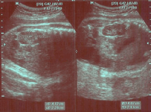
| During the course of the study, the registered women were also subjected to routine antenatal examination. Initially, the kidneys appear as hypoechogenic oval structures in the posterior mid-abdomen. As the kidneys mature, the pelvi-calyceal system becomes more apparent. The measurement of kidneys was done from outer to outer border. Kidney dimensions (length and width) were obtained during routine prenatal ultrasound examination. Ultrasonographies were taken using Medison and Aloka α10 machine. Measurements of fetal kidney length were performed by transabdominal ultrasonography in longitudinal plane and outer to outer margin (Figure 1). |
| Regarding the fact that Royan institute is an infertility center, majority of patients have the definite timing of embryo transfer or certain last menstrual period (LMP)with regular cyle. Also their gestational ages were determined by first trimester prenatal sonography through measurement of crown rump length (CRL), but at the second trimester head circumference (HC), biparietal diameter (BPD) and femur length (FL) are measured, if the gestational age which is determined by ultrasound is in agreement with LMP date, the woman was included in the study. |
| Renal length was measured from upper to lower pole. Width of the kidney was measured in a transverse section of the kidney. The transverse section is obtained at the height of the renal pelvis, if visible, otherwise at the position where the renal section is the largest as observed visually. Kidney length and width of fetus was measured in longitudinal and transverse plane. |
| Means and standard deviations of the renal length and width for consecutive gestational ages were analyzed using SPSS 16. For the comparison of the obtained values between sexes, the Student’s t test was used. Linear regression of square root of kidney length and width on gestational age was performed. |
| Results |
| Measurement of fetal kidney dimension (length and width) was obtained in 557 fetuses between 26 and 39 weeks gestation in normal pregnancy that were scanned in our institute. The mean age of pregnant women was 28.4 (± 4.6) years (ranging from 17- 44 years). |
| The mean renal length and width, as well as standard deviation of both kidneys from 26-39 gestational weeks were depicted weekly in Table 1 and fortnightly in Table 2. |
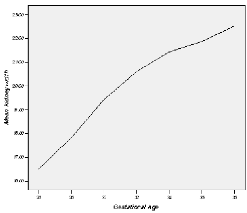
| The length and width of the right kidney compared with that of the left kidney by using the paired sample T test. There was no significant difference in right and left kidney length (PV=0.843) but right kidney width (mean: 20.56 ± 3.01) was longer than left kidney one (mean: 20.32 ± 2.72) (PV=0.004).Mean right kidney length was 37.60 ± 4.84 mm and mean left kidney length was 37.59 ± 4.89. By linear regression analysis in stepwise model, mean fetal kidney length and width increase weekly 1.07 mm and 0.5 mm respectively (Table 3). |
| There was a significant positive correlation between the gestational age and mother’s age and kidney (right and left) diameters, including length and width (Spearman r for RKL, RKW, LKL and LKW were 0.75, 0.63, 0.75 and 0.59 respectively and p<0.001for all) besides, there was relationship between LKW and fetus sex too (PV=0.005). |
| We found that the kidneys width of male fetuses is wider than the one of female fetuses (Table 4). |
| Discussion |
| Fetal kidneys reach their adult form at 12th week of gestation. By abdominal ultrasound in transverse section, they appear as hypo echogenic oval retroperitoneal structure with no distinctive borders at the second trimester [8,10-12]. Depending on fetal position and model of ultrasound machine, 90% of fetal kidneys are identified by week 20 [13-17]. |
| At first trimester, CRL (crown-rump length)is the best parameter for determining gestational age. In the second and third trimesters, estimation of gestational age is accomplished by measuring the biparietal diameter, head circumference, abdominal circumference, and femur length. In order to establish a correlation between the fetal kidney length and gestational age in 3rd trimester and detection of pathologies associated with abnormal kidney size, accurate information of gestational age is needed. |
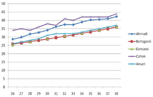
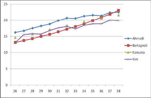
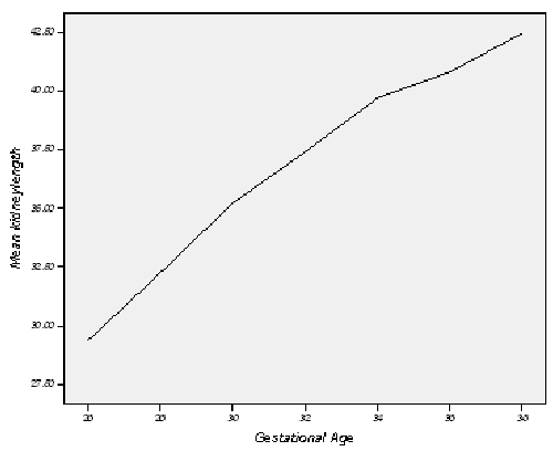
| Simple ultrasonographic measurement of renal length may be considered as a helpful tool in prenatal diagnosis of renal abnormalities [1], thus provides a new method to identify possible genetic disorders in fetus. In our study, the fetal kidney measurement began from weeks 26 to 39.Measuring the fetal kidney size is helpful in determining gestational age, especially in pregnant women who are uncertain in their LMP date [6]. Table 1 and 2 as well as chart C and D represent fetal kidney length and width, weekly and fortnightly, respectively. Also, Table 3 presents measurements of length of fetal kidney from weeks 26 to 38 done by other studies. Our gathered data confirm that the kidney length and width of fetal kidneys are related to gestational age [4-6,15]. Comparisons with other observers shown in chart A, chart B and Table 3. Except the results presented by Cohen et al., our findings revealed that length and of fetal kidney were larger than those measured by other studies [6]. Improvements in sonographic technology may be the key factor resulting in our larger measurement as compared with the earlier studies [6,7,14,16,18]. Mean renal length and width increased 1.07 and 0.50 mm per week, respectively by using regression analysis. Further study is needed for clarifying the reason of larger kidney length among Iranian fetus. |
| Conclusion |
| Kidney dimensions (length and width) are correlates well with gestational age. So, it can be concluded that kidney dimensions may be helpful in estimation of gestational age when the date of conception is uncertain [9]. In addition, it may be considered as a helpful tool in prenatal diagnosis of renal abnormalities [1]. To the best of our knowledge, this is the first study to document this relationship in Iran. |
| Financial disclosure and funding support: There are no financial interests that might inappropriately influence or interfere with research findings and there is no relevant financial or nonfinancial relationships in the products or services described, reviewed, evaluated or compared in this presentation. |
| Acknowledgements |
| The authors wish to thank imaging department staff for their assistance in figure preparation. |
| Author’s contribution |
| All the authors have participated sufficiently in the work to take public responsibility for the content. |
References
- Zalel Y,Lotan D, Achiron R, Mashiach S, Gamzu R (2002) The early development of the fetal kidney-an in utero sonographic evaluation between 13 and 22 weeks' gestation. PrenatDiagn 22: 962-965.
- Moore KL, Persaud TVN (1998) the developing human, clinically oriented embryology, 6th edn. W.B. Saunders Company, Philadelphia, pp 107, 288–293
- Filly P.A.,Feldstein V.A.. Ultrasound Evaluation of normal fetal anatomy. In: Callen PW, editor. Ultrasonography in obstetrics and gynecology. 5th ed. Philadelphia: Saunders; 2007. p. 342-3.
- Jeanty P, Dramaix-Wilmet M, Elkhazen N, Hubinont C, van Regemorter N (1982) Measurements of fetal kidney growth on ultrasound. Radiology 144: 159-162.
- Chitty LS, Altman DG (2003) Charts of fetal size: kidney and renal pelvis measurements. PrenatDiagn 23: 891-897.
- Cohen HL, Cooper J, Eisenberg P, Mandel FS, Gross BR, et al. (1991) Normal length of fetal kidneys: sonographic study in 397 obstetric patients. AJR Am J Roentgenol 157: 545-548.
- Bertagnoli L, Lalatta F, Gallicchio R, Fantuzzi M, Rusca M, et al. (1983) Quantitative characterization of the growth of the fetal kidney. J Clin Ultrasound 11: 349-356.
- Yusuf N, Moslem F, Haque J A. Fetal Kidney Length: Can be a New Parameter for Determination of Gestational Age in 3rd Trimester. TAJ: Journal of Teachers Association. 2007;20(2)
- Kim K,Park J H.Measurement of fetal kidney size and growth using ultrasonography.korean journal of nephrology:1995;14(4)
- Rosati P,Guariglia L (1996) Transvaginalsonographic assessment of the fetal urinary tract in early pregnancy. Ultrasound ObstetGynecol 7: 95-100.
- Grannum P, Bracken M, Silverman R, Hobbins JC (1980) Assessment of fetal kidney size in normal gestation by comparison of ratio of kidney circumference to abdominal circumference. Am J ObstetGynecol 136: 249-254.
- Sampaio FJ,Aragão AH (1990) Study of the fetal kidney length growth during the second and third trimesters of gestation. EurUrol 17: 62-65.
- 13-Mahony BS.Ultrasound evaluation of the fetal genitourinary system.InCallenPW,editor.Ultrasonography in Obstetrics and Gynecology .3rd ed.philadelphia:saunders,1994:389-419
- 14-JJ Kansaria, SV Parulekar. Nomogram for Foetal Kidney Length. Bombay Hospital Journal, Vol. 5, No. , 2009
- Scott JE, Wright B, Wilson G, Pearson IA, Matthews JN, et al. (1995) Measuring the fetal kidney with ultrasonography. Br J Urol 76: 769-774.
- Ansari SM,Saha M, Paul AK, Mia SR, Sohel A, et al. (1997) Ultrasonographic study of 793 foetuses: measurement of normal foetal kidney lengths in Bangladesh. AustralasRadiol 41: 3-5.
- Gloor JM,Breckle RJ, Gehrking WC, Rosenquist RG, Mulholland TA, et al. (1997) Fetal renal growth evaluated by prenatal ultrasound examination. Mayo ClinProc 72: 124-129.
- Sagi J, Vagman I, David MP, Van Dongen LG, Goudie E, et al. (1987) Fetal kidney size related to gestational age. GynecolObstet Invest 23: 1-4.
Tables and Figures at a glance
| Table 1 | Table 2 | Table 3 | Table 4 |
Figures at a glance
 |
 |
 |
 |
 |
| Figure 1 | Chart a | Chart b | Chart c | Chart d |
Relevant Topics
Recommended Journals
Article Tools
Article Usage
- Total views: 74195
- [From(publication date):
April-2015 - Aug 17, 2025] - Breakdown by view type
- HTML page views : 68124
- PDF downloads : 6071
