Hamartoma: Rare Benign Breast Tumor
Received: 20-Mar-2021 / Accepted Date: 28-Mar-2021 / Published Date: 02-Apr-2021 DOI: 10.4172/2167-7964.1000322
Abstract
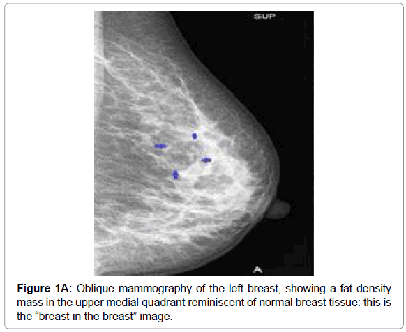
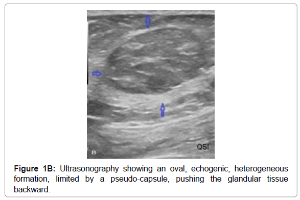
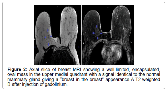
Breast hamartoma is a rare benign tumor. The diagnosis is most often made by chance during the exploration of a breast mass. The characteristic image is that of a “breast in the breast” on MRI as well as on ultrasound and mammography. We reported the case of a patient who was discovered by chance during the examination of a breast mass.
Keywords: Hamartoma, Breast, Mammography, Ultrasonography, MRI
Text
Hamartoma is a pseudotumor lesion of the breast. Benign and rare, it can appear at any age, but occurs preferentially in women over the age of 35 [1].
It is usually asymptomatic and is most often discovered incidentally. Histologically, the hamartoma contains the main components of the normal mammary gland, namely adipose, glandular and fibrous tissue. Diagnosis is mainly based on echo mammography. It may be discovered incidentally on MRI.
On mammography, the appearance of the hamartoma depends on the proportion of these constituents. It is most often a round or oval, well-limited mass of variable size, forming the image of a “breast within a breast” Figure 1A. On ultrasound, a heterogeneous, compressible echogenic lesion is found, isolated from the breast tissue by a pseudo-capsule, with no attenuation cone or posterior enhancement of the echoes Figure 1B [1,2].
They have an MRI appearance that fits well with their schematic definition of a normal breast island in the breast: a well-limited area whose contents have the appearance of the normal breast matrix (with its islands of fat), including physiological enhancement due to hormonal impregnation Figure 2 [3].
The hamartoma is often respected, except in cases of malignant transformation, when its removal is indicated.
References
- Oueslati S (2007) Harmatoma of the breast. Imaging of the woma 17: 19-25
- Boyer B, Graef C (2007) Harmatoma of the breast: a rare benign tumour of mammographic diagnosis. Press Med 36Â :1999-2000.
- Lamarque JL, Prat X, Laurent JC, Taourel P, Pujol J, et al. (2000) Magnetic résonance Imaging of the breast. Encycl Méd Chir Radiodiagnostic -Urology-Gynécology 283:810.
Citation: Thierry YTR, Omar ElA, Abdelilah MD, Hounayda J, Rachida L, Youssef O (2021) Hamartoma: Rare Benign Breast Tumor.OMICS J Radiol 10: 322. DOI: 10.4172/2167-7964.1000322
Copyright: © 2021 Thierry YTR, et al. This is an open-access article distributed under the terms of the Creative Commons Attribution License, which permits unrestricted use, distribution, and reproduction in any medium, provided the original author and source are credited.
Select your language of interest to view the total content in your interested language
Share This Article
Open Access Journals
Article Tools
Article Usage
- Total views: 3399
- [From(publication date): 0-2021 - Nov 21, 2025]
- Breakdown by view type
- HTML page views: 2479
- PDF downloads: 920


