Case Report Open Access
Hemangiopericytoma of the Greater Omentum with Pelvic Metastasis: A Very Rare Occurrence
Shemssie Shewmollo Bushira*Senior Surgical Resident at Saint Paul’s Teaching Referral Hospital, Addis Ababa, Ethiopia
- *Corresponding Author:
- Bushira SS
Senior Surgical Resident at Saint Paul’s Teaching Referral Hospital
Addis Ababa, Ethiopia
E-mail: shemssie@yahoo.com
Received date: January 17, 2017; Accepted date: February 6, 2017; Published date: February 13, 2017
Citation: Bushira SS (2017) Hemangiopericytoma of the Greater Omentum with Pelvic Metastasis: A Very Rare Occurrence. J Gastrointest Dig Syst 7:486. doi: 10.4172/2161-069X.1000486
Copyright: © 2017 Bushira SS. This is an open-access article distributed under the terms of the Creative Commons Attribution License, which permits unrestricted use, distribution, and reproduction in any medium, provided the original author and source are credited.
Visit for more related articles at Journal of Gastrointestinal & Digestive System
Abstract
Hemangiopericytoma arising from the greater omentum is very rare and only 14 case reports were found in the English literature. Here we report a case of hemangiopericytoma arising from the greater omentum with pelvic metastasis. The case was a 45 year old male patient admitted to our hospital with abdominal pain and swelling. Abdominal ultrasound and computer tomography detected a huge heterogeneously enhancing predominantly solid central abdominal mass with cystic changes. Laparotomy and excision of huge freely mobile highly vascularized mass arising from the greater omentum and multiple deposits on the anterior wall of the rectum was performed. Histological findings confirmed a diagnosis of hemangiopericytoma of the greater omentum with secondary deposits in the pelvis
Keywords
Hemangiopericytoma; Abdominal pain; Cystic changes; Laparotomy; Chemotherapy
Introduction
Hemangiopericytoma is a vascular tumor arising from pericytes that surround capillaries and can be benign or malignant. Hemangiopericytoma represents ≤ 1% of all vascular tumors. The tumor cells are surrounded by reticulin and are negative for muscles, nerve sheaths and epithelial markers and positive for CD34 [1]. Hemangiopericytoma arising from the greater omentum is a very rare tumor originating from Zimmerman’s pericyte. Stout and Murray described the first reported case of hemangiopericytoma arising from omentum [2]. Pericytes are cells that surround capillaries and have contractile behavior which enables them to regulate the flow of blood through capillaries. Even though hemangiopericytoma can arise anywhere, the predominant sites of origin are the retroperitoneum, the pelvic cavity and the lower extremity [3]. Hemangiopericytoma originating from the greater omentum is very rare limited to case reports as only 14 cases have been reported yet in the English literature. Here we report a case of hemangiopericytoma arising from the greater omentum and metastasizing to the pelvis.
Case Report
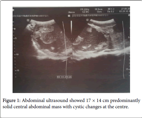
A 45 year-old Ethiopian male patient was admitted to our hospital with a complaint of abdominal pain and swelling of 1-year duration. The pain was dull aching and periumbilical whereas the swelling was initially epigastric and later progressed to involve the whole abdomen and associated with early satiety and constipation. On physical examination the abdomen was full with a palpable mass measuring 17 cm by 14 cm which is mobile, firm, irregular, non-pulsatile and palpable lower border. Abdominal ultrasound of the abdomen showed 26 cm × 21 cm × 10 cm central abdominal mass; predominantly solid with central cystic changes (Figure 1) and computer tomography of the abdomen demonstrated 16 × 13 cm heterogeneously enhancing omental mass with no ascites, liver metastasis or lymphadenopathy.
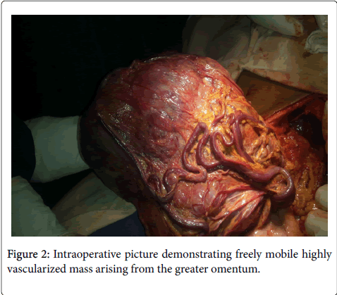
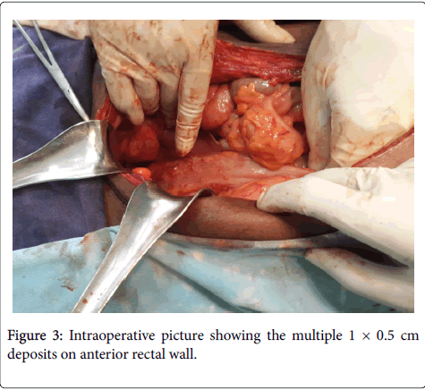
With a preoperative diagnosis of intra-abdominal mass arising from the greater omentum, laparotomy was performed and 18 cm × 15 cm × 9 cm freely mobile highly vascularized mass arising from the greater omentum and multiple 1 cm × 0.5 cm deposits on the anterior wall of the rectum was found (Figure 2 and 3). There was also a 3 cm × 2 cm lymph node on the greater omentum. No evidence of liver metastasis or ascites. The tumor was excised and the lymph node and the deposits in pelvis were biopsied. On cut section, the resected tumor was predominantly solid mass with central areas of cystic degeneration, with largest diameter of 16 cm, measured 16 cm × 13 cm × 9 cm, weighed 2.1 kg and was encapsulated with central necrosis and areas of hemorrge. On histologic examinations of both the mass and pelvic deposits, hematoxylin-eosin staining showed spindle cells growing around vascular endothelial cells. On high power field, there was high mitotic activity.
Immunohistochemical studies demonstrated that the tumor was positive CD34, factor-IIIa, and HLA-DR. These findings confirmed a diagnosis of high grade hemangiopericytoma with metastasis. The patient was transfused with 0.2 units of whole blood postoperatively and discharged improved on the 8th postoperative day. Adjuvant chemotherapy was offered and he is on his 4th month on follow-up with no evidence of recurrence.
Discussions
Hemangiopericytoma is a vascular tumor arising from pericytes that surround capillaries and can be benign or malignant (MS). Hemangiopericytoma represents ≤ 1% of all vascular tumors. The tumor cells are surrounded by reticulin and are negative for muscles, nerve sheaths and epithelial markers and positive for CD34. Malignant Hemangiopericytoma arising from the greater omentum is extremely rare and only 14 cases were reported in the English literature. Review of the reported cases showed that metastasis occurred to the liver, bones and regional lymph nodes and to our knowledge this is the first report on malignant hemangiopericytoma of the greater omentum with metastasis to the pelvis without involving the liver which makes our case unique.
Recent reports suggested that malignant hemangiopericytoma is suspected if tumor size is more than 5 cm, a high mitotic index with more than four mitoses per ten high power fields, and necrosis and hemorrhage within the tumor [4]. According to the reported cases, tumor size and mitotic index were associated with tumor recurrence after resection [5]. Our case presents after 1 year of symptoms which allows the tumor to attain such a huge size of 18 cm × 15 cm × 9 cm. The origin of the tumor was the greater omentum with such a huge size and the pelvic nodules on anterior wall of the rectum were tumor deposits.
Although effective chemotherapeutic and molecular targeting therapy is not established to date, systemic adjuvant chemotherapy is additional treatment for malignant hemangiopericytoma with peritoneal metastasis [6,7]. Even though surgical resection provides the only chance for cure for patients with low grade malignancy, additional adjuvant chemotherapy offers benefit for patients with advanced disease like ours.
References
- Chatterjee D, Sarka P, W. Gopimohan Singh (2008) Hemangiopericytoma of the greater omentum presenting as a huge abdominal lump. Saudi J Gastroenterol 14: 88-89.
- Stout AP, Murray MR (1942) Hemangiopericytoma: a vascular tumor featuring Zimmerman’s Pericytes. Ann surg 116: 26-33.
- Enzinger FM, Smith BH (1976) Hemangiopericytoma: an analysis of 106 cases. Hum Pathol 7: 61-84.
- Kempson RL, Fletcher CDM, Evans HL, Hendrickson MR, Sibley RK, et al. (1998) Atlas of tumor pathology: tumors of the soft tissue, 3rd series. Washington DC: Armed forces instate of pathology: 371-377.
- Goldberger RE, Schein CJ (1968)Hemangiopericytoma of the omentum: report of a case with unique presentation and review of the literature. Ann Surg34: 291-295.
- Imachi M, Tsukamoto N, Tsukimori K, Funakoshi K, Nakano H, et al.(1990) Malignant hemangiopericytoma of the omentum presenting as an ovarian tumor. GynecolOncol39: 208-213.
- Cajano P, Heys SD, Eremin O (1995) Hemangiopericytoma of the greater omentum. Eur J SurgOncol21: 323-324.
Relevant Topics
- Constipation
- Digestive Enzymes
- Endoscopy
- Epigastric Pain
- Gall Bladder
- Gastric Cancer
- Gastrointestinal Bleeding
- Gastrointestinal Hormones
- Gastrointestinal Infections
- Gastrointestinal Inflammation
- Gastrointestinal Pathology
- Gastrointestinal Pharmacology
- Gastrointestinal Radiology
- Gastrointestinal Surgery
- Gastrointestinal Tuberculosis
- GIST Sarcoma
- Intestinal Blockage
- Pancreas
- Salivary Glands
- Stomach Bloating
- Stomach Cramps
- Stomach Disorders
- Stomach Ulcer
Recommended Journals
Article Tools
Article Usage
- Total views: 4709
- [From(publication date):
February-2017 - Aug 18, 2025] - Breakdown by view type
- HTML page views : 3808
- PDF downloads : 901


