Hereditary Multiple Exostoses: Imaging is the Key
Received: 21-Jan-2022 / Manuscript No. roa-22-55041 / Editor assigned: 23-Feb-2022 / PreQC No. roa-22-55041(PQ) / Reviewed: 09-Mar-2022 / QC No. roa22-55041 / Revised: 14-Mar-2022 / Manuscript No. roa-22-55041(R) / Published Date: 21-Mar-2022 DOI: 10.4172/2167-7964.1000366
Image Article
Hereditary multiple exostoses HME, also known as diaphyseal aclasis, is an autosomal dominant disorder characterized by the development of several osteochondromas or exostoses, as well as accompanying bone remodeling abnormalities. Osteochondromas are cartilage-capped bony outgrowths that often occur around the metaphysis of long bones and can vary in size, shape (whether sessile or pedunculated), and number [1].
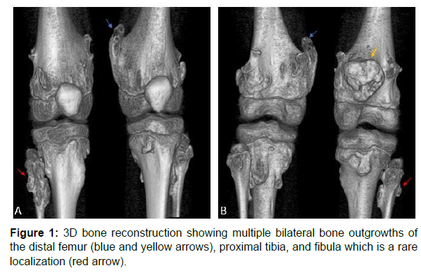
An osteochondroma can occur in any bone that originates from preexisting cartilage (enchondral ossification). The long bones of the lower extremity are the most typically affected (50% of cases) and are involved more frequently than those of the upper extremity [2] (Figures 1 and 2).
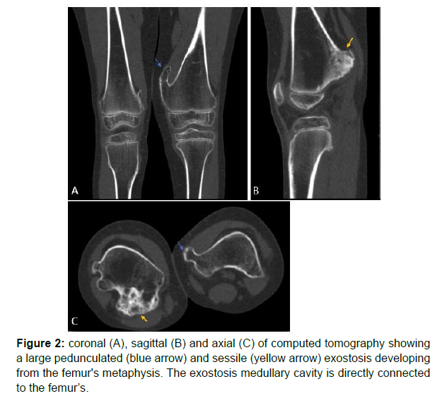
Exostosis is primarily diagnosed through radiography. Histology is not required in the standard form. It has a variety of radiological forms, but they can be divided into three categories: pedicle forms (narrow implantation and always inclined towards the diaphysis) Figure 2- blue arrow, sessile forms (broad implantation on the metaphysis and diaphysis) Figure 2- yellow arrow, and cauliflower forms [3].
The CT demonstrates cortical and marrow continuity of the lesion and parent bone, which is pathognomonic in osteochondromas Figure 2, but it is frequently absent on routine radiographs [3].
On another hand, the US enables an accurate measurement of the hyaline cartilage cap thickness [2].
In fact, a cap thickness of more than 3 cm and/or the reemergence of a latent lesion have been identified as warning indicators of malignant degeneration, which is uncommon in solitary osteochondromas, but it is more frequent in patients with hereditary multiple osteochondromatosis [2].
Finally, despite its inaccessibility, MRI is actually recognized as the best radiological tool for assessing the association of exostosis with nearby anatomical features and evaluating the cartilage [1].
References
- Kok HK, Fitzgerald L, Campbell N, Lyburn ID, Munk PL, et al. (2013) Multimodality imaging features of hereditary multiple exostoses. Br J Radiol 86: 20130398.
- Murphey MD, Choi JJ, Kransdorf MJ, Flemming DJ, Gannon FH (2000) Imaging of Osteochondroma: variants and complications with radiologic-pathologic correlation. RadioGraphics 20: 1407-1434.
- Niasse M, Kane BS, Condé K, Touré S, Sarr L, et al. (2019) Multiple Exostosis Disease. J Rare Dis Res Treat 4: 28-33.
Indexed at, Google Scholar, Crossref
Citation: Jihad B, Sahli H, Lahlou C, Idrissi N, Haddad SE, et al. (2022) Hereditary Multiple Exostoses: Imaging is the Key. OMICS J Radiol 11: 365. DOI: 10.4172/2167-7964.1000366
Copyright: © 2022 Jihad B, et al. This is an open-access article distributed under the terms of the Creative Commons Attribution License, which permits unrestricted use, distribution, and reproduction in any medium, provided the original author and source are credited.
Select your language of interest to view the total content in your interested language
Share This Article
Open Access Journals
Article Tools
Article Usage
- Total views: 9183
- [From(publication date): 0-2022 - Feb 13, 2026]
- Breakdown by view type
- HTML page views: 8311
- PDF downloads: 872


