Case Report Open Access
Idiopathic Unilateral Adrenal Hemorrhage in a 91-Year Old Woman
Rolf D Klingenberg-Noftz1*, Benthin S1, Eggers S2, Hinze R3and Gunther Weitz4
1Department for Gastroenterology and Internal Medicine, DRK-Hospital Grevesmühlen, Klützer Strasse 13-15, 23936 Grevesmühlen, Germany
2Department for Visceral and General Surgery, DRK-Hospital Grevesmühlen, Klützer Strasse 13-15, 23936 Grevesmühlen, Germany
3Department of Pathology, HELIOS Clinic Schwerin, Wismarsche Strasse 393-397, 19049 Schwerin, Germany
4Medical Department I, Gastroenterology, University Hospital of Schleswig-Holstein, Campus Lübeck, Ratzeburger Allee 160, 23538 Lübeck, Germany
- *Corresponding Author:
- Rolf D. Klingenberg-Noftz, MD
Department for Gastroenterology and Internal Medicine
DRK-Hospital Grevesmühlen
Klützer Strasse 13-15,
23936 Grevesmühlen, Germany
Tel: +49 3881 726 601
Fax: +49 3881 726 609
E-mail: rolf.klingenberg-noftz@drk-kh-gvm.de
Received date: June 6, 2015; Accepted date: June 17, 2015; Published date: June 24, 2015
Citation: Klingenberg-Noftz RD, Benthin S, Eggers S, Hinze R, Weitz G (2015) Idiopathic Unilateral Adrenal Hemorrhage in a 91-Year Old Woman. J Gastrointest Dig Syst 5:300. doi:10.4172/2161-069X.1000300
Copyright: ©2015 Klingenberg-Noftz RD, et al. This is an open-access article distributed under the terms of the Creative Commons Attribution License which permits unrestricted use distribution and reproduction in any medium provided the original author and source are credited
Visit for more related articles at Journal of Gastrointestinal & Digestive System
Abstract
A 91-year old woman was referred to our hospital for evaluation of a palpable mass in the left hemiabdomen. A marked anemia was found on admission. Imaging revealed an inhomogeneous mass of the left adrenal gland with a diameter of 19 cm. Due to a lack of perfusion in contrast enhanced ultrasound and the clear delineation to neighbouring organs on CT scan, an adrenal hemorrhage was diagnosed. A further fall of hemoglobin level prompted a resection of the mass. The mass consisted of a fibrous wall surrounding hematomas of different age. A small rest of the otherwise normal adrenal gland was found adjacent to the wall. The patient fully recovered. The patient had no underlying cause for the hemorrhage such as trauma, tumor or any coagulopathy including the intake of anticoagulants. Hence, we describe a rare case of spontaneous unilateral adrenal hemorrhage in an elderly woman that was successfully treated by surgery.
Keywords
Idiopathic; Hemorrhage
Introduction
Adrenal hemorrhage is a rare disease predominantly including bilateral involvement [1]. It typically occurs in underlying conditions such as sepsis [2,3], heparin-induced thrombocytopenia [4], antiphospholipid antibody syndrome [5], trauma [6], or adrenal tumors [7]. Moreover, it has been described in neonates [8], pregnant women [9], and patients after surgery [10,11]. We here present a 91-year old woman with a massive left-lateral idiopathic adrenal hemorrhage.
Presentation of the Case
A 91-year old woman was referred to our hospital for further evaluation of a palpable mass in the left hemiabdomen discovered by her general practitioner after complaining of light abdominal pain in this region. The patient suffered from dementia and was immobilized. Her medication included an ACE-inhibitor and haloperidol but no anticoagulants. No trauma or other conspicuous event was reported by her nursing home. A normal blood pressure had been documented during the weeks prior to admission. Physical examination revealed a pale skin and a bulging, rigid, and fixed, but indolent tumor in the left hemiabdomen.
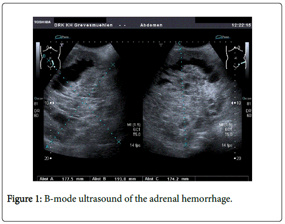
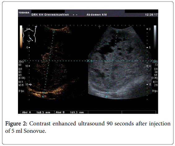
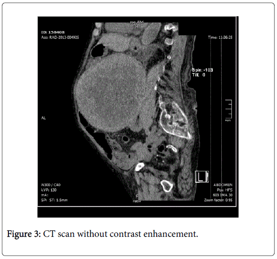
The physical examination was unremarkable otherwise. Abnormal laboratory findings were hemoglobin (8.6 g/dL) and creatinine (2.0 mg/dL), platelet count and coagulation tests were normal. Transabdominal ultrasound revealed an inhomogeneous, sharply delineated mass of 17 x 19 cm in diameter in the left hemiabdomen (Figure 1). After contrast injection (5 mL Sonovue®), the lesion did not enhance throughout the entire study (Figure 2). Due to the elevated creatinine, the CT scan was performed without contrast agent (Figure 3). A drop of hemoglobin to 6.8 g/dl on the third day after admission prompted an operation despite the patient's frailty.
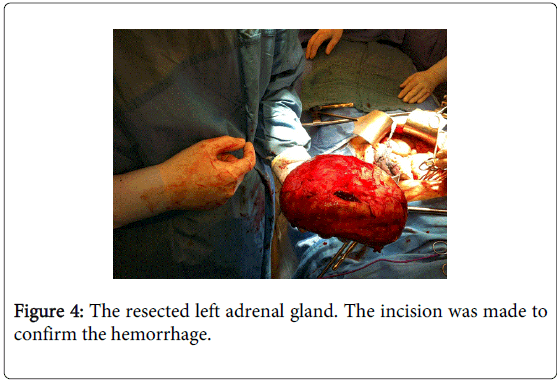
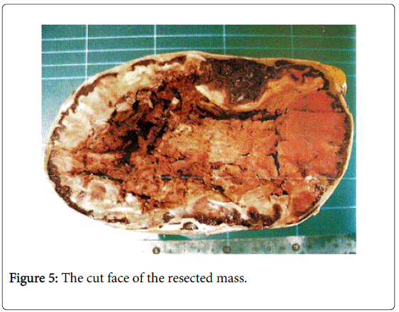
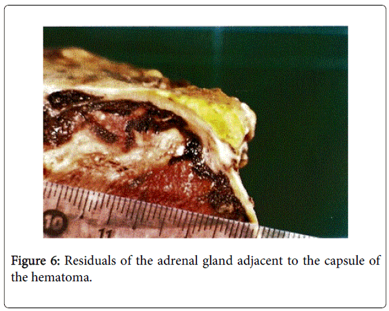
The mass was successfully removed without complications (Figure 4) and the patient was discharged one week later after full recovery to her prior status. The removed mass (17 cm in diameter) consisted of a fibrous capsule and hematomas of different ages (Figure 5). A residual of the adrenal gland was found adjacent to the capsule (Figure 6).
Discussion
Adrenal hemorrhage is a rare finding, particularly when unilateral, without underlying disease or trauma, and with normal coagulation. The precise mechanisms leading to adrenal hemorrhage are uncertain. In nontraumatic cases evidence has implicated a limited venous drain (e.g. by vein spasm or thrombosis) in contrast to the rich arterial supply of the gland [12]. As indicated by the huge capsule and the hematomas of different age within, the hemorrhage in our case was recurrent. Hemorrhage was obviously not controlled under observation leading to a significant drop in hemoglobin. Hence, in face of the patient's clinical status, we deem the decision for operation justified. Mortality of adrenal hemorrhage is estimated 15% [12]. Death can result from rupture of the capsule [13,14] or from the underlying condition. Adrenal insufficiency does not occur in unilateral hemorrhage but can be fatal in bilateral cases [15].
In our patient, primary diagnosis was made by ultrasound including a contrast enhanced study. Distinguishing an adrenal hemorrhage from a neoplasm by imaging can be difficult [16]. Contrast enhanced ultrasound (CEUS) is a valuable tool to visualize real time vascularisation and perfusion even in small adrenal masses [17]. Despite the inhomogeneous iso-hypoechogenic aspect in the baseline ultrasound, CEUS did not show any enhancement of the mass at any time. This pattern has earlier been described for adrenal hemorrhage and (in this size) strongly argues against a solid lesion [18]. To further delineate the anatomical attribution we performed a CT scan without contrast agent. The neighbouring organs were not involved. However, this test did not help to further distinguish the masses origin.
There is some disagreement over the term "hemorrhagic adrenal pseudocyst" in literature. While some authors claim this entity to be a (pseudo-)cystic degeneration of a primary adrenal neoplasm or a vascular neoplasm/malformation others hypothesize a primary hemorrhage into the adrenal gland with secondary wall formation [19,20]. The mass in our patient was surrounded by an intact fibrous wall. This may also be described as "adrenal pseudocyst". Adjacent to the wall was a small rest of the otherwise unremarkable gland. Since adrenal hemorrhage can result from a neoplasm such as pheochromocytoma [7] plasma free metanephrines should be tested before operation. Due to the urgency we failed to do so. An alternative procedure to surgery in ongoing hemorrhage can also be the embolisation of the bleeding adrenal artery [21]. Nevertheless, our patient fully recovered indicating that resection of a hemorrhagic adrenal gland can be successful in very old patients.
References
- Vella A, Nippoldt TB, Morris JC 3rd (2001) Adrenal hemorrhage: a 25-year experience at the Mayo Clinic.Mayo ClinProc 76: 161-168.
- Adem PV, Montgomery CP, Husain AN, Koogler TK, Arangelovich V, et al. (2005) Staphylococcus aureus sepsis and the Waterhouse-Friderichsen syndrome in children.N Engl J Med 353: 1245-1251.
- Tormos LM, Schandl CA (2013) The significance of adrenal hemorrhage: undiagnosed Waterhouse-Friderichsen syndrome, a case series.J Forensic Sci 58: 1071-1074.
- Ketha S, Smithedajkul P, Vella A, Pruthi R, Wysokinski W, et al. (2013) Adrenal haemorrhage due to heparin-induced thrombocytopenia.ThrombHaemost 109: 669-675.
- Ramon I, Mathian A, Bachelot A, Hervier B, Haroche J, et al. (2013) Primary adrenal insufficiency due to bilateral adrenal hemorrhage-adrenal infarction in the antiphospholipid syndrome: long-term outcome of 16 patients.J ClinEndocrinolMetab 98: 3179-3189.
- Untereiner O, Charpentier C, Grignon B, Welfringer P, Garric J, et al. (2013) Adrenal trauma: medical and surgical emergency.Emerg Med J 30: 329-330.
- Marti JL, Millet J, Sosa JA, Roman SA, Carling T, et al. (2012) Spontaneous adrenal hemorrhage with associated masses: etiology and management in 6 cases and a review of 133 reported cases.World J Surg 36: 75-82.
- Gyurkovits Z, Maróti A, Rénes L, Németh G, Pál A, et al. (2014) Adrenal haemorrhage in term neonates: a retrospective study from the period 2001-2013.J Matern Fetal Neonatal Med.
- Gavrilova-Jordan L, Edmister WB, Farrell MA, Watson WJ (2005) Spontaneous adrenal hemorrhage during pregnancy: a review of the literature and a case report of successful conservative management. ObstetGynecolSurv 60: 191-195.
- Chronopoulos E, Nikolaou VS, Masgala A, Kaspiris A, Babis GC (2014) Unilateral adrenal hemorrhage after total knee arthroplasty.Orthopedics 37: e508-511.
- Leong M, Pendyala M, Chaganti J, Al-Soufi S (2015) A case of bilateral adrenal haemorrhage following traumatic brain injury.J Intensive Care 3: 4.
- Tritos NA (2014) Adrenal hemorrhage. Medscape.
- Mahmodlou R, Valizadeh N (2011) Spontaneous rupture and hemorrhage of adrenal pseudocyst presenting with acute abdomen and shock.Iran J Med Sci 36: 311-314.
- Souiki T, Tekni Z, Laachach H, Bennani A, Zrihni Y, et al. (2014) Catastrophic hemorrhage of adrenal pheochromocytoma following thrombolysis for acute myocardial infarction: case report and literature review. World J EmergSurg 9: 50.
- McGowan-Smyth S (2014) Bilateral adrenal haemorrhage leading to adrenal crisis.BMJ Case Rep 2014.
- Vincent C, Brewster JB, Kedar V, Sundaram CP (2008) Unilateral idiopathic adrenal hematomas with a preoperative diagnosis of indeterminate adrenal tumors.J Endourol 22: 995-997.
- Dietrich CF, Ignee A, Barreiros AP, Schreiber-Dietrich D, Sienz M, et al. (2010) Contrast-enhanced ultrasound for imaging of adrenal masses.Ultraschall Med 31: 163-168.
- Cantisani V, Petramala L, Ricci P, Porfiri A, Marinelli C, et al. (2013) A giant hemorragic adrenal pseudocyst: contrast-enhanced examination (CEUS) and computed tomography (CT) features.Eur Rev Med PharmacolSci 17: 2546-2550.
- Groben PA, Roberson JB Jr, Anger SR, Askin FB, Price WG, et al. (1986) Immunohistochemical evidence for the vascular origin of primary adrenal pseudocysts.Arch Pathol Lab Med 110: 121-123.
- Laforga JB, Bordallo A, Ara FI (2000) Vascular adrenal pseudocyst: cytologic and immunohistochemical study.DiagnCytopathol 22: 110-112.
- Acar D, Gulpembe M, Akili NB, Calik AS, Koylu R, et al. (2015) Acute adrenal hemorrhage in an elderly patient. J AcadEmerg Med 6: 10-12.
Relevant Topics
- Constipation
- Digestive Enzymes
- Endoscopy
- Epigastric Pain
- Gall Bladder
- Gastric Cancer
- Gastrointestinal Bleeding
- Gastrointestinal Hormones
- Gastrointestinal Infections
- Gastrointestinal Inflammation
- Gastrointestinal Pathology
- Gastrointestinal Pharmacology
- Gastrointestinal Radiology
- Gastrointestinal Surgery
- Gastrointestinal Tuberculosis
- GIST Sarcoma
- Intestinal Blockage
- Pancreas
- Salivary Glands
- Stomach Bloating
- Stomach Cramps
- Stomach Disorders
- Stomach Ulcer
Recommended Journals
Article Tools
Article Usage
- Total views: 14761
- [From(publication date):
June-2015 - Aug 02, 2025] - Breakdown by view type
- HTML page views : 10215
- PDF downloads : 4546

