ImageJ for Counting of Labeled Bacteria from Smartphone-Microscope Images
Received: 09-Sep-2021 / Accepted Date: 23-Sep-2021 / Published Date: 30-Sep-2021 DOI: 10.4172/2329-9053.1000217
Abstract
Objective: The manual counting of gram stained bacteria examined under a microscope becomes difficult when a large number of bacterial cells exist in a microscopic field. The present study was aimed to ease this problem by applying ImageJ software to counting of gram stained bacteria.
Method: This experiment was conducted on Elmergib university, faculty of pharmacy laboratories (Al-Khoms city- Libya). In this study, a microscopic image of a gram stained bacterial cells captured using a student’s smartphone, treated and the bacterial cells were then easily and automatically counted using ImageJ.
Results: According to ImageJ reading, the total number of bacterial particles appeared in the field of a microscopic image were 332 cells.
Conclusion: Direct staining and visualization of organisms for counting can benefit greatly from the use of ImageJ software. This method is less expensive, less contamination and less laborious than other methods and is more rapid and reproducible than counting using manual microscopy methods.
Keywords: ImageJ, Bacterial cells, Automated cell counting
Introduction
The small size of bacterial cells usually makes manual counting difficult as numbers of organisms increase. This study have applied ImageJ software to counting of gram stained bacteria automatically. ImageJ is one of digital image processing softwares that are increasingly being used in many industries including medical science, food processing, particle technology, etc. In medical science field, hence with the advancements in medical and biological sciences, imaging has become an increasingly important discipline. ImageJ is a public domain Java image processing program inspired by NIH Image for the Macintosh. In this research, ImageJ was used for a bacterial cells counting where a microscopic image of the gram stained bacterial cells captured using a student’s smartphone, treated and the bacterial cells were easily automatically counted using ImageJ. In general, this method is less expensive, less contamination and less laborious than other methods and is more rapid and reproducible than counting using manual microscopy methods.
Cell counting is a general quality control analysis in research. Manual bacterial cell counting is simple but very time-consuming. Because of the human factor it is as well very subjective and can be variable. Alternatively, hence with the advancements in medical and biological sciences, imaging has become an increasingly important discipline. There are various image processing softwares available but are usually not flexible and do not allow complex manipulations on images. One of the softwares that might ease this discipline is ImageJ [1,2]. ImageJ is a very popular public domain Java image processing and analysis program that was developed at the National Institutes of Health and the Laboratory for Optical and Computational Instrumentation (LOCI, University of Wisconsin) by Wayne Rasband [1,2]. Its source code is freely available, so that users have complete freedom to run, copy, study, distribute, change and improve the software [3]. In this study, ImageJ program was applied to images that obtained from gram stained bacterial smear. The manual enumeration of gram stained bacteria examined under a microscope becomes difficult when a large number of particles exist in a microscopic field. The small size of these organisms usually makes manual counting difficult as numbers of organisms increase. Here we have applied ImageJ to counting of gram stained bacteria automatically.
Methods
Bacterial staining
Bacterial sample was taken from a volunteer’s mouth and a monolayer of the sample was spread on glass cover slips and then gram stained. In brief [4], bacterial gram staining involves three processes: staining with a water-soluble dye (crystal violet), decolorization with alcohol, and counterstaining, usually with safranin [5]. Due to peptidoglycan layer thickness differences in the cell membrane between Gram positive and Gram negative bacteria, Gram positive bacteria that have a thicker peptidoglycan layer retain crystal violet stain during the decolorization process [6], in the final staining process, while Gram negative bacteria lose the crystal violet stain and are instead stained by the safranin [7], Both Gram-positive bacteria and Gram-negative bacteria pick up the counterstain. The counterstain, however, is unseen on Gram-positive bacteria because of the darker crystal violet stain.
Digital image capture
Images of gram stained bacteria were captured by a smartphone (Samsung X7). The images were then transferred to the pc and treated using ImageJ. The method for enumeration of gram stain using ImageJ required the image file to be converted from RGB color to 8-bit grayscale.
ImageJ automated counting
Automated counting of the bacterial particles uses threshold algorithms to discriminate the features of interest from background. To set the counting threshold following opening the selected image, the following commands were used: Image > Adjust > Threshold > select algorithm to be applied > Apply. The image was converted to a binary image by selecting Process > Binary > Make binary. Bacterial particles were counted using the commands: Analyze > Analyze Particles, with the upper and lower limits for the particle size set at 0–infinity, selected to show outlines and checked box to summarize the results. Each counted particle was outlined and numbered in a new window.
Results and Discussion
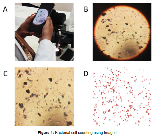
The collected image was treated and the bacterial cells were counted using ImageJ. The total number of bacterial particles was 332 cells (Figure 1). This method is less expensive, less possible contamination and less laborious than other methods and is more rapid and reproducible than counting using manual microscopy methods.
A. Microscopic image of the bacterial cells captured using a student’s smartphone, B. a collected image by the smartphone, C. zoom in from B and D. Image C is treated using ImageJ and bacterial cells was counted using ImageJ.
Conclusion
ImageJ comprises many image analysis capabilities, including functions for calculating area, measuring distances and counting. Direct staining and visualization of organisms for counting can benefit greatly from the use of ImageJ software. It can measure distances and angles. It can create density histograms and line profile plots. It supports standard image processing functions such as contrast manipulation, sharpening, smoothing, edge detection and median filtering. Because of the human factor manual bacterial cell counting is very time-consuming, very subjective and can be variable [8]. Alternatively, automated bacterial cells counting using ImageJ has the advantage that it has a lower error rate per sample and does not suffer from the subjectivity inherent to manual cell counting. Moreover, automation benefits from a high reproducibility compared to manual counting. With current setups of ImageJ, there are more detailed analysis reports available, including graphical display of the cells counted. Furthermore with cloud data storage, the operator can reanalyze data later. This may provide new results otherwise overlooked. Therefore, we –and others- [8] suggest the application of the ImageJ program as an alternative method to manual quantification of bacterial cells.
References
- Schneider CA, Rasband WS, Eliceiri KW (2012) NIH Image to ImageJ 25 years of image analysis Nature methods. 9(7): 671-675
- Admon R, Nickerson LD, Dillon DG, Holmes AJ, Bogdan R, Kumar P, Pizzagalli DA (2015) Dissociable cortico-striatal connectivity abnormalities in major depression in response to monetary gains and penalties. Psychological Medicine. 45(1): 121-131
- Leboffe MJ, Pierce BE (2019) Microbiology laboratory theory and application essentials Morton Publishing Company.
- Schoenberg E, Keller M (2021) Classic bedside diagnostic techniques. Clin Dermatol.
- Beveridge TJ, Davies JA (1983) Cellular responses of Bacillus subtilis and Escherichia coli to the gram stain. J Bacteriol 156(2): 846-858.
- Sandle T (2015) Pharmaceutical microbiology essentials for quality assurance and quality control Woodhead Publishing.
- Stolze N, Bader C, Henning C, Mastin J, Holmes AE, Sutlief AL (2019) Automated image analysis with ImageJ of yeast colony forming units from cannabis flowers. J Microbiol Methods. 164: 105681.
Citation: Al-Osta IM, Diab MS, Al-Shreef SAS (2021) ImageJ for Counting of Labeled Bacteria from Smartphone-Microscope Images. J Mol Pharm Org Process Res 9: 217. DOI: 10.4172/2329-9053.1000217
Copyright: © 2021 Al-Osta IM, et al. This is an open-access article distributed under the terms of the Creative Commons Attribution License, which permits unrestricted use, distribution, and reproduction in any medium, provided the original author and source are credited.
Select your language of interest to view the total content in your interested language
Share This Article
Recommended Journals
Open Access Journals
Article Tools
Article Usage
- Total views: 6732
- [From(publication date): 0-2021 - Dec 11, 2025]
- Breakdown by view type
- HTML page views: 5811
- PDF downloads: 921

