Inferior Lumbar Hernia: A case Report and Review of the Literature
Received: 09-Sep-2021 / Accepted Date: 23-Sep-2021 / Published Date: 29-Sep-2021 DOI: 10.4172/2167-7964.1000342
Abstract
Primary lumbar hernias are a rare; two types are described, according to the anatomical location of the hernia neck and etiology: the inferior and the posterior. While they are more lessely located within the inferior lumbar triangle. Incarceration is uncommon but represents a surgical emergency when present. We present a case of 68-year-old-woman had an inferior lumbar hernia unexpectedly discovered during an evaluation of the extent of loco regional colorectal adenocarcinoma. Abdominal CT scan demonstrated herniation of small bowel though the inferior lumbar triangle. CT scan is useful to distinguish hernia from solid mass, abscess, or other pathology, while bedside ultrasound may prompt an attempt at immediate reduction. Furthermore, CT provided the vital role in prompting surgical management. CT is now widely accepted as the imaging modality of choice for lumbar hernia, principally for its role in defining hernia contents accurately. Surgical correction is always more difficult in advanced cases; surgery must be indicated as early as possible. Surgical treatment can be performed as either an open or laparoscopic procedure with equivalent success.
Keywords: Lumbar triangle; Hernia; Inferior lumbar triangle
Introduction
Primary lumbar hernias are a rare form of posterior abdominal hernia; only around 300 cases of primary lumbar hernias have been reported in the literature, making it the rarest form of abdominal wall hernias [1]. It is the protrusion of intraperitoneal or extra peritoneal tissue through a poster lateral abdominal wall defect [2].
Case
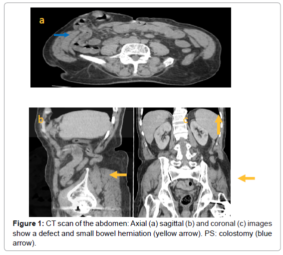
A 68-year-old woman admitted to the general surgery department for colorectal carcinoma. Her past medical history included a chronic low back pain, a cancer surgery with a colostomy. CT scan of the abdomen and pelvis with IV contrast for an evaluation of the extent of loco regional revealed a posterior left abdominal wall hernia through the inferior lumbar triangle measuring 2.6 cm containing a single loop of fluid-filled non distended small bowel in Figure. 1.
Furthermore, no flank mass was palpable. His abdomen was soft and non-tender with hypoactive bowel sounds. A therapeutic abstention was adopted.
Discussion
The first description of the inferior lumbar hernia was reported by French surgeon Jean-Louis Petit in 1738 [3]. Grynfeltt and P Lesshaft independently described superior lumbar hernias in 1866 [4] and 1870 [5]. Respectively; those two anatomical boundaries are where 95% of lumbar hernias occur, whereas the other 5% are considered to be diffuse.
It is most common in patients aged between 50 and 70 years with a male predominance. While they are more lessely located within the inferior lumbar triangle [1].
Lumbar hernias occur through defects in the lumbar muscles or the posterior fascia, below the 12th rib and above the iliac crest. Two types are described, according to the anatomical location of the hernia neck and etiology. The inferior lumbar hernia is the protrusion of tissue through the inferior lumbar triangle, also known as the Petit triangle, is an anatomical space through which inferior lumbar hernias can occur, which is bordered inferiorly by the iliac crest, anteriorly by external oblique muscle, and posteriorly latissimus dorsi muscle. It is not to be confused with the adjacent superior lumbar triangle (of Grynfeltt- Lesshaft) [2].
About 20% of lumbar hernias are congenital, discovered in infancy and are due to defects in the musculoskeletal system, it may be associated with other birth defects [6-8]. The remaining 80% are acquired and further classified as either primary or secondary.
A lumbar hernia is difficult to diagnose because the patient either is asymptomatic or presents with non specific symptoms. Lumbar hernias most commonly present as a reducible poster lateral palpable mass that increases in size with coughing and Valsalva maneuver and disappears when the patient assumes the prone position [9]. Heaviness and flank pain has also been described [6]. Reduction, when possible, is best accomplished in the decubitus position with manual compression [7]. There is a predilection for left-sided hernias. Patients with lumbar hernias can present with a bowel obstruction (if contents contain bowel), or urinary obstruction (if contents are kidney/ureter). The reported risk of bowel incarceration from lumbar hernias is about 25% [8].
Lumbar hernias may contain a number of intra- or retroperitoneal structures including: stomach, small or large bowel, mesentery, momentum ovary, spleen kidney [7].
When history and physical exam raise concern for lumbar hernia, CT scan is the preferred modality for confirming the diagnoses as well as delineating the muscular and facial layers and the contents within the hernia sac [8]. CT provided the vital role in prompting surgical management [10]. CT is also helpful in eliminating other differential diagnoses such as Lippmaz, fibromas, abscesses, hematomas, and muscle strains, none of which should cause bowel obstruction [6-8].
Ultrasound may prove a useful way of distinguishing a fluid-filled structure such as bowel from a solid mass, abscess, or hematoma, and may prompt an attempt at bedside reduction. Plain radiographs are of little use in narrowing the differential and only delay CT scan and surgical management [7].
Surgical correction is the standard treatment for a lumbar hernia. Because surgical correction is always more difficult in advanced cases, surgery must be indicated as early as possible [11]. The surgical repair can be also difficult given their location and surrounding bony structures [11,12].
A chronic or reducible hernia may be referred for elective repair while emergent repair is indicated if there is evidence of bowel obstruction or incarceration [11,12].
Conclusion
Most of lumbar hernias are non-emergent and represent a slowly enlarging mass. The remaining of hernias may develop rapidly and lead to incarceration. In these instances, imaging CT scan may prove a useful way of distinguishing hernia from other pathology, while bedside ultrasound may prompt an attempt at immediate reduction. CT is now widely accepted as the imaging modality of choice for lumbar hernia.
References
- Ankush S, Anil S, Rajesh Khullar, Vandana Soni, Manish Baijal, et al. (2019) Primary lumbar hernia: A rare case report and a review of the literature. Â Asiatique Journal Endosc Surg. Avr 12:197-200.
- Ran R, Andrew L, Makowski (2019) Inferior lumbar triangle hernia with incarceration. Am J Emerg Med 37:1218.Â
- Luis J, Petit JL Traité des maladies chirurgicales, et des opérations qui leur conviennent 1738 :257.
- Â Lesshaft P (1870) Die Lumbalgegend in Anat. Chirurgischer Hinsicht. Arch. F. Anat. u. Physiol. u. Wissensch Med. Leipzig 37:264.
- Stamatiou D, Skandalakis JE, Skandalakis LJ (2009) Lumbar hernia: surgical anatomy embryology, and technique of repair. Am Surg 75:202-207.
- Baker ME, Weinerth JL, Andriani RT, Cohan RH, Dunnick NR (1987) Lumbar hernia: diagnosis by CT. AJR 148:565-567.
- Wade P, Frey C, Lampe E (1996) Traumatic retroperitoneal hematoma followed by anuria and lumbar hernia: case report and method of repair Am. J. Surg 109: 253-9.Â
- Cavallaro G, Sadighi A, Paparelli C, Miceli M, D'Ermo G, et al. (2009) Anatomical and surgical considerations on lumbar hernias. Am Surg 75:1238-1241.
- Light B, Gopinath AB (2010) Incarcerated lumbar hernia a rare presentation, Ann Radiology Surgery Engl 92:e13-e14.
- Moreno-Egea A, Baena EG, Calle MC, Martinez JA, Albasini JL (2007) Controversies in the current management of lumbar hernias. Arch Surg 142:82-8.
- Beffa LR, Margiotta AL, Carbonell AM (2018) Flank and lumbar hernia repair. Surg Clin N Am 98:593-605.
Citation: MAHIR M, Zouine Y, Warda C, Benzalim M, Alj S (2021) Inferior Lumbar Hernia: A Case Report and Review of the Literature. OMICS J Radiol 10: 342. DOI: 10.4172/2167-7964.1000342
Copyright: © 2021 MAHIR M, et al. This is an open-access article distributed under the terms of the Creative Commons Attribution License, which permits unrestricted use, distribution, and reproduction in any medium, provided the original author and source are credited.
Select your language of interest to view the total content in your interested language
Share This Article
Open Access Journals
Article Tools
Article Usage
- Total views: 3165
- [From(publication date): 0-2021 - Nov 21, 2025]
- Breakdown by view type
- HTML page views: 2436
- PDF downloads: 729

