Intra-Articular Subtalar Osteoid Osteoma of the Talus: A Case Report
Received: 12-May-2023 / Manuscript No. joo-23- 98732 / Editor assigned: 17-Jun-2023 / PreQC No. joo-23- 98732 / Reviewed: 01-Jun-2023 / QC No. joo-23- 98732 / Revised: 06-Jun-2023 / Manuscript No. joo-23- 98732 / Published Date: 14-Jun-2023 DOI: 10.4172/2472-016X.100208
Abstract
Case: A 17-year-old boy, a competitive runner, presented with a right ankle sprain leading to progressive activitylimiting pain despite non-surgical treatment. Imaging at the initial presentation, two years before surgery, suggested a lateral talar process stress reaction. Subsequently, preoperative MRI imaging revealed an intra-articular talar mass at the anterior aspect of the subtalar joint. The patient underwent en-bloc resection of the osteoid osteoma with complete resolution of symptoms.
Conclusion: Progressive ankle and hind foot pain should be followed closely and evaluated in-depth with serial advanced imaging using either computed tomography or magnetic resonance imaging to rule out the development of neoplastic processes
Introduction
Osteoid osteomas are the third most common benign tumors, accounting for 10-14% of all benign and 2-3% of all primary bone tumors[1,2]. The tumor is generally small, measures less than 2 cm in diameter, and consists of a radiolucent nidus surrounding reactive osteosclerosis [3]. Osteoid osteomas are predominantly found in men occurring at a rate of nearly 2-3 times more often than in women [1]. 70% of osteoid osteomas cases were discovered in adults 20 years or younger in age [1,4,5]. The tumor commonly occurs in the metaphysis and diaphysis of long bones, particularly the femur and tibia. Small bones of the feet and hands are less affected, with the talus involved in 2-10% of the cases [5,6].
While the pathogenesis of osteoid osteomas has remained uncertain, some propose it is due to an inflammatory response or increased levels of prostaglandins. Others theorize a large role in neoplasm or trauma to the area [6]. This explains why patients often present with pain that is relieved with nonsteroidal anti-inflammatory drugs (NSAIDs). Patients may also exhibit local swelling and tenderness, and bony deformity [5- 9]. The typical symptoms may not always be present in patients, but a high level of clinical suspicion is necessary to direct further imaging [10,11].
Conservative treatment methods include NSAIDs, as some tumors often regress spontaneously over 2-6 years [8]. However, patients may present with severe pain or remain refractory to medical therapy; thus, surgical treatment may be necessary. Surgical treatments include open resection, arthroscopic surgical excision, or image-guided ablation of the lesion, with an 88-100% success rate [2,6,8]. Computerized tomography (CT) can detect the nidus, showing calcification and sclerosis; it is superior to plain radiography or magnetic resonance imaging (MRI) [6,8]. When the lesion presents in juxta-articular positions, the complex anatomy of the foot makes imaging interpretation even more challenging. In this paper, we present a case of a subtalar intra-articular osteoid osteoma of the talus treated successfully with en-bloc surgical excision.
Case Report
A 17-year-old male presented to the clinic for a second opinion regarding treatment for 2 years of progressive pain at the right ankle and hindfoot with associated swelling. The patient was a prior track athlete but had to relinquish all high-impact activities secondary to pain. There was no history of fever, weight loss, or loss of appetite.
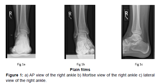
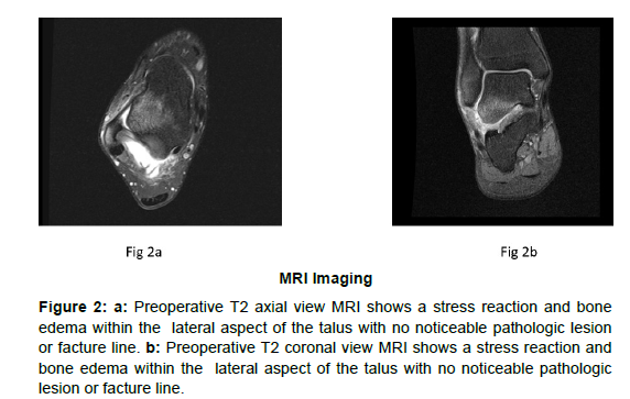
Two years before presentation, the patient was seen by his community generalist orthopaedic surgeon after an ankle sprain. Physical exam demonstrated tenderness over the lateral ankle and hindfoot. Plain films were unremarkable (Figure 1a-1c). MRI was obtained within a week of the sprain, revealing findings concerning a talar stress reaction (Figure 2a & 2b). The patient was treated with NSAIDS and a nonweightbearing cast for a month. This was followed by protective weightbearing in a Controlled Ankle Motion (CAM) boot. The patient was then transitioned to a lace-up ankle brace, a medial arch support insert, physical therapy, and discharged from care.
Figure 2: a: Preoperative T2 axial view MRI shows a stress reaction and bone edema within the lateral aspect of the talus with no noticeable pathologic lesion or facture line. b: Preoperative T2 coronal view MRI shows a stress reaction and bone edema within the lateral aspect of the talus with no noticeable pathologic lesion or facture line.
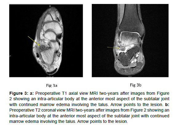
Despite these non-surgical treatment modalities, the patient continued to have progressive pain. Over the course of two years, he discontinued all high-impact activities. As a result, his surgeon ordered a repeat MRI that revealed an intra-articular body at the anterior most aspect of the posterior subtalar joint with surrounding edema (Figure 3a &3b). The patient was then referred to our clinic for management.
Figure 3: a: Preoperative T1 axial view MRI two-years after images from Figure 2 showing an intra-articular body at the anterior most aspect of the subtalar joint with continued marrow edema involving the talus. Arrow points to the lesion. b: Preoperative T2 coronal view MRI two-years after images from Figure 2 showing an intra-articular body at the anterior most aspect of the subtalar joint with continued marrow edema involving the talus. Arrow points to the lesion.
On physical exam, the patient had a subtle asymmetric cavus foot deformity on the right side with tenderness and mild swelling over the sinus tarsi. There was discomfort elicited with active and passive eversion and inversion of the hindfoot. Plain films were read as normal. Laboratory values including complete blood count, biochemical profile, and inflammatory markers were within the normal reference ranges.
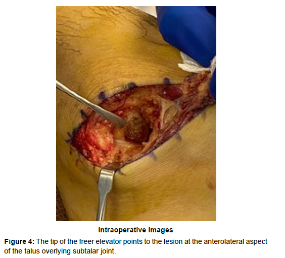
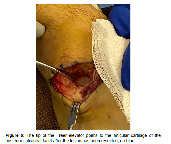
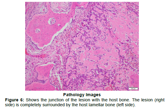
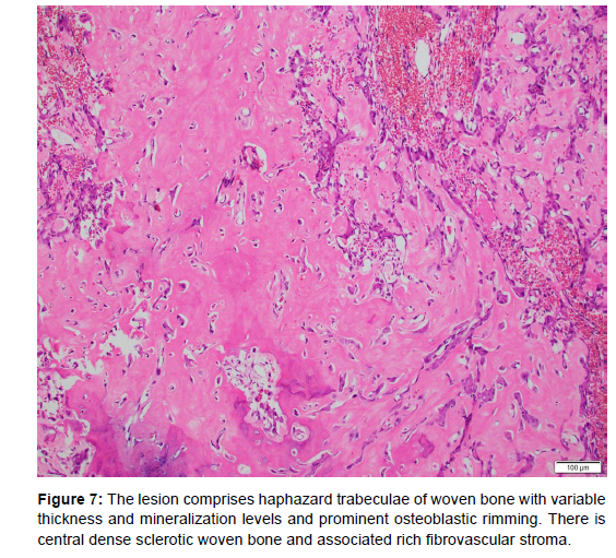
Based on the above findings, an intra-articular osteochondral body was the initial diagnosis, and the decision for surgical resection was made. A sinus tarsi approach was made under general anesthesia with a regional block for postoperative pain control. The lesion was resected en-bloc using an osteotome (Figures 4 and 5). Specimens were sent to pathology, and it was found to be an osteoid osteoma (Figures 6 and 7). The patient was instructed to be non-weight bearing for two weeks, then allowed to begin physical therapy. He was pain-free at his first postoperative visit. At three months, he was able to return to full activity, including running, without pain. At the one-year follow-up, the patient was asymptomatic and had no recurrence.
Discussion
Peri articular osteoid osteomas in the foot and ankle are rare [5,6,12- 14]. Due to the impact on the associated joint, patients can present with pain resembling arthralgia caused by many other conditions, such as juvenile rheumatoid arthritis, bony or fibrous coalition, trauma, or sinus tarsi syndrome [14]. The patient, in this case, complained of hindfoot pain emanating from the sinus tarsi area after an ankle sprain.
His symptoms did not resolve with NSAIDs. In the workup of an osteoid osteoma, CT is the modality of choice; it is superior to plain radiography or MRI [6,8].
According to several authors, the detection rate of osteoid osteomas of the hand and foot is 96.5% with CT [6]. However, an MRI was ordered for this case because there were no definitive signs of an osteoid osteoma on history, clinic exam, or plain films. In addition, an MRI is preferred to rule out more common disorders that may cause joint pain. The patient’s atypical presentation, normal labs and plain films, and lack of response to NSAIDs likely led to a definitive diagnosis and treatment delay.
In addition, initial evaluation with an MRI, in this case, revealed bony edema rather than a focal lesion. MRI fails to diagnose osteoid osteomas in 34% of cases due to its propensity only to detect edema rather than the tumor [6]. A repeat MRI approximately two years later revealed an expansion of the lesion leading to the decision for surgical treatment. Osteoid osteomas of the talus are often misdiagnosed due to the atypical presentations of the tumor. The time between presented symptoms and diagnosis of an osteoid osteoma is prolonged due to difficulties in diagnosis. A meta-analysis of 223 patients with osteoid osteoma found the average time to diagnosis was 22.1 months (range 1–120 months), with the longest period in talar lesions.
Close follow-up with serial imaging is recommended in young, healthy patients with foot and ankle pain refractory to nonsurgical treatment. In this case, a repeat MRI revealed growth of the tumor after two years, all the while, plain films remained normal. This is similar to findings reported in the literature; studies have shown lesions in certain areas, such as the talus, are difficult to evaluate by plain radiographs, showing a detection rate of only 66% of cases [6,8].
Conservative treatment methods include NSAIDs, as some osteoid osteomas often regress spontaneously over 2-6 years. [8] Invasive treatments are recommended for refractory cases. Surgical options include arthroscopic excision, image-guided ablation of the lesion, and open en-bloc resection. En-bloc surgical resection is an effective treatment for osteoid osteomas with a success rate of 88–100% of cases and reduces the risk of recurrence [2,6,8]. In this case report, en-bloc resection of the lesion was performed successfully with complete resolution of symptoms and return to normal activity within three months.
Conclusion
Intra-articular osteoid osteomas of the talus are rare benign tumors that are difficult to diagnose due to the frequently atypical presentations of the tumor. These tumors often present in young patients, so close follow-up and serial imaging are recommended for patients with persistent or progressive pain refractory to nonsurgical treatment. The intra-articular nature of these tumors requires that surgical treatment be considered to avoid further damage to the associated joint.
Acknowledgment
We thank the patient for his time, effort, and consent for this case report.
Conflict of Interest
The authors can confirm that they have no conflict of interest to declare. In addition, no funding was received for the production of this manuscript.
Statement of Informed Consent
Informed consent was obtained from the patient for the publication of this case report and any accompanying images.
References
- Zhang Y, Rosenberg AE (2017) Bone-forming tumors. Surg Pathol Clin 10:513-535.
- Boscainos PJ, Cousins GR, Kulshreshtha R, Oliver TB, Papagelopoulos PJ (2013) Osteoid osteoma. Orthopedics 36:792-800.
- Chai JW, Hong SH, Choi JY (2010) Radiologic Diagnosis of Osteoid Osteoma: From Simple to Challenging Findings. Radio Graphics 30: 737-749.
- Kitsoulis P, Mantellos G, Vlychou M (2006) Osteoid osteoma. Acta Orthop Belg 72:119–125
- Lee EH, Shafi M, Hui JH (2006) Osteoid osteoma: a current review. J Pediatr Orthop 26:695–700.
- Jordan RW, Koc T, Chapman AW, Taylor HP (2015) Osteoid osteoma of the foot and ankle -A systematic review. Foot Ankle Surg 21:228-234.
- Ghanem I (2006) The management of osteoid osteoma: updates and controversies. Curr Opin Pediatr 18:36-41.
- Tepelenis K, Skandalakis GP, Papathanakos G, Kefala MA, Kitsouli A, et al. (2021) Osteoid Osteoma: An Updated Review of Epidemiology, Pathogenesis, Clinical Presentation, Radiological Features, and Treatment Option. In Vivo 35:1929-1938.
- Pai V, Pai VS (2008) Osteoid osteoma of the talus: a case report. J Orthop Surg (Hong Kong) 16:260-262.
- He H, Xu H, Lu H (2017) A misdiagnosed case of osteoid osteoma of the talus: a case report and literature review. BMC Musculoskelet Disord 18, 35.
- Assafiri I, Sraj S (2015) Osteoid Osteoma of the Talar Neck with Subacute Presentation . Am J Orthop (Belle Mead NJ)? 44: 404-406
- Khurana JS, Mayo-Smith W, Kattapuram SV (1990) Subtalar arthralgia caused by juxtaarticular osteoid osteoma. Clin Orthop 252: 205-208.
- Gould N (1981) articular osteoid osteoma of the talus: a case report. Foot Ankle 1: 284-285.
- Gregory B Morris, Flair D Goldman (2003) Osteoid osteoma causing subtalar joint arthralgia: A case report. J Foot Ankle Surg 42:90-94.
Indexed at, Google Scholar, Crossref
Indexed at, Google Scholar, Crossref
Indexed at, Google Scholar, Crossref
Indexed at, Google Scholar, Crossref
Indexed at, Google Scholar, Crossref
Indexed at, Google Scholar, Crossref
Indexed at, Google Scholar, Crossref
Indexed at, Google Scholar, Crossref
Indexed at, Google Scholar, Crossref
Indexed at, Google Scholar, Crossref
Citation: Pierre Louis B, Belancourt P, Toussaint RJ (2023) Intra-Articular SubtalarOsteoid Osteoma of the Talus: A Case Report. J Orthop Oncol 9: 208. DOI: 10.4172/2472-016X.100208
Copyright: © 2023 Pierre Louis B, et al. This is an open-access article distributedunder the terms of the Creative Commons Attribution License, which permitsunrestricted use, distribution, and reproduction in any medium, provided theoriginal author and source are credited.
Select your language of interest to view the total content in your interested language
Share This Article
Recommended Journals
Open Access Journals
Article Tools
Article Usage
- Total views: 2871
- [From(publication date): 0-2023 - Nov 21, 2025]
- Breakdown by view type
- HTML page views: 2515
- PDF downloads: 356

