Isolated Left Superior Vena Cava: A Very Rare Venous Abnormality
Received: 08-Mar-2022 / Manuscript No. roa-22-56487 / Editor assigned: 10-Mar-2022 / PreQC No. roa-22-56487 (PQ) / Reviewed: 17-Mar-2022 / QC No. roa-22- 56487 / Revised: 23-Mar-2022 / Manuscript No. roa-22-56487 (R) / Published Date: 30-Mar-2022 DOI: 10.4172/2167-7964.1000370
Image Article
Persistent Left Superior Vena Cava (PLSVC) is the most common variant of systemic venous drainage, affecting 0.3% to 0.5 % of the general population. However, Isolated PLSVC is a very rare venous abnormality. It affects 0.09-0.13 percent of patients with congenital heart diseases [1]. It develops as a result of a failure of obliteration of the left common cardinal vein and typically drains the left subclavian and jugular veins into the right atrium via the coronary sinus [2].
Isolated PLSVC is normally asymptomatic and discovered by coincidence on imaging or during a cardiac catheterization. However, it can cause complications with central venous access, cardiothoracic surgery, and pacemaker implantation and may cause a right to left shunt if it drains to the Left Atrium (LA). For that, clinicians should be well aware of its variations and management strategies in order to avoid complications [1].
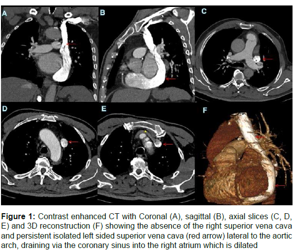
In imaging, it may be identified incidentally by echocardiography, often indirectly through recognition of a dilated coronary sinus. On cross-sectional imaging with Computed Tomography (CT) or Magnetic Resonance (MR), it is apparent as a vessel coursing vertically in the mediastinum, lateral to the aortic arch [2] (Figure 1).
Figure 1: Contrast enhanced CT with Coronal (A), sagittal (B), axial slices (C, D, E) and 3D reconstruction (F) showing the absence of the right superior vena cava and persistent isolated left sided superior vena cava (red arrow) lateral to the aortic arch, draining via the coronary sinus into the right atrium which is dilated
Note: The right brachiocephalic vein (yellow star) drains directly into the left superior vena cava
References
- Bisoyi S, Jagannathan U, Dash AK, Tripathy S, Mohapatra R, et al. (2017) Isolated persistent left superior vena cava: A case report and its clinical implications. Ann Card Anaesth 20: 104-107.
- Irwin RB, Greaves M, Schmitt M. (2012) Left superior vena cava: revisited. Eur Heart J Cardiovasc Imaging 13: 284-291.
Indexed at , Google Scholar, Crossref
Citation: Boularab J, Mandour J, Oubaddi T, Haddad S, Chat L, et al. (2022) Isolated Left Superior Vena Cava: A Very Rare Venous Abnormality. OMICS J Radiol 11: 370. DOI: 10.4172/2167-7964.1000370
Copyright: © 2022 Boularab J, et al. This is an open-access article distributed under the terms of the Creative Commons Attribution License, which permitsunrestricted use, distribution, and reproduction in any medium, provided the original author and source are credited.
Select your language of interest to view the total content in your interested language
Share This Article
Open Access Journals
Article Tools
Article Usage
- Total views: 6393
- [From(publication date): 0-2022 - Oct 18, 2025]
- Breakdown by view type
- HTML page views: 5669
- PDF downloads: 724

