Lanthanum, Yttrium Iron Oxide with Nanoparticles Structural, Magnetic Analysis, and Their Anti-Cancer Application
Received: 28-Mar-2023 / Manuscript No. ijm-23-91182 / Editor assigned: 31-Mar-2023 / PreQC No. ijm-23-91182(PQ) / Reviewed: 14-Apr-2023 / QC No. ijm-23-91182 / Revised: 21-Apr-2023 / Manuscript No. ijm-23-91182(R) / Published Date: 28-Apr-2023
Abstract
The synthesis, characterization, and biological properties of Iron Oxide with Lanthanum and Yttrium oxide nanoparticles were discussed. The samples were prepared using the microwave-accelerated technique. The products were characterized using X-ray diffraction (XRD) and Vibrating Sample Magnetometer (VSM), and also subjected to MDA – MB – 231 breast cancer and Anti-cancer activity. XRD confirmed the crystalline nature and structural changes were also noticed as a function of the size of the rare-earth ion added.The formed NPs crystalline structure and its size (18 to 23) nm have been determined using XRD analysis. The saturation magnetization, coactivity and remanence indicate the super paramagnetic behavior, but the lanthanum and yttrium have weak magnetic properties when composited of the investigated samples.The samples were safe against MDA-MB-231 breast cancer cell lines. Besides, the microbial studies, the response in human breast cancer cells exposed to Lanthanum, Yttrium oxide with iron oxide NPswere analyzed with MDA MB 231 by MTT assay.
Keywords
Microwave synthesis; XRD; Magnetic property; VSM, MDA – MB – 231 breast cancer; Anti-cancer activity
Introduction
The synthesis and characterization of NPs is asignificant area of research. Nanotechnology holds promising applications in bio-sensing, biomedical, food, drug delivery, cosmetics, cancer therapy, and other fields [1-3]. Iron Oxide blended with Lanthanum and Yttrium oxide NPs has been synthesized using multiple chemical and physical methods. But these processes involve several toxic chemicals as reducing agents, leading to environmental issues and non-biodegradable products. There is a need to adopt eco-friendly biosynthetic processes to reduce the same. Therefore, NPs fabricated for medical applicationsare currently of great interest. The bio applications based on magnetic nanoparticles have attracted considerablybecause of theiruniquenessoverother materials. The magnetic iron oxide nanoparticles are inexpensive to produce, physically and chemically stable, biocompatible, and environmentally safe. The magnetic nanoparticles by the microwave heating method revealtheir magnetic properties. Hysteresis losses during the demagnetization occur for particles that are not in the superparamagnetic state. When the particles are in a superparamagnetic state, the magnetic nanoparticles (MNPs) are introduced into the cells by endocytosis.
The blood vessels of cancerous tissue absorb larger amounts of MNPs than the normal ones [4, 5]. Further, the biomolecules such as antibodies can be easily attached to the MNPs. The iron oxides can be used as a magnetic factor in multifunctional nano construct for diagnostic imaging and drug targeting [6, 7].The present studies synthesize stable Fe2O3NPs with lanthanum and yttrium oxides in asinglestep which will be apt for investigation of cell viability in breast cancer MB 231 cells.
The properties of Y2O3 reported a high degree of hardness, unique optoelectronic properties, and chemical stability [9-11]. The carbon nanotubes (CNTs) and Y2O3 can be used to detect acetaminophen a good electrocatalyst that findsapplications such as solar energy processes [12], lithium-ion batteries [13, 14], and components for rare-earthdoped lasers [15, 16].rare earth nanomaterial, Y2O3 a highly effective and functional material was used in the form of Yttrium-stabilized zirconia films [17, 18]. Its involvement in DNA-related studies such as the purification process, DNA separation, and structuring and tumor inhibition techniques [19]. Metal oxide NPs like zinc, titanium, cerium, and yttrium oxide NPs, have been extensively studied.
Y2O3 used in LEDs of a high-spatial-resolution and multicolor imaging technique for the observation of biological cells using cathode luminescence (CL) from nano phosphors has been observed. Yttrium oxide NPs have several applications in diverse fields, including biomedicine. But the available resources on the Y2O3 synthesis methods and their emerging biomedical applications are very few. Hence, the article highlights thesynthesis of Y2O3 with iron oxide, itsbehavior, and itsbiomedical application.
Lanthanum oxide La2O3 has attractive properties, which aresuitable for applications, such as catalysts [20], optical filters [21], metal support, water treatment, and dielectric material. Nowadays, many approaches includingmicrowaves have been adopted, These elements are found in the human body at intravenous levels in the order of 1 ng/kg, which is more than 1000 times lesser than the threshold toxicity. Nano-sized magnetic particles are found to be effective for developing drug delivery vehicles based on metal-oxide nanoparticles. Studies havedescribed these NPs are cytotoxic and possess anti-cancer effects due to their unique electronic configuration which includes radio sensitization via the Auger effect, and chemotherapeutic synergy via increased apoptotic activity. The nanoparticles synthesized were then characterized using appropriate instrumentation and evaluated for their cytotoxic activities.
Experimental
Synthesis method
Iron (iii) nitrate (Fe (No3)39H2O), Lanthanum iii nitrate (La (NO3)36H2O and Yttrium iii nitrate Y(NO3)36H2O are taken with urea in equal amounts and kept in the microwave oven for 30 minutes for the removal of nitrate and water. The burnt sample are crushed to a fine powder in a mortar pestle and the resultant was dried in an oven at 100ºC for 24h. The purified Iron Oxide blended with Lanthanum Oxide and Yttrium oxide nanoparticles were then characterized.
Characterization techniques
Powder X-ray diffraction (XRD) measurements were carried out for iron oxide, erbium oxide, and erbium-doped iron oxide samples using a Bruker D8 advance diffractometer with monochromatic Cu Kα radiation (λ=1.5418 Å). The X-ray source was operated at 40 kV with a current of 40 mA. The measurements were performed by θ - 2θ scans in the 2θ range 20-80° with a step size of 0.02° and at a scan rate of 2°/min.The magnetic properties of the samples were carried out by a vibrating sample magnetometer (VSM Lakeshore model 7410) at room temperature with a maximum applied field of 20 kOe.
Cell culture and cell line maintenance
The human breast cancer cell lines MDA MB-231 were obtained. Then, these cell lines were developed as a monolayer in Dulbecco’s modified Eagle’s medium (DMEM)which was supplemented with 10% fetal bovine serum, 100 U/mL penicillin, and 100 μg/mL streptomycin (Hi-Media Laboratories Mumbai, India) at 37˚C in an incubator with 5% CO2and high humidity.
MTT assay method for evaluation of cell viability and cytotoxicity
The anticancer effect of samples on human breast cancer cell lines MDA MB-231 was determined by the MTT (3-(4, 5-dimethyl thiazol- 2yl)-2, 5-diphenyl tetrazolium bromide) assay. These cells (1 × 10⁵/ well) were plated in 0.2 ml of the cells with a concentration of 1 × 10⁵ cells/ml. The plates were incubated for 24 hrs in a 5% CO2 incubator for cytotoxicity. After incubation, normal breast (MDA MB-231) cells were cultured in a 1:1 mixture of dimethyl sulfoxide (DMSO). Then, they were added by micropipette. The percentage of viable cells was visualized by the development of purple color due to the formation of formazan crystals. The suspension was transferred to the curette of a spectrophotometer and significant instability in the optical density (OD) was observed. The concentration required for 50% inhibition of viability (IC50) was determined and used for bioassays.
Results and Discussion
Structural analysis
X-ray diffraction images of samples (Fig.1) confirmed the formation of iron oxide, lanthanum oxide, yttrium oxide, iron/lanthanum oxide, and iron/yttrium oxides, depicting the structure and polyphase material consisting of La2O3, Y2O3, and Fe2O3. Although the properties of lanthanum and yttrium are similar, their atomic radii (La3+ = 1.03Å and Y3+ = 0.90 Å) are different, which decide their occupancy in the host latticeduring the mechanism. As a result, the powders were characterized by particle size, which determined the homogeneity of phase distribution.By using the full width at half maximum (FWHW) of the broad peaks, the average particle size was calculated by Debye Scherer’s formula.
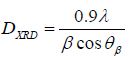
Where D is the average crystallite size of the particle, λ is the wavelength of the radiation, β is the full width at half maximum (FWHM) of the peak, and θ is Bragg’s angle.
Fig. (1.a) shows the reflection planesof pure Fe2O3(Hematite) are found to be (012), (104), and (114) of entry no. 96-210-8028 with rhomb centered. The average crystallite size of the iron oxide sample is 23 nm. The lattice constant for Fe2O3 nanoparticles was a = b = 5.040 and c = 13.74650. Fig. (1.b) shows the reflection planes are (111), (200), and (220) with entry no. 96-152-3969 of hexagonal structure. The average crystallite size for lanthanum oxide (La2O3) is 25 nm.The lattice constants are a = 4.0570 and c = 6.4300. Fig. (1.c) shows the reflection planes (200), (220), (222), (400), (420), and the entry no for this 96- 154-1744 with a cubic structure of lattice parameter a = 10.5500 and the average crystallite size of Yttrium oxide is 37 nm. Figures (1.d, e) show Iron Oxide with Lanthanum Oxide and Yttrium Oxide nanoparticles with average crystallite sizes of 15 nm and 18 nm, respectively.
Vibrating Sample MagnetometerStudies (VSM)
The M-H hysteresis loop studied at room temperature reveals the magnetic properties of iron oxide nanoparticles, which are shown in Fig.2. It confirms that synthesized iron oxide nanoparticles exhibit ferromagnetic attributeswith magnetic saturation (Ms) 34.61 emu/g, magnetic retentivity (Mr) 6.91emu/g, and coercive field (Hc) 251.72 Oe by applying a magnetic field of 15 kOe. The remnant magnetization is higher than in previous reports, which may be due to the smaller size and uniform shape of nanoparticles.
At low fields, the magnetization increases, then gradually decline. However, the plateau observed due to the magnetic saturation of the sample is replaced with a slight inclination. The M-H curve depicts the superparamagnetic behavior of the nanoparticles and determines their saturation magnetization. Further the superparamagnetic M - H curve can also be analyzed by comparing with the Langevin function and log-normal size distribution to estimate the magnetic diameter of the nanoparticles.At room temperature, Fig.3(a, b) shows a systematic change in the magnetization M versus applied field H loops of La2O3 and Fe2O3/L a2O3 samples. It can be seen from the plot the coercivity Hc for lanthanum oxide is zero Oe, the magnetic retentivity Mr = 40.873 emu/g, and the magnetic saturation Ms= 0.5098 emu/g. But when the iron is fused with lanthanum oxide, the hysteresis curve shows a change from zero coercivity to low coercivity, ie., Hc = 42.511Oe, Mr = 24.22 emu/g, and Ms = 0.44emu/g. When lanthanum with zero coercivity is composited, exhibits superparamagnetic behavior. The lanthanum oxide samples exhibit weak FM behavior and an increase in magnetization (M) with composite content of iron oxide. The observed change in the magnetic properties, i.e., the magnetic moment of pure La2O3 is zero which emphasizes the antiferromagnetic behavior of materials. The magnetization could be due to the structural distortion with a change in the Fe-O-Fe bond angle. The enhanced magnetic properties of La2O3 and Fe2O3/La2O3 are due to the increased canting effect.
The hysteresis loops (Fig 4) of yttrium oxide and composited iron/yttrium oxide are measured at room temperature (RT) and have confirmed their superparamagnetic properties with the saturation magnetization (Ms), remnant magnetization (Mr), and coercive field (Hc). The hysteresis plot for yttrium oxide shows coercivity Hc = 5.903 Oe, magnetic saturation Ms = 0.215 emu/g and magnetic retentivity Mr = 2.664 emu/g. which shows low coercive, saturation, and remanence of diamagnetic behavior, but when it is composited with iron oxide, the hysteresis loop has an excellent change in coercivity Hc = 61.44 Oe, magnetic saturation Ms = 34.416 emu/g, and magnetic retentivity Mr = 4.255 emu/g. By combining iron and yttrium, the diamagnetic behavior of yttrium was altered to superparamagnetic. If the Fe3+ ions occupy both octahedral and tetrahedral sites, the Ferri magnetic alignment will be observed. The magnetization is higher in MNPs than in bulk materials because of the formation of a partially inversed spinel. By fusing Fe3+ ions with non-magnetic Y3+ ions, the magnetization of the octahedral coordination is found to be reduced, resulting in decreasedmagnetization. The literature suggests that Y3+ prefers octahedral sites and the presence of Y3+ ions increases the size of nanoparticles, which increases blocking temperature and saturation magnetization for low concentrations.
Anti-cancer activity of Cell culture
The cytotoxicity of Fe2O3, La2O3, Y2O3, Fe2O3/La2O3, and Fe2O3/Y2O3NPs at various concentrations (5 g, 10 g, 50 g, 75 g, and 100 g/mL) against MDA-MB-231 cell lines was investigated using the MTT method, and the results are shown in Fig.5(a-d). The chosen lowest concentration (5 μg/ml or 75 μg/ml or 100 μg/ml ) of Fe2O3 NPs provided the highest cell destruction with 85% cytotoxicity, causing the least cell viability (15%). of the MDA-MB-231 cell line. The chosen lowest concentration (100 μg/ml ) of La2O3 NPs provided the highest cell destruction with 53% cytotoxicity, causing the lesser cell viability (47%.) of the MDA-MB-231 cell line. The chosen lowest concentration (100 μg/ml ) of Y2O3 NPs provided the highest cell destruction with 60% cytotoxicity, causing the lesser cell viability (40%). of the MDAMB- 231 cell line. The chosen lowest concentration (100 μg/ml ) of La2O3/Fe2O3 NPs provided the highest cell destruction with 45% cytotoxicity, causing the lesser cell viability (55%). of the MDA-MB-231 cell line.The chosen lowest concentration (100 μg/ml ) of Y2O3/Fe2O3 NPs provided the highest cell destruction with 41% cytotoxicity causing the lesser cell viability (59%.) of the MDA-MB-231 cell line. In the MTT assay, the cell morphology was captured after 48 hours to obtain the results.The graph in fig. 6 shows the cell viability for different concentrations. It shows that the cytotoxic effect on cancerous cells is excellent for iron oxide when compared to composited iron with lanthanum and yttrium oxides. It shows the cell viability for iron oxide is very low, i.e., below 20. But for lanthanum oxide, yttrium oxide, and composites of iron with lanthanum and yttrium, the reactions are mild.
Conclusion
Using microwave radiation, the iron oxide, lanthanum oxide, yttrium oxide, and composites of iron with lanthanum and yttrium oxide NPs have been prepared and their effective anticancer studies were effectively analyzed. The formed NPs crystalline structure and its size (18 to 23) nm have been determined using XRD analysis. The VSM plot for iron oxide reveals the superparamagnetic behavior, but the lanthanum and yttrium have weak magnetic properties when composited. The results of the MTT assay using the synthesized sample against MDA-MB 231 cell lines indicatea higher percentage of toxicity of iron oxide, lanthanum oxide, yttrium oxide, and composites of iron with lanthanum and yttrium oxide NPs. The study was aimed and successfully carried out toward the biological applications.
Acknowledgments
Financial support from the Tamilnadu State Council for Science and Technology under grant number, C.No.TNSCST/STP-PRG/ AR/2018-2019/9333 this part of a research programto whom we are gratefully acknowledged.
Conflict of Interest
The authors declare no conflict of interest.
Ethical approval
This article does not contain any studies with human participants or animals performed by any of the authors.
References
- Wu W, He Q G, Jiang C Z (2008) Magnetic iron oxide nanoparticles: synthesis and surface functionalization strategies. Nanoscale Res Lett 3: 397.
- Gupta A K, Gupta M (2005) Synthesis and surface engineering of iron oxide nanoparticles for biomedical applications. Biomater 26: 3995.
- Gilchrist R, Medal R, Shorey W D, Hanselman R C, Parrott J C, et al. (1957) Selective inductive heating of lymph nodes. Ann Surg 146: 596.
- Jordan A, Scholz R, Wust P, Schirra H, Schiestel T, et al. (1999) Endocytosis of dextran and silan-coated magnetite nanoparticles and the effect of intracellular hyperthermia on human mammary carcinoma cells in vitro. J Mater 194: 185-196.
- Jordan A, Scholz R, Wust P, Fähling H, Felix R (1999) Magnetic fluid hyperthermia (MFH): Cancer treatment with AC magnetic field induced excitation of biocompatible superparamagnetic nanoparticles. J Magn Mater 201: 413-419.
- Nasongkla N, Bey E, Ren J, Ai H, Khemtong C (2006) Multifunctional polymericmicellesascancer-targeted,MRI-ultrasensitivedrugdelivery systems. Nano Lett 6: 2427-2430.
- Mornet S, Vasseur S, Grasset F, Duguet E (2004) Magnetic nanoparticle design for medical diagnosis and therapy. J Mater Chem 14: 2161-2175.
- Lu A H, Salabas E L, Schuth F (2007) Magnetic nanoparticles: synthesis, protection, functionalization, and application. Angew Chem Int Edn 46: 1222.
- Rastogi AC, Sharma N (2001) Interfacial charge trapping in extrinsic Y2O3/SiO2 bilayer gate dielectric based MIS devices on Si (100). Semicond Sci Technol 16: 641-650.
- Wu CH, Chen JZ (2015) Ultrafast atmospheric-pressure-plasma-jet processed conductive plasma-resistant Y2O3/carbon-nanotube nanocomposite. J Alloy Compd 651: 357-362.
- Xu YN, Gu Zq, Ching WY (1997) Electronic, structural, and optical properties of crystalline yttria. Phys Rev B 56: 14993-15000.
- Kenyon AJ (2002) Recent developments in rare-earth-doped materials for optoelectronics. Prog Quant Electron 26: 225-284.
- Wen W, Yang X, Wang X, Shu LGH (2015) Improved electrochemical performance of the spherical LiNi0.5Mn1.5O4 particles modified by nano-Y2O3 coating. J Solid State Electrochem 19: 1235-1246.
- Wu F, Wang M, Su Y, Chen S (2009) Surface modification of LiCo1/3Ni1/3Mn1/3O2 with Y2O3 for lithium-ion battery. J Power Sources 189: 743-747.
- Kong J, Tang DY, Zhao B, Lu J, Ueda K (2005) 9.2 W diode end pumped Yb:Y2O3 ceramic laser. Appl Phys Lett 86: 161116.
- Baytak AK, Teker T, Duzmen S, Aslanoglu M (2016) A composite material based onnanoparticles of yttrium (III) oxide for the selective and sensitive electrochemical determination of acetaminophen. Mater Sci Eng C 66: 278-284.
- Verhiest K, Almazouzi A, Wispelaere N, Petrov R, Claessens S (2009) Development of oxides dispersion strengthened steels for high-temperature nuclear reactor applications. J Nucl Mater 385: 308.
- Wang ZC, Kim KB (2008) Fabrication of YSZ thin films from suspension by electrostatic spray deposition. Mater Lett 62: 425.
- Salata OV (2004) Applications of nanoparticles in biology and medicine. J Nanobiotechnol 2: 3.
- Ghiasi M, Malekzadeh A (2015) Synthesis, characterization and photocatalytic properties of lanthanum oxycarbonate, lanthanum oxide and lanthanum hydroxide nanoparticles. Superlattices Microstruct 77: 295-304.
- Shinde VG, Gaikwad VB, Deore MK (2018) Synthesis of Lanthanum Oxide (La2O3) Nanoparticles by Hydrothermal method and studies it "s Physical Properties. Int J Chem Phys 7: 669-674.
Indexed at, Google Scholar, Crossref
Indexed at, Google Scholar, Crossref
Indexed at, Google Scholar, Crossref
Indexed at, Google Scholar, Crossref
Indexed at, Google Scholar, Crossref
Indexed at, Google Scholar, Crossref
Indexed at, Google Scholar, Crossref
Indexed at, Google Scholar, Crossref
Indexed at, Google Scholar, Crossref
Indexed at, Google Scholar, Crossref
Indexed at, Google Scholar, Crossref
Indexed at, Google Scholar, Crossref
Indexed at, Google Scholar, Crossref
Indexed at, Google Scholar, Crossref
Indexed at, Google Scholar, Crossref
Indexed at, Google Scholar, Crossref
Indexed at, Google Scholar, Crossref
Indexed at, Google Scholar, Crossref
Indexed at, Google Scholar, Crossref
Citation: Umadevi P, Baskaran I, Sathyaseelan B, Senthilnathan K, Manikandan E (2023) Lanthanum, Yttrium Iron Oxide with Nanoparticles Structural, Magnetic Analysis, and Their Anti-Cancer Application. Int J Inflam Cancer Integr Ther, 10: 214.
Copyright: © 2023 Umadevi P, et al. This is an open-access article distributed under the terms of the Creative Commons Attribution License, which permits unrestricted use, distribution, and reproduction in any medium, provided the original author and source are credited.
Select your language of interest to view the total content in your interested language
Share This Article
Recommended Journals
Open Access Journals
Article Usage
- Total views: 3949
- [From(publication date): 0-2023 - Dec 19, 2025]
- Breakdown by view type
- HTML page views: 3520
- PDF downloads: 429