Make the best use of Scientific Research and information from our 700+ peer reviewed, Open Access Journals that operates with the help of 50,000+ Editorial Board Members and esteemed reviewers and 1000+ Scientific associations in Medical, Clinical, Pharmaceutical, Engineering, Technology and Management Fields.
Meet Inspiring Speakers and Experts at our 3000+ Global Conferenceseries Events with over 600+ Conferences, 1200+ Symposiums and 1200+ Workshops on Medical, Pharma, Engineering, Science, Technology and Business
Case Report Open Access
Mandibular Ameloblastoma Giant
| Fahd El Ayoubi El Idrissi*, Azeddine Lachkar, Mohammed Chouai, Ahmed Aabach and Mohamed Rachid Ghailan | |
| Service d’ORL, hôpital des spécialités de Rabat, CHU Ibn Sina, avenue Hafiane-Cherkaoui, Rabat, Morocco | |
| Corresponding Author : | Fahd El Ayoubi El Idrissi Service d’ORL, hôpital des spécialités de Rabat CHU Ibn Sina, avenue Hafiane-Cherkaoui, 10100 Rabat Morocco Tel: 00212661285208 E-mail: fahd.el@hotmail.fr |
| Received: December 29, 2014; Accepted: June 29, 2015; Published: July 03, 2015 | |
| Citation: Ayoubi El Idrissi FEl, Lachkar A, Chouai M, Aabach A, Ghailan MR (2015) Mandibular Ameloblastoma Giant. J Preg Child Health 2:173. doi: 10.4172/2376-127X.1000173 | |
| Copyright: © 2015 Ayoubi El Idrissi FEl, et al. This is an open-access article distributed under the terms of the Creative Commons Attribution License, which permits unrestricted use, distribution, and reproduction in any medium, provided the original author and source are credited. | |
Visit for more related articles at Journal of Pregnancy and Child Health
Abstract
The ameloblastoma is a benign odontogenic tumor, locally aggressive and recidivism determines prognosis and monitoring. We report a unique case of a mandibular ameloblastoma giant on a Patient of 12 years referred for mandibular swelling with important functional gene. The scanner objectified picture unilocular osteolytic extent of the right ramus. Mandibulectomy subtotal interrupter with reconstruction plate was made.
| Keywords |
| Mandibular; Ameloblastoma; Mandibulectomie |
| Introduction |
| The ameloblastoma is a benign odontogenic tumor, locally aggressive and recidivism determines prognosis and monitoring. We report a unique case of a mandibular ameloblastoma giant. |
| Observation |
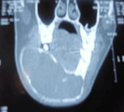
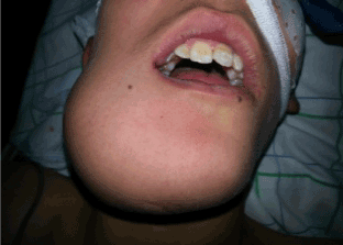
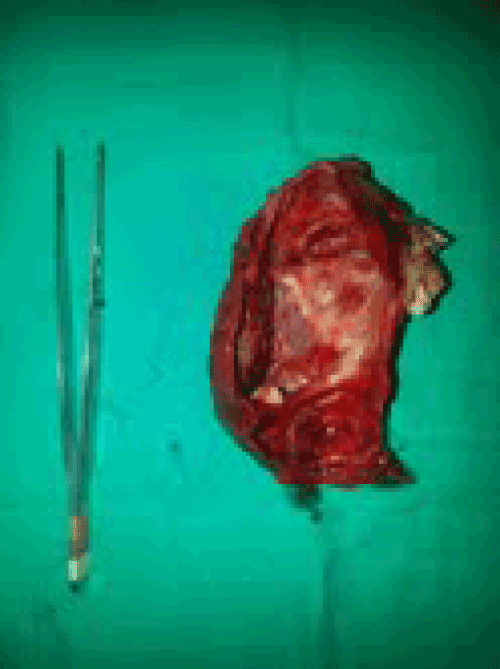
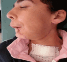
| Patient of 12 years referred for mandibular swelling with important functional gene. The review found a hard swelling integral with the mandible with dental displacement in (Figure 1). The scanner objectified picture unilocular osteolytic extent of the right ramus to the tooth 36, with thinning of cortical bone (Figure 2). Mandibulectomy subtotal interrupter with reconstruction plate was made (Figure 3,4). Pathological examination confirmed the diagnosis of ameloblastoma Benin mixed, the suites were simple. |
| Discussion |
| Relatively frequent among odontogenic tumors but rare considering tumors and cysts of maxillary ameloblastoma accounts for only 1% of bone tumors and 25.2 to 58.6% of benign odontogenic tumors according to the authors. The frequency is much higher in the black African population, it can reach up to 80.1% in some series, it mainly occurs between the 3rd and 4th decade with a slight predisposition for subjects 20 to 35 years. The ameloblastoma mainly interested mandible (80% of cases). The angular region represents the most common location, with extension to the horizontal branch (70%), then followed by the parasymphysaires regions (20%) and symphysis (10%) [1,2]. The ameloblastoma is characterized by a long latency, patients most commonly see at the stage of mandibular swelling. Dental signs most often found in the literature are mostly tooth mobility and displacement. |
| In all cases, two negative signs are highlighted: the lack of satellite nodes and absence of impaired muco-cutaneous sensitivity. Medical imaging is an essential consideration for the diagnosis of ameloblastoma. It uses conventional examinations or even CT or MRI that allow further study. The radiological picture is not unequivocal and none is specific, it only helps to guide the diagnosis. The latter is based on a set of arguments: clinical, epidemiological and histological above. |
| Two types of images can be observed: multilocular pictures (2/3 cases) separated or unilocular Images (1/3 of cases) [2]. |
| The radical treatment mandibulectomy subtotal is not discussed in the case of mandibular ameloblastoma giant. It is no longer possible to perform a simple curettage or enucleation. Recidivism is higher after conservative treatment as radical. The risk of recurrence is highest in the first three years but recurrence may occur much later, after 15 to 30 years [3]. |
| Conclusion |
| Benignity of the giant ameloblastoma is uncertain, the treatment should be radical, long-term monitoring is essential. |
References
- Olgac V, Koseoglu BG, Aksakalli N (2006) Odontogenic tumours in Istambul: 527 cases. Br J Oral Maxillofac Surg 44:386-388.
- Fernandez AM (2005) Odontogenic tumors: a study of 340 cases in Brazilian population. J Oral Pathol Med.
- Ruhin B, Guilbert F, Fouret P, Ghoul S, Berdal A, et al (2005) Tumeurs des maxillaires améloblastome: données actuelles et perspectives. Rev Stomatol Chir Maxillofac 106: 1s64.
Figures at a glance
 |
 |
 |
 |
| Figure 1 | Figure 2 | Figure 3 | Figure 4 |
Post your comment
Relevant Topics
Recommended Journals
Article Tools
Article Usage
- Total views: 14142
- [From(publication date):
August-2015 - Aug 28, 2025] - Breakdown by view type
- HTML page views : 9507
- PDF downloads : 4635
