Case Report Open Access
Neonatal Congenital Pancreatic Cyst: A Report of Three Cases
| Chahed Jamila1*, Kechiche Nahla1, Hidouri Saida1, Aloui Sameh1, Ksia Amine1, Mekki Mongi1, Sahnoun Lassad1, Krichene Imed1, Belghith Mohsen1, Zakhama Abdelfatteh2, Salem Randa3 and Nouri Abdellatif1 | |
| 1Paediatric Surgery Department of EPS Fattouma Bourguiba, Tunisia | |
| 2Department of Histopathology of EPS Fattouma Bourguiba, Tunisia | |
| 3Department of Radiology of EPS Fattouma Bourguiba, Tunisia | |
| Corresponding Author : | Chahed Jamila Pediatric Surgery Department of EPS Monastir CP 5000 Research laboratory LR 12SP13, School of medicine of Monastir University of Monastir, Tunisia Tel: +21673461144 Fax: +21673460678 E-mail: j.chahed@voila.fr |
| Received June 11, 2014; Accepted July 15, 2015; Published July 20, 2015 | |
| Citation: Jamila C, Nahla K, Saida H, Sameh A, Amine K, et al. (2015) Neonatal Congenital Pancreatic Cyst: A Report of Three Cases. J Preg Child Health 2:179. doi: 10.4172/2376-127X.1000179 | |
| Copyright: © 2015 Jamila C, et al. This is an open-access article distributed under the terms of the Creative Commons Attribution License, which permits unrestricted use, distribution, and reproduction in any medium, provided the original author and source are credited. | |
Visit for more related articles at Journal of Pregnancy and Child Health
Abstract
Neonatal congenital pancreatic cyst is rare. It can be isolated or associated to other malformations. Early management is recommended because of possible severe complications. The aim of our work is to report our experience with three newborns with congenital pancreatic cyst. A five year (2010-2015) retrospective study is carried. Data were collected from medical files of three different patients. Diagnosis was prenatal in two patients. The Cyst was associated to Ivemark syndrome in two cases and solitary in one. Cystic complications were infection in one case and pancreatitis with liver cirrhosis in another case. These pancreatic cysts were successfully managed by surgery. One patient succumbed as a result of heart failure. To our knowledge we report the first case of congenital pancreatic cyst complicated with liver cirrhosis. We concluded that prompt diagnosis is important to prevent severe complications. Prognosis is dependent on early management and on life threatening associated malformations.
| Keywords |
| Pancreatic cyst; Congenital; Polymalformative syndrome; IVEMARK syndrome; Complications |
| Abbreviations |
| CPC: Congenital Pancreatic Cyst; WG: Weeks of Gestation |
| Introduction |
| Neonatal congenital pancreatic cyst (CPC) is rare in new-borns. It can be isolated or associated to other malformations. Prompt diagnosis and management is crucial to prevent complications. We report three different cases of neonatal pancreatic cyst associated to Ivemark syndrome in two and isolated in one. |
| Materials and Methods |
| Our work is a 5 year (2010-2015) retrospective study in the paediatric surgery department of Fattouma Bourguiba Teaching hospital of Monastir-Tunisia. Data were obtained from medical files of the three patients. |
| Results |
| Case 1 |
| A 2.3 kg twin, full-term male infant, aged of 45 days with no familial history, was hospitalized in paediatric department at day 13 of life for vomiting, hypothermia along with weight loss and altered general health. |
| A suspected post-natal infection was managed by Cefotaxime- Aminoside association. The evolution was marked by abdominal distension with bilious vomiting during oral feeding. At physical examination a mobile, smooth mass was palpated at the left abdominal side. |
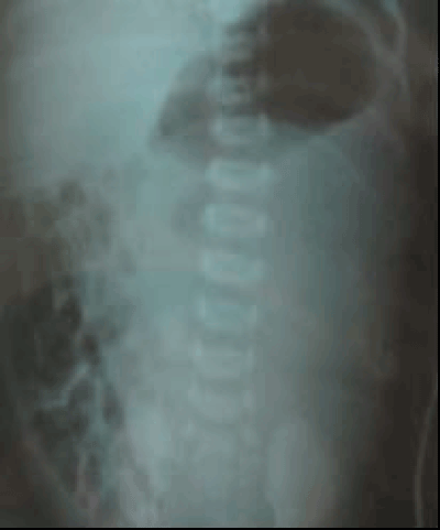
| Plain thoraco-abdominal x-ray showed a dextro-cardia and a left abdominal opacity without calcifications (Figure 1). This opacity corresponded at the ultra-sonography to a 7x6x6 cm size-anechoic cystic mass, occupying the left abdominal side, against the anterior side of the stomach which was pushed back and to the abdominal right side along with the left kidney and the liver. CT-scan revealed this cystic mass with asplenia. |
| An echocardiography demonstrated a complex heart disease with single ventricle and pulmonary atresia. A preliminary diagnosis of digestive duplication, choledochal cyst, pancreatic cyst and polycystic kidney disease was made. |
| During surgical exploration an intestinal malrotation with ectopic development of the pancreas and asplenia were observed. Cyst punction with an internal drainage were performed. Bilious aspect of the cyst content suggested a cystic communication with biliary duct. A total cystectomy was not possible because of intimate contact with other organs. Histopathological examination found a pancreatic cyst with undefined epithelium origin. Cyst content revealed a high amylase activity with presence of acinetobacter. Evolution was initially eventless and enteral feeding was started at 6th day post-surgery. The patient died at the age of two months and a half due to heart failure. |
| Case 2 |
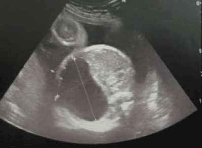
| A 3.2 kg full-term female, aged of 2 months with no familial history, was hospitalised in our paediatric surgery department for exploration of two intra-peritoneal cystic malformations. She was a product of a normal vaginal delivery with APGAR 9/10. Parents were not consanguineous. Antenatal ultrasonography (28 WG) showed two anechoic cystic malformations (1.7x4.5 cm), which seemed to be dependent on the left kidney (Figure 2). A preliminary diagnosis of a polycystic kidney disease, a digestive duplication, a Cystic lymphangioma and pancreatic cyst was made. |
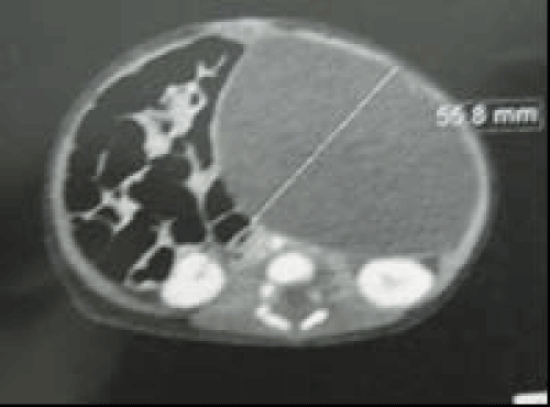
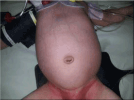
| A control ultrasonography (at the 22nd day of life) showed a normal left kidney with two cystic malformations of the left quadrant. At the abdomino-pelvic scan there were two cystic malformations (2.3x2.2 and 4.9x4.5 cm) with multiple other micro cysts at the pancreatic head and dilatation of the intrahepatic bile ducts with no individualisation of the pancreas tail (Figure 3). At physical examination, the infant was eutrophic: 5 kg (+1DS) with a mild icterus. A renitent, smooth mass was palpated at the left abdominal quadrant (Figure 4). Biology showed a moderate cholestatic icterus (total bilirubinemia: 67 μom/l, direct bilirubinemia: 40 μmol/l), a moderate cytolysis (ASAT/ALAT: 199/105 ui/l) and a normal hemostasea et amylasemia. |
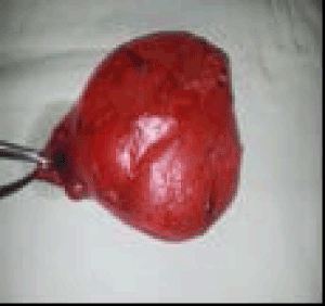
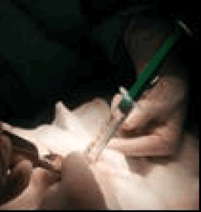
| An echocardiography demonstrated a small inter-auricular communication. Surgical exploration via laparotomy showed two retro-peritoneal cysts (5 and 3.5 cm) dependant on the pancreatic body and tail. The liver had a cirrhotic aspect, the spleen was bilobated with an extra small spleen. The patient had body and tail pancreatectomy (Figure 5) including the two cysts with excision of the extra spleen and liver biopsy. Cyst punction drew out a white yellowish liquid (Figure 6) with high amylase and lipase levels. |
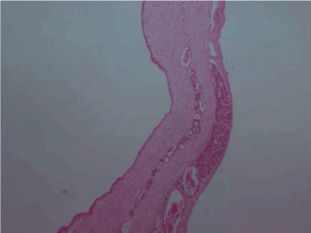
| Histopathological examination found pancreatic cysts (Figure 7) with chronic pancreatitis. Also, the liver was cirrhotic with cholestasis and the spleen was effectively an extra one. The association of pancreatic cysts, multiple spleens, liver and spleen heterotaxia was compatible with Ivemark I syndrome. Evolution was eventless, with disappearance of icterus and normalisation of bilirubinemia. Nevertheless, a moderate liver cytolysis persisted. The patient is now aged of three years and has a normal weight (4 kg) with normal psychomotor development. She is under regular clinical and radiological control of the pancreas and the liver. |
| Case 3 |
| A 3.15 kg full term female, was born by cesarean delivery for hydramnios with an APGAR 7/9. At 22 WG, an intra-abdominal cystic structure (6.4x4.6 cm) of the left upper quadrant was identified by ultrasonography. The prenatal ultrasonography follow up didn’t show any significant modification of the cyst size. |
| At birth, physical examination revealed a palpable smooth mass at the left upper quadrant. No further anomalies were noticed. Biology was normal. Plain abdominal radiograph showed a left side abdominal opacity with intestinal gas deviation to the opposite side. |
| Abdominal CT scan showed a 9.8x7.5x4.3 cm, unilocular cyst occupying the left abdominal side with intestinal structure deviation. It was anterior between the stomach, the pancreas and the left kidney. The patient had laparotomy (day 16 of life) via Pfannentiel incision. Ovaries and uterus were normal. The cystic mass was surrounded by the stomach, the spleen and the pancreatic tail. The content had a serous aspect. The cyst was completely excised. Enzymatic analysis of the cyst fluid was not performed. Histopathologic examination revealed a cuboidal epithelial cells uni- and bi- stratified. Postoperative course was eventless and the child was discharged at day 11 after surgery. The child is one year old and is healthy but with a weak weight gain 6.8 kg. |
| Discussion |
| Congenital pancreatic cysts in paediatric population are rare. Their incidence is less than 1% of all abdominal cysts [1,2]. These cysts can be single or multiple as to transform the pancreas into a cystic mass as it is the case for our 2nd observation. Transition from one to the other form has been also reported [1]. A pubmed search showed 26 cases of solitary CPC [2,3]. CPC may or may not possess an epithelial lining or elevated amylase levels; the presence of either supports the diagnosis. Microscopic examination may show pancreatic acini or ductules between the cystic locules [4-6]. Embryologically, it is accepted that true cysts occur as a result of developmental anomalies related to the sequestration of primitive pancreatic ducts [4]. |
| As it is reported in literature, a female predominance is found as our patients are 2 females and one male [1]. CPC may exist alone or in association with systemic diseases such as Von Hippel-Lindeau syndrome or polycystic kidney disease [4,5,7], Ivemark syndrome. Other associated malformations have been reported. Our first case is a congenital pancreatic cyst associated to Ivemark II which includes: intestinal malrotation, asplenia, ectopic pancreas and a complex heart disease. Whereas the second case; is an Ivemark I syndrome with association of multilocular pancreatic cysts, multiple spleens, liver and spleen heterotaxia. It was complicated by chronic pancreatitis and cholestatic liver cirrhosis. The third case was a non-complicated solitary one. |
| Diagnosis can be ante-natally or post-natally. Prenatal diagnosis is extremely rare. To our knowledge only 11 cases of prenatal diagnosis were reported [1,8,9]. Two of our three cases were diagnosed prenatally. Post-natal presentation has included an asymptomatic abdominal mass, abdominal distension, vomiting caused by gastric mass effect and jaundice from biliary obstruction [10,11]. |
| A prompt diagnosis is important to avoid the development of serious or fatal complications such as infection, cholangitis, pancreatitis, cyst rupture and peritonitis [6]. Unfortunately, two of our patients had severe complications: infection of the cyst in the first case and pancreatitis with cholestatic liver cirrhosis in the second case. |
| Pancreatitis results either from congenital anomalies of the duct system or from visceral compression by the cyst [4,12]. To our knowledge, we report the first case of prenatal CPC complicated by liver cirrhosis due to chronic cholestasis which can be explained by compression of the biliary system. Imaging investigations consist of a plain abdominal x-ray which usually shows a right opacity of a soft tissue mass. An ultra-sound and a computed tomography are necessary to evaluate this mass before surgery. It may be difficult to distinguish the origin of the cyst and the differential diagnosis can include renal cysts, choledochol cysts, enteral duplications, omental or mesenteric cysts and lymphatic malformations among others. Endoscopic retrograde cholangio pancreatography (ERCP) is useful to rule out communication with pancreatic duct or biliary tree [7]. Cysts near the tail are best treated by distal pancreatectomy. Cysts at the head of the pancreas have been treated by cystenterostomy or by complete resection. |
| Conclusion |
| We report three cases of congenital pancreatic cysts, two of them are rare cases of CPC with Ivermark II and Ivemark I syndrome. Complications of the pancreatic cysts are important to prevent by early diagnosis and treatment. |
References
- Gerscovich EO, Jacoby B, Field NT, Sanchez T, Wootton-Gorges SL, et al. (2012) J ultrasound Med 31:811-813.
- Kazez A, Akpolat N, Kocakoç E, Parmaksiz ME, Köseogullari AA (2006) Congenital true pancreatic cyst: A rare case. DiagnIntervRadiol12:31-33.
- Casadei R, Campione O, Greco VM (1996) Congenital true pancreatic cysts in young adults: Case report and literature review. Pancreas 12:419-421.
- Auringer ST, Ulmer JL, Summer TE (1993)Congenitalcyst of the pancreas. J PediatrSurg 12: 1570-1571.
- Fremond B, Poulain P, Odent S (1997)Prenatal detection of a congenital pancreatic cyst and Beckwith-Wiedmann syndrome. PrenatDiagn17: 276-280.
- Kebapci M, Aslan O, Kaya T (2000)Prenatal diagnosis of giant congenital pancreatic cyst of a neonate. AJR Am J Roentgenol175: 1408-1410.
- Boulanger Scott C, BorowitzDrucy S, Fisher John F (2003)Congenital pancreatic cysts in children. J PediatrSurg7: 1080-1082.
- Baker LL, Hartman GE, Northway WH (1990)Sonographicdetection of congenital pancreatic cysts in the newborn: Report of a case and review of the literature. PediatrRadiol20:488-490.
- Jae Hee Chung, Gye-Yeon Lim, Young Tack Song (2007)Congenital true pancreatic cyst detected prenatally in neonate: a case report. J PediatrSurg42: E27-E29.
- Miles RM (1959)Pancreatic cyst in the new-born: a case report. Ann Surg 149: 576-81.
- Pilot LMJR, Gooselaw JG, Isaacson PG. Obstruction of the common bile duct in the new-born by a pancreatic cyst. Lancet 84: 204-205.
- Boulanger SC, Gosche JR (2007)Congenital pancreatic cyst arising from occlusion of the pancreatic duct. PediatrSurgInt23: 903-905.
Figures at a glance
 |
 |
 |
 |
| Figure 1 | Figure 2 | Figure 3 | Figure 4 |
 |
 |
 |
| Figure 5 | Figure 6 | Figure 7 |
Relevant Topics
Recommended Journals
Article Tools
Article Usage
- Total views: 14328
- [From(publication date):
August-2015 - Aug 30, 2025] - Breakdown by view type
- HTML page views : 9659
- PDF downloads : 4669
