Case Report Open Access
Serious Influence of Yersinia Enterocolitis on Pregnancy in a Woman Complicated With Chronic Hypertension and Gestational Diabetes Mellitus: A Case Report
| Hirobumi Asarkua*, Takehiko Fukami, Tomoko Inagaki and Naoko Tateyama | |
| Nippon Medical School, Musashikosugi Hospital, Obstetrics and gynecology, Kawasaki city, Kanagawa Prefecture, Japan | |
| Corresponding Author : | Hirobumi Asarkua Nippon Medical school, Musashikosugi Hospital Obstetrics and gynecology, 1-396 Kosugicho Nakaharaku Kawasaki city, Kanagawa Prefecture 211-8533, Japan Tel: 088 044 733 5182 E-mail: morgen@nms.ac.jp |
| Received November 11, 2014; Accepted February 12, 2015; Published February 16, 2015 | |
| Citation: Asarkua H, Fukami T, Inagaki T, Tateyama N (2015) Serious Influence of Yersinia Enterocolitis on Pregnancy in a Woman Complicated With Chronic Hypertension and Gestational Diabetes Mellitus: A Case Report. J Preg Child Health 2:135. doi: 10.4172/2376-127X.1000135 | |
| Copyright: © 2015 Asarkua H, et al. This is an open-access article distributed under the terms of the Creative Commons Attribution License, which permits unrestricted use, distribution, and reproduction in any medium, provided the original author and source are credited. | |
Visit for more related articles at Journal of Pregnancy and Child Health
Abstract
A 40-year-old pregnant woman admitted into hospital to treat hypertension (HT) and gestational diabetes at 19 weeks of gestation. She contracted enterocolitis due to the foodborne pathogen Yersinia enterocolitica (YE) at 22 weeks of gestation. Hypoalbuminemia and ascites developed due to prolonged intractable diarrhea and vomiting. Since 25 weeks of gestation, marked albuminuria appeared and blood pressure gradually elevated. Fetal growth was found to be retarded. Cesarean section for non-reassuring fetal status was performed at 27 weeks 2 days, delivering a 641 gr boy (-2.9 SD from averaged neonatal body weights compatible with the gestational weeks, Apgar score 3/8 at1/5min.). Gastrointestinal symptoms and ascites resolved within 1 week of delivery. This represents the first report of YE infection seriously affecting perinatal prognosis.
| Keywords |
| Yersinia enterocolitica; Fetal growth retardation; Hypoalbuminemia; Preeclampsia |
| Introduction |
| Yersinia are anaerobic, Gram-negative, rod-shaped bacteria and Y. enterocolitica (YE) is a common foodborne pathogen. Infection is characterized by acute gastroenteritis and YE isolation rates in patients with sporadic diarrhea are higher among children than among adults [1] YE infection commonly presents as acute enterocolitis. Transmission is oral with an incubation period of 3–7 days. Main symptoms include diarrhea, low-grade fever, abdominal pain, nausea, and vomiting. To the best of our knowledge, no previous reports have described effects of YE infection on perinatal prognosis such as occurrence of preeclampsia and fetal growth retardation (FGR). We report herein a case of YE enterocolitis leading to massive ascites caused by hypoalbuminemia. As a result, the perinatal prognosis of the present case was seriously affected by YE enterocolitis. |
| Case Report |
| A 39-year-old gravida 2, para 2 (height, 153 cm; weight, 92 kg; body mass index, 36.6; body type, obese) was examined at our institution at 5 weeks of gestation with a spontaneous pregnancy. Past obstetric history comprised two cesarean sections, each performed due to severepregnancy induced hypertension at 38 weeks of gestation, at 27 and 35 years old. Hypertension (HT) was diagnosed at 35 years old. Methyldopa of 750 mg wasinitiated.Examination at 10 weeks of gestation revealed HT (185/122 mmHg), high concentration of fasting blood glucose (144 mg/dL) and hemoglobin A1c (7.0%). She was diagnosed with HT and gestational diabetes mellitus (GDM). Termination was presented as an option in consideration of the risk of continued pregnancy due to severe HT. However, she strongly wished to continue with the pregnancy. Diet (1600 Kcal/ day) and insulin therapies(6IU regular insulin per day) were initiated for GDM. However, at 19 weeks 6 days of gestation she was hospitalized due to exacerbatedHT (220/120 mmHg). After admission,GDM was controlled by diet therapyand insulin injection(Humalog 24IU and human regular insulin12 IU per day). HT was treated with bed rest and furosemide (20mg/day), azelnidipine (16 mg/day) and hydralazine (30 mg/day). Since blood pressure subsequently decreased to around 140/90 mmHg,furosemide and hydralazine were stopped on hospital day 8. |
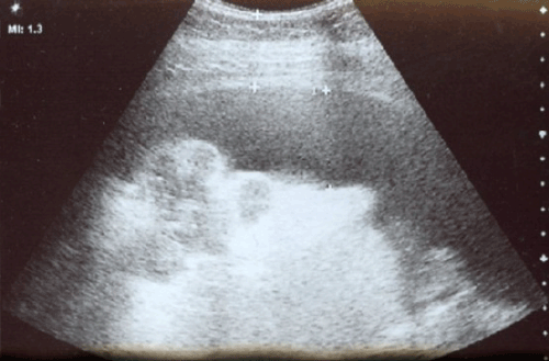
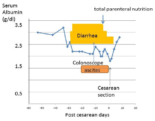
| On hospital day 20 (22 weeks 4 days of gestation), the patient suddenly experienced extreme epigastric pain, frequent vomiting and diarrhea. Bacterial enterocolitis was suspected and daily intravenous treatment with fluid replacement and Beta-lactam antibiotics of fulomox (FOMX) (2 g/day) was initiated. From stool culture YE were detected and became negative at 6 days after treatment. However, diarrhea subsequently persisted and marked ascites was identified at24 weeks 1 day of gestation (Figure 1). Her weight gain of 10 Kg in a week was found. To reduce ascites furosemide of 40mg/day was used for a week but itwas not effective.Ascites was presumably associated with malnutrition since concentration of serum albumin became low to 2.2 g/dLfrom 3.2 g/dL on admission (Figure 2 and Table 1). |
| Colonoscopy on 26 weeks 2 days of gestation revealed no coarse lesions in the intestine, but cecal biopsy demonstrated infectious colitis, explaining the intractable diarrhea. Due to difficulty with dietary intake, persistent vomiting and diarrhea, hypoalbuminemia, and poorly controlled ascites, oral food intake was discontinued from 26 weeks 4 days of gestation and high-calorie total parenteral nutrition (1600 kcal) was implemented. By the treatment frequency of diarrhea and vomiting decreased tofew episodes per day (Figure 2). |
| She was negative for albuminuria on admission, but spot urine testing on 25 weeks 3 days of gestation showed high urine protein(664 mg/dL) with granular and hyaline casts in the urinary sediment. Superimposed preeclampsia was diagnosed. Continuous intravenous infusion of nicardipine was needed from 26 weeks 5 days of gestation because of sustained HT.Due to the patient’s obesity and marked ascites, continuous recording of a fetal heart rate (FHR) monitor was difficult, so fetal well-being was evaluated using a biophysical profilesobtained through ultrasonography. Biophysical profile scores had been full marks except a finding of FHR monitoring. Fetal growth retardation (FGR) was foundat 26 weeks of gestation although fetal growth was proportional with gestational age at admission. On 27 weeks 2 days of gestation, pulsed Doppler findings of the blood flow velocitywaveforms in the umbilical artery revealed an absence of end-diastolic flow velocity. Emergency cesarean section was performed for non-reassuring fetal status probablydue to superimposed preeclampsia and FGR.The male baby was born weighing 641 g (-2.9 S D from averaged Japanese neonatal body weights compatible with the gestational weeks). Apgar scores at 1 and 5 min after birth were 3 and 8, respectively. Umbilical artery blood pH was 7.15. The placenta weighed 290 g and showed marked ischemic findings associated with preeclampsia, such as syncytial knots and accelerated maturation. A total of 5,400 ml of ascites fluid was found intra operatively. As cites rapidly disappeared after delivery. HT and blood glucose control also improved rapidly. |
| As shown in Table 1, there were no abnormal laboratory findings except serum concentrations of albumin and total protein during the course of pregnancy. Disappearance of ascites and diarrhea after delivery was associated with recovery of serum levels of albumin (Figure 2).The source of the YE infection was believed to be contaminated food brought into the hospital for the patient from outside. She has not disputed this theory. The newborn was admitted to the neonatal intensive care unit immediately after birth. Artificial ventilation was necessary due to respiratory distress syndrome from birth until 26 days of age. Afterwards, breathing condition became stable and brain MRI revealed no abnormal neurological findings.The neonate had no infectious complications. He discharged from NICU at 107 days of age. |
| Discussion |
| YE infection commonly presents as acute enterocolitis. Transmission is oral with an incubation period of 3–7 days and symptomatic infection such as food poisoning is common in children, but rare in adults. Main symptoms include diarrhea, low-grade fever, abdominal pain, nausea, and vomiting. Mass outbreaks sometimes occur when commercial milk or food products were contaminated. As the present case was sporadic, the patient was believed to have eaten YE-contaminated food brought in from outside the hospital.In cases of intestinal infection, YE enters through the Peyer’s patches in the small intestine, causing enteritis, and is then disseminated via the lymph, leading to mesenteric lymphadenitis and potentially sepsis if hematogenous dissemination occurs. Patients with underlying conditions such as DM, anemia, hemochromatosis, and liver cirrhosis are more susceptible to sepsis. While children are prone to developing acute gastroenteritis, adults tend to present with ileocecal lesions such as mesenteric lymphadenitis and terminal ileitis [1]. Colonoscopic findings for the present case support this tendency. |
| The majority of patients show spontaneous recovery, with symptoms improving within several days [1]. Even in the absence of sepsis, in cases involving severe infection with a prolonged recovery such as in the present patient, administration of antibacterial drugs is indicated. |
| A search of MEDLINE for the period from 1995 to 2013 revealed no reports of YE infection contracted after conception significantly affecting the course of pregnancy. The severity of YE enterocolitis in the present patient was believed to be due to increased susceptibility to infection due to poor control of the underlying GDM. |
| Reports have described residual terminal ileal and cecal lesions such as small ulcers observed on colonoscopy at 1 month or longer after YE enterocolitis onset [2]. The present patient continued to experience intractable diarrhea even after stool cultures came back negative for YE. Colonoscopy performed on hospital day 45 found signs of enterocolitis in the region of the cecum, and this was considered the cause of the intractable diarrhea. Diarrhea persisted until cesarean section, indicating that considerable time is required for ileal and cecal inflammation to subside. |
| DM and obesity are known risk factors for pregnancy-induced HT (PIH) [3-6] while pregnant women with severe HT are at considerable (50%) risk of superimposed preeclampsia [7]. A mean blood pressure of ≥90 mmHg in the second trimester of pregnancy is associated with significantly increased rates of perinatal death, PIH and FGR. The incidence of FGR also increases in women with superimposed preeclampsia [8]. The present patient had pre-existing HT, obesity and DM, and the sudden fluctuations in plasma volume caused by additional severe diarrhea and vomiting due to YE enterocolitis may have further exacerbated uteroplacental hemodynamics. The incidences of large-volume ascites (≥2 L) in patients with severe PIH or HELLP (hemolysis, levated liver enzymes and lower platelet) syndrome have been reported as 2.2% and 10% [9], respectively. The exact pathophysiological mechanisms underlying ascites in PIH remain unclear, but renal retention of sodium and water and retention of water in the extravascular space are involved. In PIH, there is extensive vascular endothelial malfunction throughout the body, encouraging water leakage from the capillaries and unpredictable protein leakage causing decreased intravascular colloid oncotic pressure and exacerbated ascites [10]. Association of hypoprotenemia as well as hypoalbuminemia with massive ascites in severe preeclampsia was reported [9,11]. Malnutrition (hypoalbuminemia) due to YE enterocolitis could potentially have played a pathogenic role and exacerbated the ascites (Figure 1). From experience of 23 PIH patients with ascites it was concluded that termination of pregnancy is thought as the only treatment for large-volume ascitesas it cannot be cured by medical treatment [11]. This was same as the present case.Ascites rapidly disappeared after cesarean section. It is likely to assume that malnutrition and PIH were also influential in FGR. |
| Conclusion |
| To the best of our knowledge, this represents the first report of Yersenia infection seriously affecting pregnancy. In high-risk pregnancies in mothers with complications of HT and GDM, gastrointestinal conditions such as prolonged severe diarrhea and vomiting may cause PIH and further complications of large-volume ascites. |
References
- Grant LC and David TD (1998) Plague and yersinia infections. In Fauci A, Braunwald E, Isselbacher KJ, Wilson JD, MartinTB et al.(eds). Harrison’s Prinsiples of Internal Medicine 14th ed. MacGraw-Hill.Co.975-983.
- Matsmoto T, Iida M, Matsui T, Sakamoto K, Fuchigami T, et al. (1990) Endoscopic findings in Yersinia enterocolitica enterocolitis. GastrointestEndosc 36:583-587.
- Duckitt K and Hamington D (2005) Risk factors for preeclampsia at antenatal booking: systemic review of controlled studies. BJM :330:565.
- Matzger BE, Lower LP, Dver AR, Trimble ER (2008) Hyperglcemia and adverse pregnancy outcome. New Engl J Med 19: 1991-2002.
- Athukorala C, Rumbold AR,Wilkar KJ, Crawther CA (2010) The risk of adverse pregnancy outcomes in women who are overweight or obese. BMC Pregnancy and childbirth , 10: 56.
- Nohr BA, Vaeth M, Beker JL, SÃ?Â?rensen Tia, Olsen J et al.(2008) Combined association of prepregnancy body mass index and gestational weight gain with the outcome of pregnancy. Am J Clin Nutr 87:1750-9.
- American Coollage of Obstetricians and Gynecology. ACOG practical bulletin, Chronic hypertension in pregnancy.ObstetGynecol 2012,119:396-407.
- Report of the National High Blood Pressure Education Program Working Group on High Blood Pressure in Pregnancy. Am J ObstetGynecol 2000; 183:S1-S22
- Vajjyanath AM, Navar B, Malhotra N, Deka D(2002) Massive ascites in severe-preeclampsia: a rare complication. J Obstet Gynecol Res 28:199-202
- Brown MA, Zammit VC, Lowe SA (1989) Capillary permeability and extracellular fluid volumes in pregnacy-induced hypertension. ClinSci77:599-604.
- Cong KJ, Wang TT (1994) Complication of ascites in pregnancy-induced hypertension. Chang-Hua-Fu-Chan-Ko-Tsa-Chin 29:7-9.
Tables and Figures at a glance
| Table 1 |
Figures at a glance
 |
 |
| Figure 1 | Figure 2 |
Relevant Topics
Recommended Journals
Article Tools
Article Usage
- Total views: 15952
- [From(publication date):
February-2015 - Aug 30, 2025] - Breakdown by view type
- HTML page views : 11340
- PDF downloads : 4612
