The Evaluation of Shoulder Muscle Fatigue in Volleyball Players
Received: 08-Mar-2018 / Accepted Date: 10-Apr-2018 / Published Date: 17-Apr-2018 DOI: 10.4172/2165-7025.1000388
Abstract
Objective: To identify the difference in infraspinatus, posterior deltoid, and teres minor muscle fatigability between the dominant and non-dominant side in elite volleyball players and to examine the differences between three sEMG signal processing methods used in assessment of shoulder muscle imbalance due to fatigue in volleyball players.
Methods: In 18 male volleyball players (21-26 years; 186.6 ± 8.4 cm; 85.7 ± 9.8 kg) with no previous shoulder injury the bioelectrical activity of the right and left infraspinatus, posterior deltoid, and teres minor muscles was measured during 60 seconds of isometric contraction in prone position with the shoulder in external rotation. Fatigue related changes as mean frequency shift were calculated from the RAW sEMG signal using 3 processing methods: FFT (Fast Fourier Transform), STFT (Short Time Fourier Transform) and CWT (Morlet Continues Wavelet Transform).
Results: There were no statistically significant differences (p>0.05) in the values of the mean frequency slope, intercept and difference between dominant and non-dominant sides in all the evaluated muscles. There were no significant differences between FFT and STFT sEMG signal processing methods in mean frequency slope, intercept values and difference. The sEMG signal processing using CWT showed the significantly higher values of mean frequency slope for infraspinatus and teres minor muscles. Significantly lower values of mean frequency intercept were observed for the infraspinatus, posterior deltoid and the teres minor muscles. There were no significant differences observed in mean frequency difference for all the evaluated muscles.
Conclusions: In elite volleyball players without previous shoulder injury, the fatigue indices in muscles of the shoulder region were similar on both the dominant and non-dominant sides. Therefore, we have hypothesized that asymmetric shoulder loading during volleyball training should not be considered as an obvious factor increasing the risk of shoulder injury. Muscle fatigue indices measured by sEMG may be a sensitive and objective method of evaluation, but may reach different values depending on the used signal processing method. Consequently, the clinical interpretation and any comparison between different measurements, without knowledge of how those values were calculated, may be misleading and be the reason for misdiagnosis.
Keywords: Volleyball; Muscle fatigue; sEMG; Shoulder injury
Introduction
Overhead sports such as volleyball are closely related to shoulder pain and strain injuries of the shoulder region [1,2]. The specifics of this discipline require the transfer of high energy through the shoulder in large ranges of motion and with high precision [1]. It was reported that repetitive movements during sport activities may lead to cumulative tissue loading, muscle fatigue and strain injuries [3,4]. It was demonstrated that during repetitive movements muscle fatigue may be accompanied by changes in movement patterns and by changes in joint proprioception [4,5]. The alterations in proprioception due to fatigue are very important especially in sport activities where optimal movement patterns and appropriate motor control are required [5]. It was reported that changes in movement patterns due to muscle fatigue may contribute to acute or overuse injuries and to many musculoskeletal disorders, particularly in the shoulder region [3,6,7]. This process is especially devastating when including the postural and stabilizing muscles of the shoulder complex, specifically, the rotator cuff muscles [7,8]. Studies have shown that altered position and motion of the scapula are considered as potential risk factors for shoulder pain and shoulder injury [2,4,8]. It was reported that scapular dyskinesis is observed in a 43% of overhead athletes and is influenced by acute and chronic fatigue [1].
It has been reported that fatigue during exercise is accompanied by changes in electromyographic muscle activity [9-11]. This leads to an increase in signal amplitude and to a higher fatigue index [11,12]. The sEMG signal analysis in muscle fatigue assessment usually includes changes in average sEMG amplitude and changes in sEMG spectral frequency [11-13]. Muscle fatigue is usually evaluated by sEMG signal spectral frequency analysis. But signal processing methods may be different even if the final parameters are the same [13-15]. Therefore, the differences in clinical usefulness of different methods of sEMG signal processing in the evaluation of muscle fatigue due to physical effort needs comprehensive investigation.
These small differences in sEMG signal spectral frequency due to signal processing method may be crucial in muscle imbalance assessment [16,17]. The appropriate assessment of shoulder muscle fatigability is crucial in prevention, diagnosis and management of strain and acute injuries which is especially important in sport performance [18]. Because the shoulder strain and acute injuries in overhead athletes are important and common problems, there is a need to verify the factors which may be the source of misdiagnosis.
The aim of this investigation was to identify the difference in infraspinatus, posterior deltoid, and teres minor muscle fatigability between the dominant and non-dominant side in elite volleyball players. We have hypothesized that if the differences in muscle fatigability between dominant (more loaded) and non-dominant (less loaded) side are visible immediately following isolated fatigue, this may suggest the presence of shoulder muscle chronic overloading and higher predisposition of more fatigued muscles to strain injury.
The secondary aim of this study was to examine the differences between three sEMG signal processing methods used in assessment of shoulder muscle imbalance due to fatigue in volleyball players.
Materials and Methods
Participants
18 male volleyball players (21-26 years; 186.6 ± 8.4 cm; 85.7 ± 9.8 kg) participated in this study. The athletes belonged to a regional team, and all were healthy, with no previous shoulder injury. They did not perform any high-intensity physical activity for 2 days before the test to avoid the effects of cumulative muscular fatigue. They were asked which arm is their dominant one – the right or left. All measurements were performed by one examiner. The study participants were informed in detail about the research protocol and gave their written informed consent to participate in the study.
The EMG measurement
The bioelectrical activity of the right and left infraspinatus, posterior deltoid, and teres minor muscles was recorded according to the SENIAM guidelines [13,19]. Prior to electrode placement, the skin was cleaned and degreased with alcohol. Surface electrodes (Ag/AgCl) (Sorimex, Poland) with a 2 cm center-to-center distance were attached along the direction of the muscle fibers on the muscle bellies. The signals were registered with 16-bit accuracy at a sampling rate of 1500 Hz and stored for subsequent analysis using Noraxon G2 TeleMyo 2400 unit (Noraxon USA).
The sEMG signal from the evaluated muscles was measured during 60 seconds of isometric contraction with the 4 kg resistance attached to the wrist. The measurement was performed in prone position with the shoulder in external rotation (Weight Prone External Rotation). Fatigue related changes as mean frequency shift were calculated from the RAW sEMG signal using 3 processing methods [14,15,20-22].
FFT (Fast Fourier Transform)-unfiltered RAW sEMG signal was analysed step-wise in 1,000 ms increments over 60-second static contractions. Mean frequency was calculated for each 1 second step and then regression line (slope) and intercept (Hz) values were computed. The mean frequency difference (%) was calculated as the difference between average first 3 and last 3 period values.
STFT (Short Time Fourier Transform)-unfiltered RAW sEMG signal was analysed with a 512 point window over 60 second’s static contractions. Mean frequency was calculated for each window and the regression line (slope) and intercept (Hz) values were computed. The mean frequency difference (%) was calculated as the difference between the average first 3 seconds and last 3 last seconds.
CWT (Morlet Continues Wavelet Transform) - Unfiltered RAW sEMG signal was analysed over 60-second static contractions (frequency 12.5-400 Hz, 40 voices, 200% voice bandwidth). From mean instantaneous frequency the regression line (slope) and intercept (Hz) values were computed. The mean frequency difference (%) was calculated as the difference between the average first 3 seconds and the last 3 seconds.
Statistical Analysis
Statistical analysis was performed using the STATISTICA 12.0 Pl software. The Shapiro-Wilk test was conducted to assess normality of data. The paired t-test was used to determine the differences in muscle fatigue variables between the dominant and non-dominant side. The ANOVA test for independent samples was used to determine the significance of the differences between sEMG signal processing methods. The differences were considered as statistically significant if the level of test probability was lower than the assumed level of significance (p<0.05).
Results
Comparison of dominant and non-dominant sides
There were no statistically significant differences (p>0.05) in the values of the mean frequency slope, intercept and difference between dominant and non-dominant sides in all the evaluated muscles (Figures 1-3).
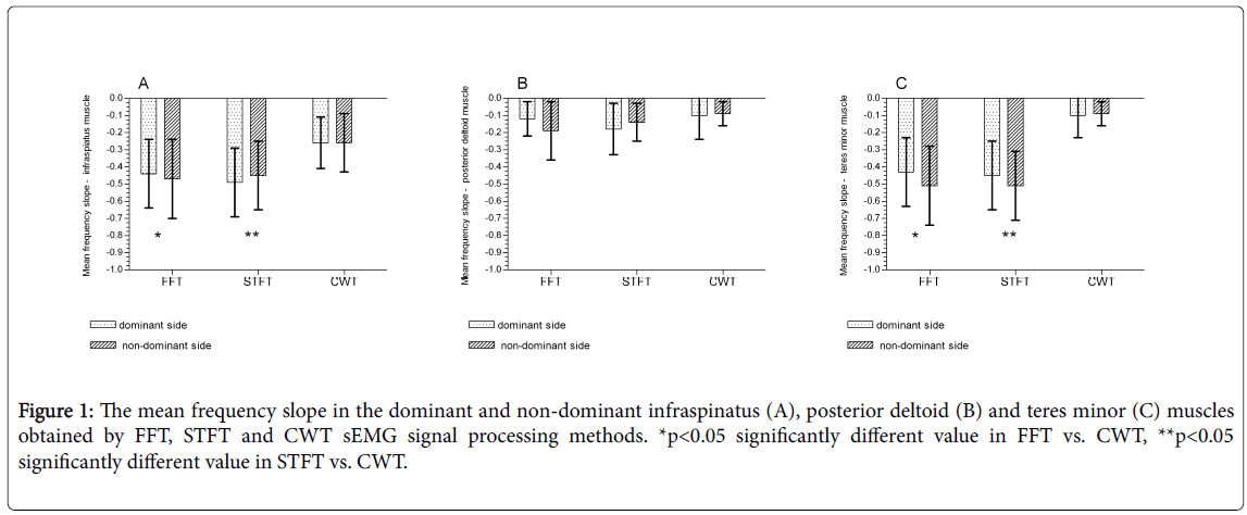
Figure 1: The mean frequency slope in the dominant and non-dominant infraspinatus (A), posterior deltoid (B) and teres minor (C) muscles obtained by FFT, STFT and CWT sEMG signal processing methods. *p<0.05 significantly different value in FFT vs. CWT, **p<0.05 significantly different value in STFT vs. CWT.
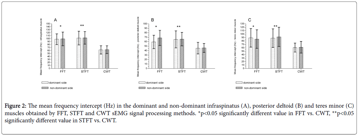
Figure 2: The mean frequency intercept (Hz) in the dominant and non-dominant infraspinatus (A), posterior deltoid (B) and teres minor (C) muscles obtained by FFT, STFT and CWT sEMG signal processing methods. *p<0.05 significantly different value in FFT vs. CWT, **p<0.05 significantly different value in STFT vs. CWT.
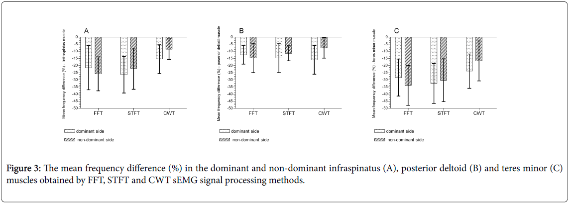
Figure 3: The mean frequency difference (%) in the dominant and non-dominant infraspinatus (A), posterior deltoid (B) and teres minor (C) muscles obtained by FFT, STFT and CWT sEMG signal processing methods.
Comparison of sEMG signal processing methods
FFT and STFT showed similar values in almost all the evaluated muscles. There were no significant differences between sEMG signal processing methods in mean frequency slope values (Figure 1), in mean frequency intercept values (Figure 2) and in mean frequency difference (Figure 3). The lack of significant differences in muscle fatigue indices obtained with FFT and STFT allow considering them as equal.
The sEMG signal processing using CWT showed different results in comparison to FFT and STFT. The significantly higher values of mean frequency slope was observed for infraspinatus (Figure 1A) and teres minor (Figure 1C) muscles. Significantly lower values of mean frequency intercept were observed for the infraspinatus (Figure 2A), posterior deltoid (Figure 2B) and the teres minor (Figure 2C) muscles. There were no significant differences observed in mean frequency difference for all the evaluated muscles (Figure 3).
Discussion
The most important information obtained in this study is the observation that in elite volleyball players, the fatigue indices in muscles of the shoulder region were similar on both the-dominant and non-dominant sides. The lack of significant differences in fatigability between shoulder muscles which are more loaded during training (dominant) and less loaded (non-dominant) may suggest that muscles on both sides were similarly resistant to fatigue. It may indicate that intensive training without additional risk factors of shoulder strain injury does not lead to chronic changes in muscle fatigability. In other words, we have hypothesized that asymmetric shoulder loading during volleyball training should not be considered as an obvious factor increasing the risk of shoulder injury. Based on our results, we have suggested that this fatigue was similar on both sides sin volleyball play, therefore, the risk of injury due to this fatigue is also similar on both sides, and the more loaded, dominant shoulder may not be more prone to injury than the non-dominant one.
The other important observation from our study is that muscle fatigue indices measured by sEMG may reach different values depending on the used signal processing method. If the parameters describing muscle fatigability (e.g. mean frequency slope, intercept or difference) are calculated via different processing methods, the results obtained may greatly vary. Therefore, the comparison between measurements (e.g. between different subjects), without knowledge how those values were calculated, may be misleading and clinically, such comparisons may lead to misdiagnosis. The significant differences shown in our study between values of a single parameter (e.g. mean frequency slope) calculated by different signal processing is the example of such a situation. In other words, due to signal processing, we may obtain significantly different values of mean frequency slope, even if they are calculated from the same sEMG record. Consequently, if we do not know the details of how this value was calculated, we should not compare data from two different subjects or results from different studies. This difference is clinically important because we may obtain conflicting results, even from the same subject e.g. if s/he is be evaluated at two different clinics during recovery from shoulder injury. Because volleyball is an asymmetric sports discipline, the potential implications of asymmetric training loads in relation to high prevalence of strain and acute shoulder injury occurrence should be addressed.
Postural asymmetry is considered a risk factor for injuries [23]. Some authors have suggested that the dominant shoulder of volleyball players is biomechanically and morphologically different to their nondominant shoulder [2,24]. They have reported the occurrence of adaptive changes in the dominant shoulder due to asymmetric training and have indicated that those adaptations may be associated with shoulder injury and pain [7,24,25].
Some studies have reported scapulohumeral rhythm asymmetry in overhead athletes between their dominant and non-dominant shoulder [24,26]. However, they have suggested that some scapular asymmetry may be common in overhead athletes and it should not be considered as pathology but as adaptation to extensive use of the dominant upper limb [23]. Also, Hosseinimehr et al. [24] have indicated that in unilateral overhead athletes, the asymmetric scapular posture between the dominant and non-dominant sides may be normal and might not be related to injury.
Those observations stay in line with the results of our study in which the lack of differences between dominant and non-dominant shoulder muscle fatigability have suggested that asymmetric training in volleyball players may not lead to pathological changes in the more loaded - dominant shoulder. Even if some biomechanical changes appear as an adaptation to asymmetric loads it probably does not cause muscle overload. It was also suggested that muscle fatigue may lead to an increase in movement variability which is a strategy decreasing the risk of overuse injury [6]. Some authors have reported that increased upper extremity kinematic variability and altered motor coordination associated with muscle fatigue may be present without any visible alterations in task performance [3,6].
The asymmetry in athletes without previous history of shoulder pain or injury may be considered as natural and not related to increased risk of injury, but the situation in previously injured individuals appears differently. Burkhat et al. [25] have noted the asymmetry in injured overhead athletes and they reported increased scapular protraction, anterior tilting and internal rotation of the scapula in the symptomatic side. Other authors have reported decreased dominant shoulder internal rotation range of motion, deficits in external rotator eccentric peak torque and higher rotator fatigability in volleyball players. In those studies, all changes were correlated with previous shoulder pain or injury [2,27,28].
Many authors have reported that repetitive shoulder movements may lead to cumulative biomechanical loading and to muscle fatigue [3,29,30]. It was suggested that this fatigue may lead to changes in muscle activation patterns and in shoulder kinematics [18,31,32]. Joshi et al. [31] have reported that fatigue-induced alterations in the lower trapezius might predispose the infraspinatus to injury through chronically increased activation.
However, none of the previous studies have described the differences in muscle fatigability between the dominant and nondominant shoulder in overhead athletes. They have reported asymmetries in muscle strength [28,31] or in shoulder and scapular kinematics [32,33] but there is a lack of studies analyzing asymmetries in muscle bioelectrical activity measured with sEMG. As was reported, surface electromiography is a sensitive and appropriate method for evaluation of muscle imbalance or acute and chronic fatigue [9-11,16,17]. The changes in muscle bioelectrical activity due to fatiguing effort are considered as a reliable diagnostic method of muscles performance [16,17]. Because we did not observe any significant differences between dominant and non-dominant shoulder muscle fatigability in our study, we think that both arms of the evaluated volleyball players reacted similarly to the fatiguing effort. Based on this observation, we may suggest that in overhead athletes without previous shoulder injury, even if some potential asymmetries due to training loads are present, they do not probably influence the muscle fatigability.
Mc Donald et al. [8] reported that shoulder complex and rotator cuff muscles require a long recovery time after fatigue. Therefore, during the intensive training period with inadequate rest opportunities, chronic fatigue may cause shoulder strain injuries [8,34,35]. Also, the importance of muscle fatigue as a risk factor of shoulder injury in overhead athletes was underlined by Andrade et al. [36] They have reported a higher incidence of injuries late in the game than during the early period and suggested that this fatigue can affect muscular strength balance and consequently, shoulder joint stabilization.
With sEMG, we may quantify neuromuscular fatigue during muscular work [11,12]. Its advantages are: non-invasiveness and realtime fatigue monitoring during performance [13]. It was reported that in evaluation of muscles imbalance due to chronic or acute fatigue, the small alterations in muscle bioelectrical activity may be considered as a sensitive and objective sign of muscle overloading [9,16]. Thus, the differences between contralateral muscle fatigability may suggest some muscles imbalance, which may be a potential factor increasing the risk of strains or acute injury.
The sEMG is widely used in diagnosis of muscle disorders and as a reliable evaluation method of muscle recovery efficacy during training and post-injury rehabilitation [10,11]. The crucial element of sEMG accuracy in muscle evaluation is the use of appropriate signal processing method. There are some studies showing that the value of muscle fatigue parameters derived from sEMG signal may vary due to the used signal processing method [16,17,21,22]. This appears to be an important limitation when we need to compare data from two other bioelectrical signal measurements. Very often, our diagnosis is based on muscle activity clinical reports containing only final values of fatigue variables, without any information on how they were calculated. The clinicians usually do not realize that if the same sEMG record is processed by two different methods, the results may be different. The comparison of two reports with the same final variables, but obtained by different sEMG signal processing methods, is not possible, and such comparison may cause misdiagnosis. The results of our study showed that muscle fatigue assessed with continuous wavelet transform (CWT) signal processing was significantly different from the other two methods used.
There are some limitations of this study that need to be addressed. First, the study population consisted of volleyball players without previous shoulder injury, thus, it may not be possible to extrapolate these findings to overhead athletes with a history of shoulder injury. Additionally, the present study involved only volleyball players, so we think that future studies should also include other overhead athletes.
The most important information obtained in this study is the observation that in elite volleyball players without previous shoulder injury, the fatigue indices in muscles of the shoulder region were similar on both the dominant and non-dominant sides. Therefore, we have hypothesized that asymmetric shoulder loading during volleyball training should not be considered as an obvious factor increasing the risk of shoulder injury. The other important observation from our study is that muscle fatigue indices measured by sEMG may be a sensitive and objective method of evaluation, but may reach different values depending on the used signal processing method. Consequently, the clinical interpretation and any comparison between different measurements, without knowledge of how those values were calculated, may be misleading and be the reason for misdiagnosis.
References
- Hickey D, Solvig V, Cavalheri V, Harrold M, Mckenna L (2018) Scapular dyskinesis increases the risk of future shoulder pain by 43% in asymptomatic athletes: a systematic review and meta-analysis. Br J Sports Med 52: 102-110.
- Tonin K, Stražar K, Burger H, Vidmar G (2013) Adaptive changes in the dominant shoulders of female professional overhead athletes: mutual association and relation to shoulder injury. Int J Rehabil Res 36: 228-235.
- Qin J, Lin JH, Faber GS, Buchholz B, Xu X (2014) Upper extremity kinematic and kinetic adaptations during a fatiguing repetitive task. J Electromyogr Kinesiol 24: 404-411.
- Tse CT, McDonald AC, Keir PJ (2016) Adaptations to isolated shoulder fatigue during simulated repetitive work. Part I: Fatigue. J Electromyogr Kinesiol 29: 34-41.
- Spargoli G (2017) The acute effects of concentric versus eccentric muscle fatigue on shoulder active repositioning sense. Int J Sports Phys Ther 12: 219-226.
- Cowley JC, Gates DH (2017) Proximal and distal muscle fatigue differentially affect movement coordination. PLoS One 12: e0172835.
- Challoumas D, Stavrou A, Dimitrakakis G (2017) The volleyball athlete's shoulder: biomechanical adaptations and injury associations. Sports Biomech 16: 220-237.
- McDonald AC, Tse CT, Keir PJ (2016) Adaptations to isolated shoulder fatigue during simulated repetitive work. Part II: Recovery. J Electromyogr Kinesiol 29: 42-49.
- Farina D (2006) Interpretation of the surface electromyogram in dynamic contractions. Exerc Sport Sci Rev 34: 121-127.
- Ament W, Bonga GJJ, Hof AL, Verkerke GJ (1996) Electromyogram median power frequency in dynamic exercise at medium exercise intensities. Eur J Appl Physiol Occup Physiol 74: 180-186.
- DeLuca CJ (1984) Myoelectrical manifestations of localized muscular fatigue in human. Crit Rev Biomed Eng 11: 251-279.
- Gandevia SC (2001) Spinal and supraspinal factors in human muscle fatigue. Physiol Rev 81: 1725-1789
- Merletti R, Parker P (2004) Electromyography: Physiology, Engineering, and Non-Invasive Applications. Wiley-IEEE Press, USA.
- Cifrek M, Medved V, Tonković S, Ostojić S (2009) Surface EMG based muscle fatigue evaluation in biomechanics. Clin Biomech 24: 327-340.
- Coorevits P, Danneels L, Cambier D, Ramon H, Druyts H, et al. (2008) Correlations between short-time Fourier- and continuous wavelet transforms in the analysis of localized back and hip muscle fatigue during isometric contractions. J Electromyogr Kinesiol 18: 637-644.
- Shair EF, Ahmad SA, Marhaban MH, Mohd Tamrin SB, Abdullah AR (2017) EMG Processing Based Measures of Fatigue Assessment during Manual Lifting. Biomed Res Int 2017: 3937254.
- Dantas JL, Camata TV, Brunetto MA, Moraes AC, Abrão T, et al. (2010) Fourier and wavelet spectral analysis of EMG signals in isometric and dynamic maximal effort exercise. Conf Proc IEEE Eng Med Biol Soc 2010: 5979-5982.
- Kibler WB, Wilkes T, Sciascia A (2013) Mechanics and pathomechanics in the overhead athlete. Clin Sports Med 32: 637-651.
- Hermens HJ, Freriks B, Disselhorst-Klug C, Rau G (2000) Development of recommendations for SEMG sensors and sensor placement procedures. J Electromyogr Kinesiol 10: 361-374.
- Coorevits P, Danneels L, Cambier D, Ramon H, Druyts H, et al. (2008) Test-retest reliability of wavelet - and Fourier based EMG (instantaneous) median frequencies in the evaluation of back and hip muscle fatigue during isometric back extensions. J Electromyogr Kinesiol 18: 798-806.
- Costa MV, Pereira LA, Oliveira RS, Pedro RE, Camata TV, et al. (2010) Fourier and wavelet spectral analysis of EMG signals in maximal constant load dynamic exercise. Conf Proc IEEE Eng Med Biol Soc 2010: 4622-4625.
- MacIsaac D, Parker PA, Scott RN (2001) The short-time Fourier transform and muscle fatigue assessment in dynamic contractions. J Electromyogr Kinesiol 11: 439-449.
- Oyama S, Myers JB, Wassinger CA, Daniel Ricci R, Lephart SM (2008) Asymmetric resting scapular posture in healthy overhead athletes. J Athl Train 43: 565-570.
- Hosseinimehr SH, Anbarian M, Norasteh AA, Fardmal J, Khosravi MT (2015) The comparison of scapular upward rotation and scapulohumeral rhythm between dominant and non-dominant shoulder in male overhead athletes and non-athletes. Man Ther 20: 758-762.
- Burkhart SS, Morgan CD, Kibler WB (2003) The disabled throwing shoulder: spectrum of pathology Part I: pathoanatomy and biomechanics. Arthroscopy 19: 404-420.
- Ozunlu N, Tekeli H, Baltaci G (2011) Lateral scapular slide test and scapular mobility in volleyball players. J Athl Train 46: 438-444.
- Myers JB, Guskiewicz KM, Schneider RA, Prentice WE (1999) Proprioception and neuromuscular control of the shoulder after muscle fatigue. J Athl Train 34: 362-367.
- Lee HM, Liau JJ, Cheng CK, Tan CM, Shih JT (2003) Evaluation of shoulder proprioception following muscle fatigue. Clin Biomech 18: 843-847.
- Jensen BR, Laursen B, Sjogaard G (2000) Aspects of shoulder function in relation to exposure demands and fatigue – a mini review. Clin Biomech (Bristol, Avon) 15: S17-S20.
- van Rijn RM, Huisstede BM, Koes BW, Burdorf A (2010) Associations between work-related factors and specific disorders of the shoulder – a systematic review of the literature. Scand J Work Environ Health 36: 189-201.
- Joshi M, Thigpen CA, Bunn K (2011) Shoulder external rotation fatigue and scapular muscle activation and kinematics in overhead Athletes. J Athl Train 46: 349-357.
- Maenhout A, Dhooge F, Van Herzeele M (2015) Acromiohumeral distance and 3-dimensional scapular position change after overhead muscle fatigue. J Athl Train 50: 281-288.
- Ebaugh DD, McClure PW, Karduna AR (2006) Effects of shoulder muscle fatigue caused by repetitive overhead activities on scapulothoracic and glenohumeral kinematics. J Electromyogr Kinesiol 16: 224-235.
- Karduna AR, Williams GR, Williams JL, Iannotti JP (1996) Kinematics of the glenohumeral joint: influences of muscle forces, ligamentous constraints, and articular geometry. J Orthop Res 14: 986-993.
- Elliott MB, Barr AE, Kietrys DM, Al-Shatti T, Amin M, et al. (2008) Peripheral neuritis and increased spinal cord neurochemicals are induced in a model of repetitive motion injury with low force and repetition exposure. Brain Res 1218: 103-113.
- Andrade MS, de Carvalho Koffes F, Benedito-Silva AA, da Silva AC, de Lira CA (2016) Effect of fatigue caused by a simulated handball game on ball throwing velocity, shoulder muscle strength and balance ratio: a prospective study. BMC Sports Sci Med Rehabil 8: 13.
Citation: Oleksy L, Czarny W, Bajorek W, Król P, Mika A, et al. (2018) The Evaluation of Shoulder Muscle Fatigue in Volleyball Players. J Nov Physiother 8: 388. DOI: 10.4172/2165-7025.1000388
Copyright: © 2018 Oleksy L, et al. This is an open-access article distributed under the terms of the Creative Commons Attribution License, which permits unrestricted use, distribution, and reproduction in any medium, provided the original author and source are credited.
Select your language of interest to view the total content in your interested language
Share This Article
Recommended Journals
Open Access Journals
Article Tools
Article Usage
- Total views: 7312
- [From(publication date): 0-2018 - Nov 25, 2025]
- Breakdown by view type
- HTML page views: 6232
- PDF downloads: 1080