The Laser and Ultrasound: The Ultra Laser like Efficient Treatment to Fibromyalgia by Palms of Hands - Comparative Study
Received: 19-Nov-2020 / Accepted Date: 03-Dec-2020 / Published Date: 10-Dec-2020
Abstract
Fibromyalgia, a chronic disease, affects skeletal muscles and soft tissues, causing disabling pain. The most used physiotherapeutic treatments are the resources of therapeutic ultrasound and therapeutic LASER, responsible for the anti-inflammatory and analgesic action. Currently, the synergistic action of these resources has emerged as an alternative for treatment with application on the palms. The conceptual basis of treatment occurs through the stimulation of neuroreceptors close to the blood vessels located in the palms of the hands, an area that is extremely rich in nerve endings. The aim of this study was to evaluate variations in technological parameters, forms of application in the hypotenar and tenar areas of the palms and the influence on the variability in the number of weekly sessions. The evaluation was based on the Fibromyalgia Impact Questionnaire and Visual Analgue Scale, allowing evaluating the application on Trigger Points versus Palms, different ultrasound frequencies, different LASER wavelengths, and different application regions on the palms of the hands, number of weekly sessions and percentage of improvement through the proposed treatment. The results show that the treatment used on the palms is more effective, confirming 1 MHZ of ultrasound intensity, 660 nm of wavelength, Hypotenar plus Tenar regions of the palms and 3 times a week as a protocol. It points out that 70% of the patients obtained 50% improvement in the pain of fibromyalgia, laying the foundations for a new methodology, the Photosonic treatment, reducing pain and increasing the quality of life of patients affected by fibromyalgia.
Keywords: Fibromyalgia; Ultralaser; Palms of the hands; Quality of life; Decreased pain
Introduction
Fibromyalgia, a chronic disease, with an inflammatory, hyperalgesic and disabling characteristic, is considered a contemporary disease, which appears in high growth among the population, due to the improvement of the clinical diagnosis based solely on symptoms and signs. The main evaluated points called trigger points are the clinical examination points that allow clinically diagnosing the disease, which in all are 18 trigger points [1].
Its prevalence occurs in a great majority among women. However, there is also the occurrence in men, but in a smaller amount. There are estimates that fibromyalgia may affect 3% to 10% of the population considered to be adults. Over time, there were several names of fibromyalgia. The first record of the disease was found in 1924, in England, where cases of patients who had tender points, with greater sensitivity to brief manual pressure, were reported.
All these characteristics mentioned lead fibromyalgia to interfere widely in the patient’s lifestyle and daily routine (both personal and professional) [2]. Thus, there is an immense impact on personal, social, professional patient environments [3], causing an extremely negative condition in the quality of life of the patient and their family [4,5].
The treatments proposed for this disease are pharmacological and non-pharmacological. In pharmacological intervention, there is a wide use of analgesics, anti-inflammatories, antidepressants, anxiolytics and anticonvulsants, both to reduce inflammation and to control pain attacks [6,7]. Among the non-pharmacological treatment options, exercise and diet can be included for the clinical improvement of fibromyalgia patients [8-12]. Still, as means of non-pharmacological treatments, there are technological resources, such as therapeutic ultrasound and therapeutic laser, which have a great analgesic condition.
Ultrasound, a technological resource widely used, allow to reduce muscle pain due to its analgesic and anti-inflammatory power, as well as, its thermal action, conditioning a scenario of vasodilation and increased signaling speed [13]. Another technological resource, the therapeutic laser, is efficient in anti-inflammatory and analgesic action, where through a modulatory process of mitochondrial enzymes, greater production of ATP (Adenosine Triphosphate) occurs. Still, its antiinflammatory action is done through the activation of transcriptional factors, as well as synthesis of new proteins and cells proliferation [14].
Recently, equipment was developed by the São Carlos Institute of Physics, University of São Paulo, capable of simultaneously promoting therapeutic ultrasound and therapeutic laser, enabling a synergistic action of therapies used in previous studies not only in the treatment of fibromyalgia but also in the treatment of osteoarthritis. The equipment, called Ultralaser, allow it to occur the overlapping fields of the therapeutic resource, potentiating the analgesic and anti-inflammatory action, observed in the resources when separately [15-18]. Clinical observation points to an improvement in the daily functionality of patients in their activities, with a great reduction in pain, which was previously disabling.
Another important feature is the innovation in the treatment methodology, which transcends the treatment performed at pain points and addresses the treatment through the patients’ palms. In analyzed studies, the palms of patients affected by fibromyalgia have a greater amount of sensory nerve fibers close to blood vessels [19]. There is also the possible condition that neurovascular disorders cause changes in the patient’s pain threshold, providing generalized muscle fatigue due to the lower oxygen supply, causing severe sleep disturbances through hyperalgia [20]. Another study points to changes in cerebral and peripheral blood flow that can influence the hyperalgia of the disease, affecting the organism as a whole [21].
The aim of this study was to evaluate variations in technological parameters, forms of application in hypotenar and tenar areas of the palms and the influence on the variability in the number of weekly sessions.
Materials and Methods
Approval and Location
This study was approved by the Research Ethics Committee and the National Research Ethics Committee through CAAE 13789319.5.0000.8148, following resolution 466/2012. The study was carried out on the premises of the Photodynamic Therapy Unit of Santa Casa de Misericórdia de São Carlos, under the coordination of the São Carlos Physics Institute, University of São Paulo, São Carlos, São Paulo, Brazil and at the MultFISIO Brasil Clinics in São Carlos, São Paulo, Brazil and Ribeirão Preto, São Paulo, Brazil.
Patients and Research Groups
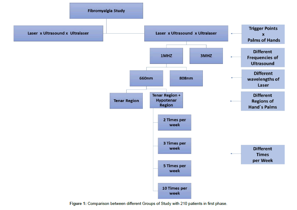
In this study, 450 female patients aged between 30 and 65 years were selected. The following comparisons were made in first phase: applications to pain points versus application to the palms of hands (6 groups, n=15); applications on the palms of hands with variation between 1 MHZ versus 3 MHZ ultrasound frequencies (2 groups, n=10); applications on the palms of hands with variation between wavelengths 660 nm versus 808 nm (2 groups, n=10); applications on the palms of hands with variation between application only in the Hypotenar region versus application in the Hypotenar plus Tenar region (2 groups, n=10); applications on the palms of hands, Tenar plus Hipotenar region, with variation in the number of weekly sessions, being 2 sessions, 3 sessions, 5 sessions and 10 weekly sessions (Table 1), considering the total number of sessions, regardless of the treatment time, 10 sessions in total (4 groups, n=15). These comparions are observed in Figure 1. The following comparison was made in second phase: application of the best option between the comparisons of variables, in a standard protocol (Table 2), considering the applications on the palms of hands, Hypotenar plus Tenar region, ultrasound frequency of 1 MHZ and wavelength 660 nm, variation in the number of weekly sessions, (n=240), separating the various percentages of response to treatment (10% ranges in the 0 to 100% response range).
| Weekly Sessions | Time of Treatment | Intervals Between Sessions |
| 2 sessions | 5 weeks | 84 hours (3.5 days) |
| 3 sessions | 3.1 weeks | 48 hours (2 days) |
| 5 sessions | 2 weeks | 24 hours (1day) |
| 10 sessions | 1 week | 4 hours (0 day) |
All session frequency variations have a total of 10 sessions.
Table 1: Number of weekly sessions, treatment time and interval between sessions.
| Groups | Protocols |
| Laser Protocol | Wavelength of 660 nm, continuous mode, power of 100 mW. Time of 3 minutes per point of each palm of hands. |
| Ultrasound Protocol | Pulsed mode, 1 MHz frequency, 100 Hz, 50% duty cycle and average space time of 0.5 w/cm2 (SATA). Time of 3 minutes per point of each palm of hands. |
| Ultralaser Protocol | Wavelength of 660 nm, continuous mode, power of 100 mW. Pulsed mode, 1 MHz frequency, 100 Hz, 50% duty cycle and average space time of 0.5 w/cm2 (SATA). Time of 3 minutes per point of each palm of hands. |
Table with parameters used.
Table 2: Protocols of Treatment.
Application Points
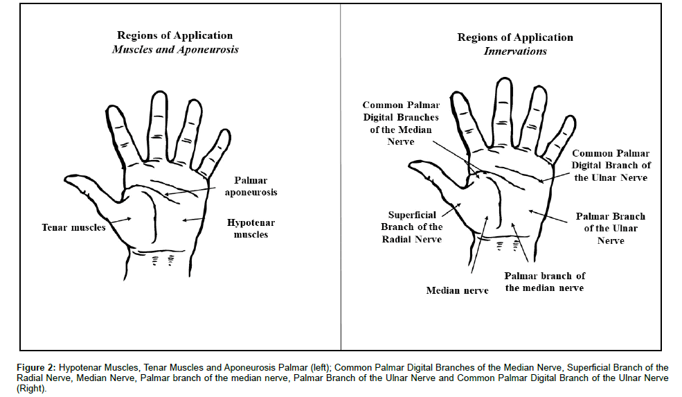
The application of therapy in the so-called trigger points was carried out in the region of the trapezius and arms, both in the posterior region, totaling 4 points of application, performed in a rotating manner. The application on the palms of the hands was performed in the Hypotenar Muscles, Tenar Muscles and Aponeurosis Palmar, in both hands, totaling 2 points of application and performed in a rotating manner. In this area, there are large numbers of innervations, being: Common Palmar Digital Branches of the Median Nerve, Superficial Branch of the Radial Nerve, Median Nerve, Palmar branch of the median nerve, Palmar Branch of the Ulnar Nerve and Common Palmar Digital Branch of the Ulnar Nerve (Figure 2).
Figure 2: Hypotenar Muscles, Tenar Muscles and Aponeurosis Palmar (left); Common Palmar Digital Branches of the Median Nerve, Superficial Branch of the Radial Nerve, Median Nerve, Palmar branch of the median nerve, Palmar Branch of the Ulnar Nerve and Common Palmar Digital Branch of the Ulnar Nerve (Right).
Equipment and Patent
The present study used a phase 3 prototype developed by the São Carlos Physics Institute (University of São Paulo), with the Technical Support Laboratory (LAT), patent number BR102014007397-3 A2. The current prototype was developed by MMOptics, São Carlos, São Paulo, Brazil. The equipment has a synergistic capacity for simultaneous application of Therapeutic Laser and Therapeutic Ultrasound, including the formation of an “overlap of therapeutic fields”.
Questionnaire and Scale
Patients were analyzed, regardless of the comparison made, in the initial situation and after the treatment period. As a form of evaluation, the Fibromyalgia Impact Questionnaire was carried out, or what is the possibility of measuring the improvement in the patient’s quality of life. In addition, the Visual Analogue Scale was performed, a visual scale that allows to measure the use of the questionnaire with the patient [15,16,22]. In addition, as a way to normalize the data, a percentage form of Delta [23] was used, expressed in:
“Delta Value (Δ) = (Initial value - Final value) / Initial value × (100)”
Used to data of Fibromyalgia Impact Questionnaire, and:
“Delta Value (Δ) = (final value - initial value) / final value × (100)”
Used to data of Visual Analogue Scale.
Statistical Treatment
Statistical analysis was performed using Instat 3.0 softwares for Windows 7 (Graph Pad, San Diego, CA, USA, 1998). All data were expressed as mean and standard deviation. The level of significance was set at p<0.05. The Kolmogorov-Smirnov test was used to analyze the normality of the data. Subsequently, a one-way ANOVA with a posttest was performed, using Student-Newman-Keuls for parametric data in comparison between protocols and post-hoc-Tukey-Kramer for comparison between therapeutic resources.
Results
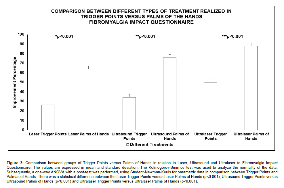
The present study made several comparisons, considering several variables. Figure 3 shows the comparison of the application on pain points in relation to the application on the palms, considering the treatment performed only with Laser or Ultrasound and the combined treatment with Ultralaser in relation to the Fibromyalgia Impact Questionnaire. In this figure, where the groups have n=15, it is observed that in all comparisons performed, there was a better result for the treatment performed on the palms when compared to the pain points, in relation to the therapeutic resources. This difference is seen in the comparison Laser Trigger Points versus Laser Palms of Hands (p<0.001), Ultrasound Trigger Points versus Ultrasound Palms of Hands (p<0.001) and Ultralaser Trigger Points versus Ultralaser Palms of Hands (p<0.001).
Figure 3: Comparison between groups of Trigger Points versus Palms of Hands in relation to Laser, Ultrasound and Ultralaser to Fibromyalgia Impact Questionnaire. The values are expressed in mean and standard deviation. The Kolmogorov-Smirnov test was used to analyze the normality of the data. Subsequently, a one-way ANOVA with a post-test was performed, using Student-Newman-Keuls for parametric data in comparison between Trigger Points and Palmas of Hands. There was a statistical difference between the Laser Trigger Points versus Laser Palms of Hands (p<0.001), Ultrasound Trigger Points versus Ultrasound Palms of Hands (p<0.001) and Ultralaser Trigger Points versus Ultralaser Palms of Hands (p<0.001).
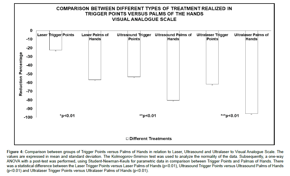
The Figure 4 illustrates the comparison of the application at the pain points in relation to the application on the palms, considering the treatment performed only with Laser or Ultrasound and the combined treatment with Ultralaser in relation to the Visual Analogue Scale. In this figure, where the groups have n=15, it is observed that in all comparisons performed, there was a better result for the treatment performed on the palms when compared to the pain points, in relation to the therapeutic resources. This difference is seen in the comparison Laser Trigger Points versus Laser Palms of Hands (p<0.01), Ultrasound Trigger Points versus Ultrasound Palms of Hands (p<0.01) and Ultralaser Trigger Points versus Ultralaser Palms of Hands (p<0.01).
Figure 4: Comparison between groups of Trigger Points versus Palms of Hands in relation to Laser, Ultrasound and Ultralaser to Visual Analogue Scale. The values are expressed in mean and standard deviation. The Kolmogorov-Smirnov test was used to analyze the normality of the data. Subsequently, a one-way ANOVA with a post-test was performed, using Student-Newman-Keuls for parametric data in comparison between Trigger Points and Palmas of Hands. There was a statistical difference between the Laser Trigger Points versus Laser Palms of Hands (p<0.01), Ultrasound Trigger Points versus Ultrasound Palms of Hands (p<0.01) and Ultralaser Trigger Points versus Ultralaser Palms of Hands (p<0.01).
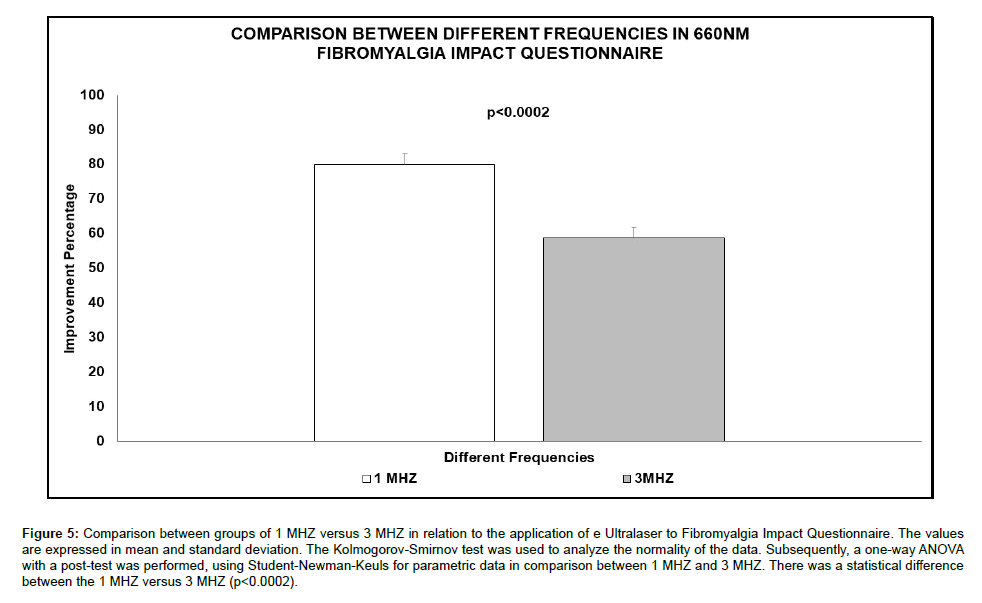
Figure 5 illustrates the use of Ultralaser by comparing the different ultrasound frequencies (1 MHZ and 3 MHZ), when the 660 nm wavelength is used in the application on the palms, in relation to the Fibromyalgia Impact Questionnaire (n=10). It is observed that in the comparison between the frequencies, the frequency 1 MHZ was more efficient in relation to the frequency of 3MHZ (p<0.01).
Figure 5: Comparison between groups of 1 MHZ versus 3 MHZ in relation to the application of e Ultralaser to Fibromyalgia Impact Questionnaire. The values are expressed in mean and standard deviation. The Kolmogorov-Smirnov test was used to analyze the normality of the data. Subsequently, a one-way ANOVA with a post-test was performed, using Student-Newman-Keuls for parametric data in comparison between 1 MHZ and 3 MHZ. There was a statistical difference between the 1 MHZ versus 3 MHZ (p<0.0002).
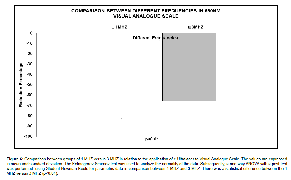
Figure 6 shows the result of using Ultralaser in the comparison of the different ultrasound frequencies (1 MHZ and 3 MHZ), when the 660 nm wavelength is used in the application on the palms, in relation to the Visual Analogue Scale (n=10). It is possible to observe in the comparison between the frequencies, the frequency 1 MHZ was more efficient in relation to the frequency of 3 MHZ (p<0.01).
Figure 6: Comparison between groups of 1 MHZ versus 3 MHZ in relation to the application of e Ultralaser to Visual Analogue Scale. The values are expressed in mean and standard deviation. The Kolmogorov-Smirnov test was used to analyze the normality of the data. Subsequently, a one-way ANOVA with a post-test was performed, using Student-Newman-Keuls for parametric data in comparison between 1 MHZ and 3 MHZ. There was a statistical difference between the 1 MHZ versus 3 MHZ (p<0.01).
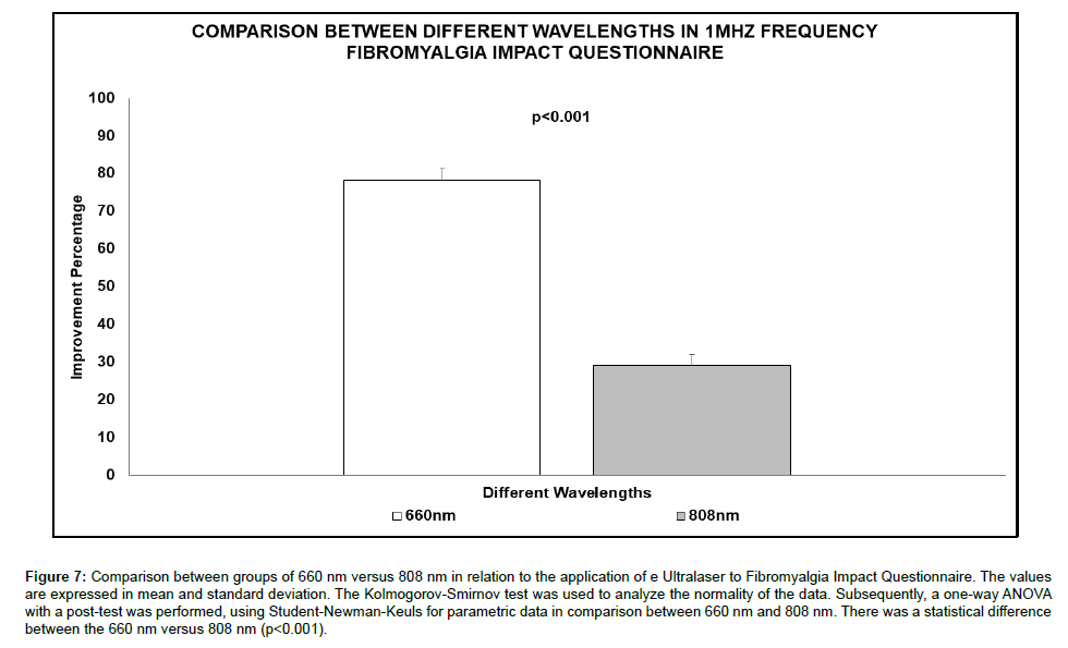
Figure 7 shows the use of Ultralaser by comparing the different wavelengths (660 nm and 808 nm), when the frequency of 1 MHZ is used in the application on the palms, in relation to the Fibromyalgia Impact Questionnaire (n=10). It is observed that in the comparison between the wavelengths, the 660 nm wavelength proved to be more efficient in relation to the 808 nm (p<0.001).
Figure 7: Comparison between groups of 660 nm versus 808 nm in relation to the application of e Ultralaser to Fibromyalgia Impact Questionnaire. The values are expressed in mean and standard deviation. The Kolmogorov-Smirnov test was used to analyze the normality of the data. Subsequently, a one-way ANOVA with a post-test was performed, using Student-Newman-Keuls for parametric data in comparison between 660 nm and 808 nm. There was a statistical difference between the 660 nm versus 808 nm (p<0.001).
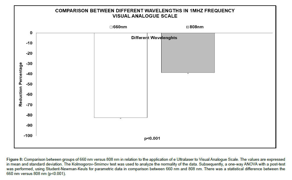
In Figure 8, it is possible to observe the Ultralaser by comparing the different wavelengths (660 nm and 808 nm), when using the frequency of 1 MHZ in the application on the palms, in relation to the Visual Analogue Scale (n=10). It is shown that in the comparison between wavelengths, the 660 nm wavelength proved to be more efficient compared to 808 nm (p <0.001).
Figure 8: Comparison between groups of 660 nm versus 808 nm in relation to the application of e Ultralaser to Visual Analogue Scale. The values are expressed in mean and standard deviation. The Kolmogorov-Smirnov test was used to analyze the normality of the data. Subsequently, a one-way ANOVA with a post-test was performed, using Student-Newman-Keuls for parametric data in comparison between 660 nm and 808 nm. There was a statistical difference between the 660 nm versus 808 nm (p<0.001).
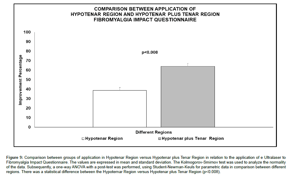
In Figure 9, it is possible to observe the Ultralaser applied to different regions of the palms (Hypotenar Region and Hypotenar more Tenar Region). The comparison of the application to different regions of the palms used an ultrasound frequency of 1 MHZ and 660 nm wavelengths, when the Fibromyalgia Impact Questionnaire (n=10) was evaluated. It is observed that in the comparison, the sum of the Hypotenar plus Tenar Region was more efficient in relation to the application only in the Tenar Region (p<0.008).
Figure 9: Comparison between groups of application in Hypotenar Region versus Hypotenar plus Tenar Region in relation to the application of e Ultralaser to Fibromyalgia Impact Questionnaire. The values are expressed in mean and standard deviation. The Kolmogorov-Smirnov test was used to analyze the normality of the data. Subsequently, a one-way ANOVA with a post-test was performed, using Student-Newman-Keuls for parametric data in comparison between different regions. There was a statistical difference between the Hypoternar Region versus Hypotenar plus Tenar Region (p<0.008).
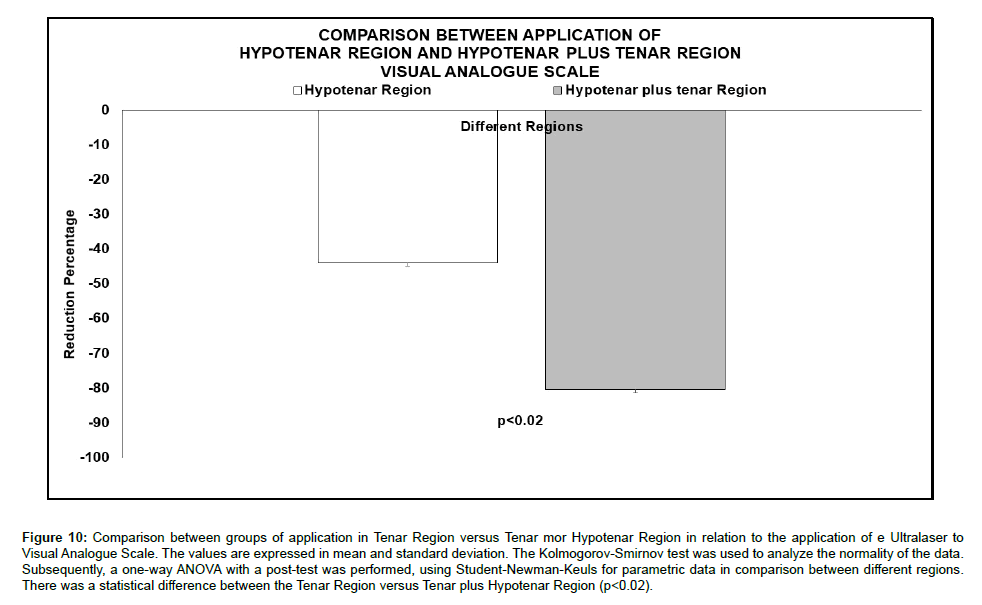
In Figure 10, the Ultralaser applied to different regions of the palms (Hypotenar Region and Hypotenar more Tenar Region) is shown. The comparison of the application of the different regions of the palms used an ultrasound frequency of 1 MHZ and 660 nm wavelengths, with the Visual Analogue Scale being evaluated (n=10). It is observed that in the comparison, the sum of the Hypotenar plus Tenar Region was more efficient in relation to the application only in the Tenar Region (p<0.02).
Figure 10: Comparison between groups of application in Tenar Region versus Tenar mor Hypotenar Region in relation to the application of e Ultralaser to Visual Analogue Scale. The values are expressed in mean and standard deviation. The Kolmogorov-Smirnov test was used to analyze the normality of the data. Subsequently, a one-way ANOVA with a post-test was performed, using Student-Newman-Keuls for parametric data in comparison between different regions. There was a statistical difference between the Tenar Region versus Tenar plus Hypotenar Region (p<0.02).
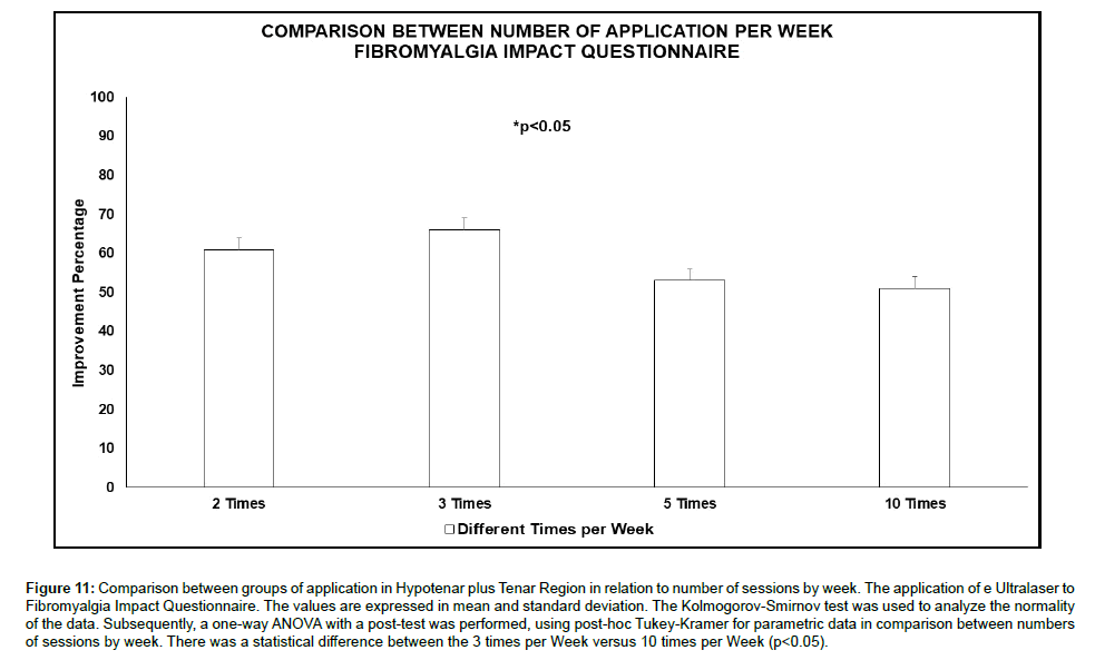
Figure 11 shows the Ultralaser applied to the palms of the hands, in Hypotenar and Tenar Region, considering the number of weekly sessions, ranging from 2 to 10 weekly sessions. The comparison of the weekly frequency of sessions used an ultrasound frequency of 1 MHZ and 660 nm wavelengths, with the Fibromyalgia Impact Questionnaire (n=15) being evaluated. It is shown that when comparing the different weekly frequencies, there is only a significant difference for 3 weekly sessions (p<0.05) in relation to 10 weekly sessions. However, all other frequencies show an improvement equal to or greater than 50%.
Figure 11: Comparison between groups of application in Hypotenar plus Tenar Region in relation to number of sessions by week. The application of e Ultralaser to Fibromyalgia Impact Questionnaire. The values are expressed in mean and standard deviation. The Kolmogorov-Smirnov test was used to analyze the normality of the data. Subsequently, a one-way ANOVA with a post-test was performed, using post-hoc Tukey-Kramer for parametric data in comparison between numbers of sessions by week. There was a statistical difference between the 3 times per Week versus 10 times per Week (p<0.05).
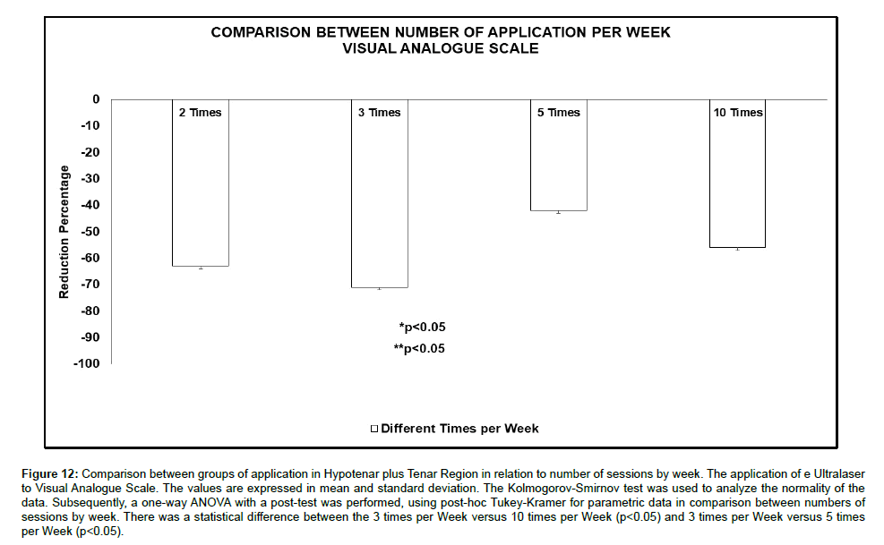
Figure 12 shows the application of Ultralaser, applied to the palms of the hands, in Hypotenar and Tenar Region, considering the number of weekly sessions, ranging from 2 to 10 weekly sessions. The comparison of the weekly frequency of sessions used as parameters of application of ultrasound frequency of 1 MHZ and wavelengths 660 nm, being evaluated the Visual Analogue Scale (n=15). It is shown that when comparing the different weekly frequencies, there is a significant difference for 3 weekly sessions (p<0.05) in relation to 5 weekly sessions and 10 weekly sessions. However, the frequency of 10 weekly sessions showed an improvement equal to or greater than 60%.
Figure 12: Comparison between groups of application in Hypotenar plus Tenar Region in relation to number of sessions by week. The application of e Ultralaser to Visual Analogue Scale. The values are expressed in mean and standard deviation. The Kolmogorov-Smirnov test was used to analyze the normality of the data. Subsequently, a one-way ANOVA with a post-test was performed, using post-hoc Tukey-Kramer for parametric data in comparison between numbers of sessions by week. There was a statistical difference between the 3 times per Week versus 10 times per Week (p<0.05) and 3 times per Week versus 5 times per Week (p<0.05).
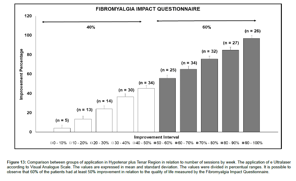
In Figure 13, the fragmentation in percentage ranges of the use of Ultralaser, applied to the palms of the hands, in Hypotenar and Tenar Region, regardless of the number of weekly sessions, is shown. The application of 1 MHZ ultrasound frequency and 660 nm wavelengths, with the Fibromyalgia Impact Questionnaire (n=240) being evaluated. It is shown that when comparing the fragmentation of percentage ranges that at least 60% of patients have at least 50% improvement in relation to their ability to perform daily activities. It is important to note that only 2.08% (n=5) are in the range of 0 to 10% improvement.
Figure 13: Comparison between groups of application in Hypotenar plus Tenar Region in relation to number of sessions by week. The application of e Ultralaser according to Visual Analogue Scale. The values are expressed in mean and standard deviation. The values were divided in percentual ranges. It is possible to observe that 60% of the patients had at least 50% improvement in relation to the quality of life measured by the Fibromyalgia Impact Questionnaire.
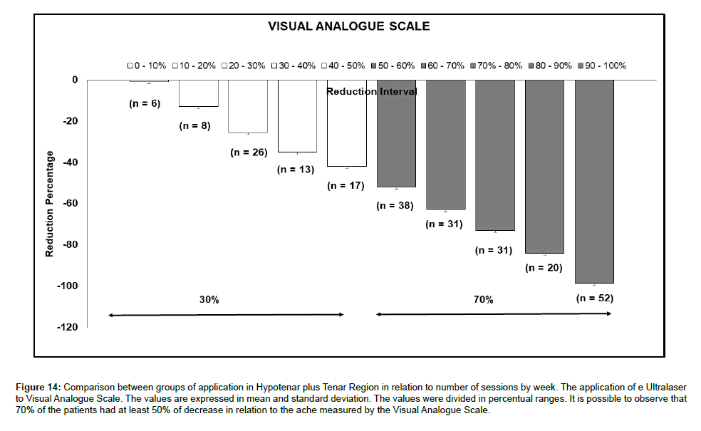
In Figure 14, the selection of percentage ranges for using the Ultralaser, applied to the palms of the hands, in Hypotenar and Tenar Region, regardless of the number of weekly sessions, is shown. The application of 1 MHZ ultrasound frequency and 660 nm wavelengths, being assessed using the Visual Analogue Scale (n=240). It is shown that when comparing the fragmentation of percentage ranges that at least 70% of patients have at least 50% improvement in relation to their ability to perform daily activities. It is important to note that only 2.5% (n=8) are in the range of 0 to 10% improvement.
Figure 14: Comparison between groups of application in Hypotenar plus Tenar Region in relation to number of sessions by week. The application of e Ultralaser to Visual Analogue Scale. The values are expressed in mean and standard deviation. The values were divided in percentual ranges. It is possible to observe that 70% of the patients had at least 50% of decrease in relation to the ache measured by the Visual Analogue Scale.
Discussion
A disease where the main characteristic is chronic disabling pain that is associated with a severe inflammatory condition, capable of resulting in a marked decrease in quality of life. So is Fibromyalgia. The physiotherapeutic treatments used to treat this disease are based on the action of therapeutic laser or therapeutic ultrasound, applied in isolation to the so-called trigger points, often achieving only brief relief from suffering. Thus, in our study, we aim to use a new technology that combines the application of therapeutic laser resources and therapeutic ultrasound, allowing the synergy of these resources in simultaneous application, called Ultralaser. Still, supported by Albrecht [19], where in his study he pointed out the existence of a large number of nerve endings close to blood vessels in the palms, in the hypotenar region, of fibromyalgia in relation to non-fibromyalgia, allowed the formation of the hypothesis of treatment via palms, as a possible gateway to the new therapy. Previous studies carried out by our group [15,16,22] have shown, both in case studies and in group comparisons, the effectiveness of the new proposal. In the present study, several comparisons were made, first determining the greater effectiveness of treatment when the resources of therapeutic laser and therapeutic ultrasound were performed synergistically, as well as in isolation (Figure 3 and 4). This technological innovation was tested both in the treatment of Fibromyalgia [15,16,22], and in Osteorthrosis [17,18,24,25]. Still, it was shown that the treatment on the palms has a better result than when applied to trigger points (Figure 3 and 4).
When analyzing the ultrasound frequency variables, it was observed that the frequency of 1 MHZ was more efficient than the frequency of 3 MHZ, showing that the range of greater tissue penetration depth is better received by the organism (Figure 5 and 6). It should be noted that in this case, in addition to the anti-inflammatory and analgesic action, there is the cavitation action, in addition to ionic and vascular permeability and stimulation of angiogenesis [26,27]. In addition, when comparing the laser wavelength, there was a better action of 660 nm compared to 808 nm, given that the action of the red light wavelength, through greater anti-inflammatory and analgesic action [28] was able to act better synergistically with ultrasound (Figure 7 and 8).
When comparing the application to areas determined by the anatomical differentiation found by Albercht [19], hypotenar region in relation to a broader application, with coverage in the hypotenar, tenar and palmar aponeurosis regions (Figure 1), greater efficiency is indicated when applied to greater coverage, not limiting the palmar region of application (Figure 9 and 10). The recommended action in this case is established by the large number of nerve endings present in the region (Figure 1). Thus, according to the hypothesis established by Franco [22], where the fibromyalgia patient’s organism can modulate the nervous system and peripheral blood flow, causing severe changes in the pain threshold, the use of nerve endings existing in the palms of the hands proved to be the ideal gateway for the recommended treatment. Thus, the nerve endings (Figure 1), Common Palmar Digital Branches of the Median Nerve, Superficial Branch of the Radial Nerve, Median nerve, Palmar branch of the median nerve, Palmar Branch of the Ulnar Nerve and Common Palmar Digital Branch of the Ulnar Nerve are the gateway to Ultralaser treatment.
The frequency of application of the new treatment, recommended in a 10-session protocol, was extensively tested, reducing the interval time (Table 1), allowing the 10 sessions to be applied twice a week, 3 times a week, 5 times a week and 10 times a week, enabling treatment of the same intensity, but with a shorter duration (Figure 11 and 12).
When we question the effectiveness of the treatment in relation to the percentage of improvement of the patients, either in the quality of life, measured by the Fibromyalgia Impact Questionnaire or in relation to the decrease in pain, measured by the Visual Analogue Scale, we see, in figures 13 and 14, that 60 % of patients obtained at least 50% improvement in quality of life, while 70% of patients obtained at least 50% reduction in pain. It is important to remember that these values are observed in 240 patients treated using the best treatment option observed in the comparative phase.
Thus, our hypothesis that the promotion of systemic homeostasis achieved is done through the application of the Ultralaser equipment on the palms, where in the hypotenar, tenar and palmar aponeurosis regions, we find a vast innervation, which acts as a receiver of the ultrasonic stimulus -light emitted by the ultralaser. Consequently, it is possible to restore the normality of peripheral and cerebral blood flow [22], restoring the positive action of excitatory neurotransmitters and inflammatory cytokines in the cerebrospinal fluid [29,30], as well as modulating the return to normal the pain threshold. As a positive cycle, homeostasis of metabolic abnormalities occurs, thus, improving the sleep of the fibromyalgia patient, the consequent gradual reduction in fatigue, in addition to normalizing the action of deep tissue nociceptors [31,32], contributing to thermoregulation.
The proposed treatment proved to be efficient for reducing pain and consequently improving patients’ quality of life.
Conclusion
Thus, the new treatment proposal for fibromyalgia, through innovative technology and methodology, made it possible to reduce the pain of fibromyalgia patients and consequently improve the quality of life. In this way, through the possibility of reducing disabling pains, intense fatigue and a consequent return to family and professional activities, this new treatment is efficient for the treatment of fibromyalgia, until then a neglected disease.
Ethical Approval
This study was approved by the Research Ethics Committee and the National Research Ethics Committee through CAAE 13789319.5.0000.8148.
Conflicts of Interest
All authors confirm that there is no conflict of interest.
References
- Gomez-Arquelles JM, Maestu-Unturbe C, Gomez-Aguilera EJ (2018) Neuroimaging in fibromyalgia. Rev Neurol 67: 394-402.
- Berber JSS, Kupek E, Berber SC (2005) Prevalence of depression and its relationship with quality of life in patients with fibromyalgia syndrome. Rev Bras Reumatol 45: 47-54.
- Spaeth M (2009) Epidemiology, costs, and the economic burden of fibromyalgia. Arthritis Res Ther 11: 117.
- Kaplan RM, Schmidt SM, Cronan TA (2000) Quality of well-being in patients with fibromyalgia. J Rheumatol 27: 785-789.
- Martinez J, Ferraz MB, Fontana AM, Atra E (1995) Psychological Aspects of Brazilian Women with Fibromyalgia. J Psychosom Res 39: 167-174.
- Borchers AT, Gershwin ME (2015) Fibromyalgia: A critical and comprehensive review. Clin Rev Allergy Immunol 49: 100-151.
- Skaer TL (2014) Fibromyalgia: Disease synopsis, medication cost effectiveness and economic burden. Pharmacoeconomics 32: 457-466.
- Matsutani LA, Marques AP, Ferreira EA, Assumpção A, Lage LV, et al. (2007) Effectiveness of muscle stretching exercises with and without laser therapy at tender points for patients with fibromyalgia. Clin Exp Rheumatol 25: 410-415.
- Gur A, Karakoc M, Nas K, Cevik R, Saraç J, et al. (2002) Efficacy of low power laser therapy in fibromyalgia: a single - blind, placebo-controlled trial. Lasers Med Sci 17: 57-61.
- Hamblin MR (2017) Mechanisms and applications of the antiinflammatory effects of photobiomodulation. AIMS Biophysics 4: 337-361.
- Çitak-Karakaya II, Akbayrak T, Demirtürk F, Ekici G, Bakar Y (2006) Short and Long-Term results of Connctive Tissue Manipulation and Combined Ultrassound Therapy in Patients with Fibromyalgia. J Manipulative Physiol Ther 29: 524-528.
- Amaral J, Franco DM, de Aquino AE Jr, Bagnato VS (2018) Fibromyalgia Treatment: A New and Efficient Proposal of Technology and Methodological – A Case Report. J Nov Physiother 8.
- Bruno JSA, Franco DM, Ciol H, Zanchin AL, Bagnato VS, et al. (2018) Could Hands be a New Treatment to Fibromyalgia? A Pilot Study. J Nov Physiother 8: 393.
- Jorge AES, Simão MLd, Fernades AC, Chiari A, de Aquino Jr AE, et al. (2018) Ultrasound conjugated with Laser Therapy in treatment of osteoarthritis: A case study. J Sports Med Ther 3: 024-027.
- Jorge AES, Simão MLS, Fernades AC, Chiari A, de Aquino Junior AE, et al. (2017) Can Combined Ultrasound and Laser Therapy Potentiate the Treatment of a Symptomatic Osteoarthritis? A Case Report. J Nov Physiother 7.
- Albrecht PJ, Hou Q, Argoff CE, Storey JR, Wymer JP, et al. (2013) Excessive peptidergic sensory innervation of cutaneous arteriole-venule shunts (AVS) in the palmar glabrous skin of fibromyalgia patients: implications for widespread deep tissue pain and fatigue. Pain Med 4: 895-915.
- Vierck CJA (2012) A Mechanism-Based Approach to Prevention of and Therapy for Fibromyalgia. Pain Res Treat 2012.
- Kaya A, Akgöl G, Gülkesen A, Poyraz AK, Yildirim T, et al. (2018) Cerebral Blood Flow Volume Using Color Duplex Sonography in Patients With Fibromyalgia Syndrome. Arch Rheumatol 33: 66-72.
- Franco DM, Amaral Bruno JS, Zanchin AL, Ciol H, Bagnato VS, de Aquino Junior AE (2018) Therapeutic Ultrasound and Photobiomodulation Applied on the Palm of Hands: A New Treatment for Fibromyalgia–A Man Case Study. J Nov Physiother 8.
- de Aquino Junior AE, Carbinatto FM, Tan Moriyama L, Bagnato VS (2018) Regression of Non-Alcoholic Fatty Liver by Metabolic Reduction: Phototherapy in Association with Aerobic Plus Resistance Training In Obese Man–A Pilot Study J Obes Weight Loss Ther 8.
- De Souza Simão ML, Fernandes AC, Casarino RL, Zanchin AL, Ciol H, et al. (2018) Sinergic Effect of Therapeutic Ultrasound and Low-Level Laser Therapy in the Treatment of Hands and Knees Ostheoarthritis. J Arthritis 7.
- De Souza Simão ML, Fernandes AC, Ferreira KR, De Oliveira LS, Mário EG, et al. (2019) Comparison between the Singular Action and the Synergistic Action of Therapeutic Resources in the Treatment of Knee Osteoarthrosis in Women: A Blind and Randomized Study. J Nov Physiother 9.
- Maxwell L (1992) Therapeutics ultrasound: Its effects on the cellular and molecular mechanisms of inflammation and repair. Physiotherapy 78: 421-426.
- Dyson M, Pond JB (1973) The effects of ultrasound on circulation. Physiotherapy 59: 284-287.
- Hamblin MR (2017) Mechanisms and applications of the anti-inflammatory effects of photobiomodulation. AIMS Biophys 4: 337-361.
- Russell IJ, Larson AA (2009) Neurophysiopathogenesis of fibromyalgia syndrome: a unified hypothesis. Rheum Dis Clin North Am 35: 421-435.
- Staud R, Robinson ME, Price DD (2007) Temporal summation of second pain and its maintenance are useful for characterizing widespread central sensitization of fibromyalgia patients. J Pain 8: 893-901.
- Staud R, Weyl EE, Price DD, Robinson ME (2012) Mechanical and heat hyperalgesia highly predict clinical pain intensity in patients with chronic musculoskeletal pain syndromes. J Pain 13: 725-735.
- Morf S, Amann-Vesti B, Forster A, Franzeck UK, Koppensteiner R, et al. (2005) Microcirculation abnormalities in patients with fibromyalgia - measured by capillary microscopy and laser fluxmetry. Arthritis Res Ther 7: 209-216.
Select your language of interest to view the total content in your interested language
Share This Article
Recommended Journals
Open Access Journals
Article Usage
- Total views: 4705
- [From(publication date): 0-2021 - Dec 15, 2025]
- Breakdown by view type
- HTML page views: 3745
- PDF downloads: 960

