Short Communication Open Access
The Mercedes-Benz Sign of Ischemic Colitis
Akira Hokama1*, Akane Fujita1, Tetsuya Ohira1, Masatoshi Kaida1, Tetsu Kinjo2 and Jiro Fujita2
1Department of Endoscopy, University of the Ryukyus, Okinawa, Japan
2Department of Infectious, Respiratory, and Digestive Medicine, University of the Ryukyus, Okinawa, Japan
- *Corresponding Author:
- Akira Hokama
Department of Endoscopy, 207 Uehara
Nishihara, Okinawa 903-0215, Japan
Tel: 81-98-895-1144
Fax: 81-98-895-1414
Email: hokama-a@med.u-ryukyu.ac.jp
Received date: May 7, 2015; Accepted date: May 23, 2015; Published date: May 29, 2015
Citation: Hokama A, Fujita A, Ohira T, Kaida M, Kinjo T, et al. (2015) The Mercedes-Benz Sign of Ischemic Colitis. J Gastrointest Dig Syst 5:289. doi:10.4172/2161-069X.1000289
Copyright: © 2015 Hokama A, et al. This is an open-access article distributed under the terms of the Creative Commons Attribution License, which permits unrestricted use, distribution and reproduction in any medium, provided the original author and source are credited.
Visit for more related articles at Journal of Gastrointestinal & Digestive System
Abstract
Ischemic colitis (IC) appears differently in computed tomography scans relative to the slice axis, including target sign on cross section, and the accordion sign in longitudinal image. The Mercedes-Benz sign is originally a triradiate radiolucent shadow, representing gas accumulation within a gallstone. The Mercedes-Benz sign of IC, which we call, is presented by hyperemic mucosa and the triradiate form is produced presumably by tenia coli. We present a case of IC, presenting this interesting radiological finding. Although nonspecific, this sign may allow us to predict the presence of IC.
Keywords
Computed tomography; Colonoscopy; Ischemic colitis; Mercedes-benz sign
Introduction
In imaging studies, a sign is usually a marker or indicator of the presence of a particular disease and many signs are related to clinical features. In relation to ischemic colitis (IC), only a few signs have been described that may assist with the interpretation of radiological examination.
Case Presentation
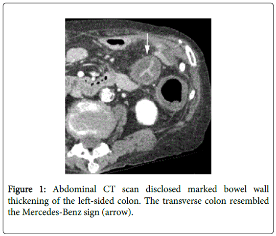
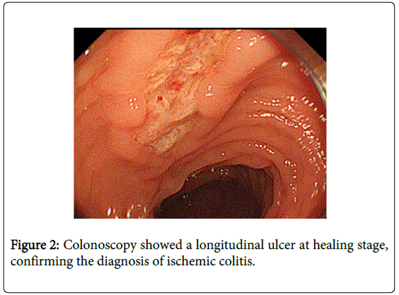
A 75 years old woman presented with acute onset of severe abdominal pain. She denied diarrhea or hematochezia. Medical history included polymyositis and interstitial pneumonia. On physical examination, there was tenderness and rebound tenderness of the left abdomen without bowel sounds. Laboratory examination showed that white blood cell count was elevated at 14,300/mm3. Abdominal CT scan disclosed marked bowel wall thickening of the left-sided colon. The transverse colon resembled the Mercedes-Benz sign (Figure 1). IC with peritoneal sign was diagnosed and she was treated conservatively. Stool culture was negative. Colonoscopy on day 8 showed longitudinal ulcers at healing stage in the affected colon (Figure 2), confirming the diagnosis of IC. She gradually improved and discharged on day 9.
IC is caused by hypoperfusion or vasospasm of the splanchnic arteries. Left-sided IC is typically occurred in the elderly. IC appears differently in CT scans relative to the slice axis, including target sign on cross section, and the accordion sign in longitudinal image, which is the CT analog of thumbprinting on radiographs [1]. In target sign, hyperemia in the mucosa, the muscularis propria and serosa appear the high attenuation, whereas, submucosal edema and thickening present the lower attenuation [2]. The Mercedes-Benz sign is originally a triradiate radiolucent shadow, representing gas accumulation within a gallstone [3].
The Mercedes-Benz sign of IC, which we call, is presented by hyperemic mucosa and the triradiate form is produced presumably by tenia coli. In conclusion, this case illustrates the importance of this sign which may allow us to predict the presence of IC.
References
- Thoeni RF, Cello JP (2006) CT imaging of colitis. Radiology 240: 623-638.
- Ahualli J (2005) The target sign: bowel wall. Radiology 234: 549-550.
- Longley JD (2002) The Mercedes-Benz sign. CMAJ 167: 172.
Relevant Topics
- Constipation
- Digestive Enzymes
- Endoscopy
- Epigastric Pain
- Gall Bladder
- Gastric Cancer
- Gastrointestinal Bleeding
- Gastrointestinal Hormones
- Gastrointestinal Infections
- Gastrointestinal Inflammation
- Gastrointestinal Pathology
- Gastrointestinal Pharmacology
- Gastrointestinal Radiology
- Gastrointestinal Surgery
- Gastrointestinal Tuberculosis
- GIST Sarcoma
- Intestinal Blockage
- Pancreas
- Salivary Glands
- Stomach Bloating
- Stomach Cramps
- Stomach Disorders
- Stomach Ulcer
Recommended Journals
Article Tools
Article Usage
- Total views: 18556
- [From(publication date):
June-2015 - Aug 17, 2025] - Breakdown by view type
- HTML page views : 13914
- PDF downloads : 4642


